



GSK854

Based on 1 Customer Validation
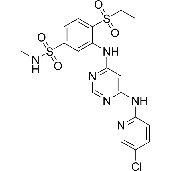
GSK854 is a TNNI3K inhibitor. GSK854 binds to the DFG-out conformation of the ATP-binding site and forms hydrogen bonds with the hinge and gatekeeper. GSK854 reduces mitochondrial superoxide production, ROS, and p38 MAPK activation, and protects mitochondrial membrane potential and cardiac function. GSK854 can be used in research on ischemia/reperfusion cardiac injury, heart failure, acute coronary syndrome, and ischemic heart disease.
For research use only. We do not sell to patients.
- Purity : 98.40%
- CAS No.: 1316059-00-3
- Formula: C18H19ClN6O4S2
- Molecular Weight:482.96
-
Storage:Powder -20°C, 3 years , 4°C, 2 years ; In solvent -80°C, 6 months , -20°C, 1 month

To place orders, for customer services and technical support, please contact: MedChemExpress USA
Tel: 609-228-6898 E-mail: [email protected] [email protected]


-
 Biological Activity
-
Chemical Information
-
Solvent & Solubility
- Protocols
- Purity & Documentation
- References
-
 Help & FAQs


All MAP3K Isoforms
More

Biological Activity
Description
IC50 & Target
[1]|
TNNI3K/HH498 |
In Vitro
GSK854 is a potent inhibitor of TNNI3K with a biochemical IC50 of ≤10 nM[1].
GSK854 demonstrates potent cellular activity against TNNI3K with an IC50 of 8 nM and an AMP of 74 nm/s[1].
GSK854 is a highly potent inhibitor of TNNI3K with an IC50 of ≤10 nM and demonstrates significant selectivity over B-Raf and EGFR[2].
GSK854 rescues ROS elevation in NRVMs but does not act as a non-specific ROS scavenger[3].
MedChemExpress (MCE) has not independently confirmed the accuracy of these methods. They are for reference only. Further protocols information, click here.
In Vivo
GSK854 (2.75 mg/kg; i.p.; at reperfusion and again 6 hours after reperfusion, followed by ad libitum chow for 6 weeks) administered at reperfusion and via chow for 6 weeks preserves cardiac function, limits adverse ventricular remodeling, reduces hypertrophy, and decreases fibrosis in a mouse model of reperfused myocardial infarction[3].
MedChemExpress (MCE) has not independently confirmed the accuracy of these methods. They are for reference only.
-
Animal Model:C57BL/6 (wild-type)[3]
-
Dosage:2.75 mg/kg
-
Administration:i.p.; single dose at reperfusion
-
Result:Significantly reduced infarct size versus vehicle control after I/R with no effect on area at risk.
Blunted superoxide production in the ischemic LV 30 min after reperfusion.
Significantly reduced p38 phosphorylation in the ischemic LV during reperfusion.
-
Animal Model:C57BL/6 (wild-type)[3]
-
Dosage:2.75 mg/kg
-
Administration:i.p.; at reperfusion and again 6 hours after reperfusion, followed by ad libitum chow for 6 weeks
-
Result:Significantly ameliorated the decline in LV ejection fraction (%EF) at 2 weeks after MI/R.
Maintained functional benefit at 4 weeks with higher %EF and smaller LV end-systolic dimension versus vehicle-treated mice.
Protected against adverse ventricular remodeling with significantly smaller LV end-diastolic dimension at 4 weeks.
Markedly decreased plasma pro-ANP levels at 6 weeks.
Slightly attenuated heart weight/body weight ratio increase at 6 weeks.
Significantly smaller cardiomyocyte cross-sectional area at 6 weeks.
Reduced cardiac fibrosis versus vehicle-treated mice at 6 weeks.


Chemical Information
-
CAS No. 1316059-00-3
-
Appearance Solid
-
Molecular Weight 482.96
-
Formula C18H19ClN6O4S2
-
Color Off-white to light yellow
-
SMILES
O=S(C1=CC=C(C(NC2=NC=NC(NC3=NC=C(C=C3)Cl)=C2)=C1)S(=O)(CC)=O)(NC)=O
-
Shipping
Room temperature in continental US; may vary elsewhere.
-
Storage
Powder -20°C 3 years 4°C 2 years In solvent -80°C 6 months -20°C 1 month


Solvent & Solubility
In Vitro:
DMSO : 250 mg/mL (517.64 mM; Need ultrasonic; Hygroscopic DMSO has a significant impact on the solubility of product, please use newly opened DMSO)
Please refer to the solubility information to select the appropriate solvent. Once prepared, please aliquot and store the solution to prevent product inactivation from repeated freeze-thaw cycles.
Storage method and period of stock solution: -80°C, 6 months; -20°C, 1 month. When stored at -80°C, please use it within 6 months. When stored at -20°C, please use it within 1 month.
Please refer to the solubility information to select the appropriate solvent. Once prepared, please aliquot and store the solution to prevent product inactivation from repeated freeze-thaw cycles.
Storage method and period of stock solution: -80°C, 6 months; -20°C, 1 month. When stored at -80°C, please use it within 6 months. When stored at -20°C, please use it within 1 month.
Concentration (start) × Volume (start) = Concentration (final) × Volume (final)
In Vivo:
Select the appropriate dissolution method based on your experimental animal and administration route.
- For the following dissolution methods, please ensure to first prepare a clear stock solution using an In Vitro approach and then sequentially add co-solvents:
- To ensure reliable experimental results, the clarified stock solution can be appropriately stored based on storage conditions. As for the working solution for In Vivo experiments, it is recommended to prepare freshly and use it on the same day.
- The percentages shown for the solvents indicate their volumetric ratio in the final prepared solution. If precipitation or phase separation occurs during preparation, heat and/or sonication can be used to aid dissolution.
Add each solvent one by one: 10% DMSO 40% PEG300 5% Tween-80 45% Saline
Solubility: ≥ 2.08 mg/mL (4.31 mM); Clear solution
This protocol yields a clear solution of ≥ 2.08 mg/mL (saturation unknown).
Taking 1 mL working solution as an example, add 100 μL DMSO stock solution (20.8 mg/mL) to 400 μL PEG300, and mix evenly; then add 50 μL Tween-80 and mix evenly; then add 450 μL Saline to adjust the volume to 1 mL.
Preparation of Saline: Dissolve 0.9 g sodium chloride in ddH₂O and dilute to 100 mL to obtain a clear Saline solution.
In Vivo Dissolution Calculator
Please enter the basic information of animal experiments:
-
-
-
-
Recommended: Prepare an additional quantity of animals to account for potential losses during experiments.
Please enter your animal formula composition:
-
%DMSO +
Recommended: Keep the proportion of DMSO in working solution below 2% if your animal is weak.
-
%+
-
+%Tween-80 + +
-
%Saline +
The co-solvents required include: DMSO, . All of co-solvents are available by MedChemExpress (MCE). , Tween 80. All of co-solvents are available by MedChemExpress (MCE).
Working solution concentration: 0.22 mg/mL
Method for preparing stock solution: mg drug dissolved in μL DMSO. Stock solution concentration: mg/mL.
1. Take μL DMSO stock solution;
2. Add μL .
μL , mix evenly;
3. Then add μL Tween 80, mix evenly;
4. Then add μL
Please ensure that the stock solution in the first step is dissolved to a clear state, and add co-solvents in sequence. You can use ultrasonic heating (ultrasonic cleaner, recommended frequency 20-40 kHz), vortexing, etc. to assist dissolution.

Protocols
-
Mitochondrial membrane-potential fluorescent assay
Mitochondrial membrane potential fluorescent assays estimate ΔΨm in living cells using lipophilic cationic dyes such as TMRM, TMRE, rhodamine 123, and JC-1, which accumulate in mitochondria according to membrane polarization; loss of signal after FCCP or CCCP treatment is interpreted as mitochondrial depolarization. TMRM/TMRE and rhodamine 123 are commonly used for semi-quantitative live-cell microscopy or flow cytometry, while JC-1 can report a shift from red aggregate fluorescence to green monomer fluorescence during depolarization; interpretation requires controls because dye concentration, quenching mode, cell type, dye efflux, and mitochondrial mass can affect fluorescence independently of ΔΨm.
-
ROS/oxidative-stress fluorescent staining
ROS/oxidative-stress fluorescent staining uses cell-permeant fluorogenic probes that become fluorescent after oxidation inside cells or tissues; commonly used examples include DCFH-DA/DCFDA for broad cellular oxidant detection, DHE for superoxide-related signal detection, MitoSOX for mitochondrial superoxide-related signal detection, and CellROX probes for oxidative-stress-associated fluorescence readouts. The assay detects probe oxidation rather than a single ROS species unless the probe and analysis method have been chemically validated for that species. DCFH-DA enters cells, is deacetylated by intracellular esterases to DCFH, and produces fluorescent DCF after oxidation, so the readout is used as an operational measure of total cellular oxidative stress rather than a species-specific ROS measurement. DHE and MitoSOX can report superoxide-related oxidation, but red fluorescence alone can include non-specific ethidium-like oxidation products; HPLC or optimized spectral approaches are
-
Fluorescent plasma-membrane potential dye assay
Fluorescent plasma-membrane potential dye assays measure changes in cell membrane potential using voltage-sensitive dyes whose fluorescence changes when cells depolarize or hyperpolarize. Anionic bis-oxonol dyes such as DiBAC4(3) enter depolarized cells more readily and show increased fluorescence after intracellular binding, while hyperpolarization reduces dye accumulation and fluorescence. FMP/FLIPR membrane-potential dyes are used for faster, homogeneous microplate assays of ion-channel or receptor-mediated membrane-potential changes.
-
Mitochondrial membrane-potential and mitochondrial mass staining
Mitochondrial membrane potential staining measures the electrochemical polarization across the mitochondrial inner membrane in live cells using lipophilic cationic fluorescent probes; early rhodamine-based work showed that selective mitochondrial dye accumulation is lost when the mitochondrial transmembrane potential is dissipated. JC-1 reports mitochondrial polarization by shifting from green monomer fluorescence to red J-aggregate fluorescence as dye concentration increases within energized mitochondria; therefore, the red/green fluorescence ratio is used as a relative readout of mitochondrial membrane potential. TMRE or TMRM staining provides a single-channel relative readout because these cationic rhodamine esters accumulate in polarized mitochondria, and lower fluorescence indicates reduced mitochondrial polarization when acquisition and dye-loading conditions are controlled. Mitochondrial mass staining is commonly performed with MitoTracker Green FM or related MitoTracker dyes as
-
Research Protocol for Cardiovascular Diseases
Cardiovascular disease can be modeled as maladaptive cardiac remodeling, where ischemic injury or pressure overload activates inflammatory signaling, fibroblast activation, extracellular-matrix deposition, cardiomyocyte hypertrophy, vascular remodeling, and progressive ventricular dysfunction. The TGF-β/SMAD axis is a central profibrotic pathway after myocardial injury and pressure overload, while innate immune and cytokine pathways regulate leukocyte recruitment, scar formation, and adverse remodeling. Key unresolved questions include which inflammatory signals are reparative versus harmful, when fibrosis is protective versus maladaptive, and whether pathway inhibition improves function without weakening necessary infarct healing or compensatory remodeling.


Purity & Documentation
-
Data Sheet (293 KB)
-
SDS (251 KB)
- English - EN (251 KB)
- Français - FR (251 KB)
- Deutsch - DE (251 KB)
- Norwegian - NO (251 KB)
- Español - ES (251 KB)
- Swedish - SV (251 KB)
- Italian - IT (251 KB)
- Korean - KR (251 KB)
- Portuguese - PT (251 KB)
-
Handling Instructions (2659 KB)
References
[1]. Patterson JR, et al. Identification of Diarylurea Inhibitors of the Cardiac-Specific Kinase TNNI3K by Designing Selectivity Against VEGFR2, p38α, and B-Raf. Journal of medicinal chemistry. 2021 Nov 11;64(21):15651-15670. [Content Brief]
[2]. Philp J, et al. 4,6-Diaminopyrimidines as Highly Preferred Troponin I-Interacting Kinase (TNNI3K) Inhibitors. Journal of medicinal chemistry. 2018 Apr 12;61(7):3076-3088. [Content Brief]


Complete Stock Solution Preparation Table
Please refer to the solubility information to select the appropriate solvent. Once prepared, please aliquot and store the solution to prevent product inactivation from repeated freeze-thaw cycles.
Storage method and period of stock solution: -80°C, 6 months; -20°C, 1 month. When stored at -80°C, please use it within 6 months. When stored at -20°C, please use it within 1 month.
| Optional Solvent | Concentration Solvent Mass | 1 mg | 5 mg | 10 mg | 25 mg |
|---|---|---|---|---|---|
| DMSO | 1 mM | 2.0706 mL | 10.3528 mL | 20.7056 mL | 51.7641 mL |
| 5 mM | 0.4141 mL | 2.0706 mL | 4.1411 mL | 10.3528 mL | |
| 10 mM | 0.2071 mL | 1.0353 mL | 2.0706 mL | 5.1764 mL | |
| 15 mM | 0.1380 mL | 0.6902 mL | 1.3804 mL | 3.4509 mL | |
| 20 mM | 0.1035 mL | 0.5176 mL | 1.0353 mL | 2.5882 mL | |
| 25 mM | 0.0828 mL | 0.4141 mL | 0.8282 mL | 2.0706 mL | |
| 30 mM | 0.0690 mL | 0.3451 mL | 0.6902 mL | 1.7255 mL | |
| 40 mM | 0.0518 mL | 0.2588 mL | 0.5176 mL | 1.2941 mL | |
| 50 mM | 0.0414 mL | 0.2071 mL | 0.4141 mL | 1.0353 mL | |
| 60 mM | 0.0345 mL | 0.1725 mL | 0.3451 mL | 0.8627 mL | |
| 80 mM | 0.0259 mL | 0.1294 mL | 0.2588 mL | 0.6471 mL | |
| 100 mM | 0.0207 mL | 0.1035 mL | 0.2071 mL | 0.5176 mL |


GSK854 Related Classifications
HY-119608 Related Classifications
 Powered by Bioz