



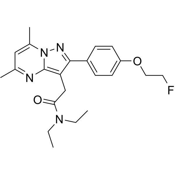
DPA-714
Based on 1 publication(s) in Google Scholar
DPA-714 is a high affinity translocator protein (TSPO) ligand (Ki=7 nM), which is designed with a fluorine atom in its structure, allowing labelling with fluorine -18 and in vivo imaging using positron emission tomography. 18FDPA-714 successfully evaluates for the specific imaging of inflammation in various models of neuroinflammation and in a brain tumor model.
For research use only. We do not sell to patients.
- Purity : 99.82%
- CAS No.: 958233-07-3
- Formula: C22H27FN4O2
- Molecular Weight:398.47
-
Storage:Powder -20°C, 3 years , 4°C, 2 years ; In solvent -80°C, 2 years , -20°C, 1 year

To place orders, for customer services and technical support, please contact: MedChemExpress USA
Tel: 609-228-6898 E-mail: [email protected] [email protected]


-
 Biological Activity
-
Chemical Information
-
Solvent & Solubility
- Protocols
- Purity & Documentation
- References
-
 Help & FAQs


Publications Citing Use of MedChemExpress (MCE) DPA-714
More

Biological Activity
Description
IC50 & Target
Ki: 7 nM (TSPO)[1]
In Vivo
MedChemExpress (MCE) has not independently confirmed the accuracy of these methods. They are for reference only.
Clinical Trial
| NCT Number | Sponsor | Condition | Start Date |
Phase

|
|---|---|---|---|---|
| NCT01329991 | Plexxikon| | 2011-05 | PHASE1 |


Chemical Information
-
CAS No. 958233-07-3
-
Appearance Solid
-
Molecular Weight 398.47
-
Formula C22H27FN4O2
-
Color White to off-white
-
SMILES
O=C(N(CC)CC)CC1=C2N=C(C)C=C(C)N2N=C1C3=CC=C(OCCF)C=C3
-
Shipping
Room temperature in continental US; may vary elsewhere.
-
Storage
Powder -20°C 3 years 4°C 2 years In solvent -80°C 2 years -20°C 1 year


Publications (1)
-
Journal Impact Factor
-
Most Recent
-
J Neuroinflammation
Multi-dimensional decoding of the TSPO signal in animal models of neuroinflammation using fluorescence-conjugated DPA714. [Abstract]2026 Jun 24;23(1):212. PMID: 42343311


Solvent & Solubility
In Vitro:
DMSO : 33.33 mg/mL (83.64 mM; Need ultrasonic; Hygroscopic DMSO has a significant impact on the solubility of product, please use newly opened DMSO)
Please refer to the solubility information to select the appropriate solvent. Once prepared, please aliquot and store the solution to prevent product inactivation from repeated freeze-thaw cycles.
Storage method and period of stock solution: -80°C, 2 years; -20°C, 1 year. When stored at -80°C, please use it within 2 years. When stored at -20°C, please use it within 1 year.
Please refer to the solubility information to select the appropriate solvent. Once prepared, please aliquot and store the solution to prevent product inactivation from repeated freeze-thaw cycles.
Storage method and period of stock solution: -80°C, 2 years; -20°C, 1 year. When stored at -80°C, please use it within 2 years. When stored at -20°C, please use it within 1 year.
Concentration (start) × Volume (start) = Concentration (final) × Volume (final)
In Vivo:
Select the appropriate dissolution method based on your experimental animal and administration route.
- For the following dissolution methods, please ensure to first prepare a clear stock solution using an In Vitro approach and then sequentially add co-solvents:
- To ensure reliable experimental results, the clarified stock solution can be appropriately stored based on storage conditions. As for the working solution for In Vivo experiments, it is recommended to prepare freshly and use it on the same day.
- The percentages shown for the solvents indicate their volumetric ratio in the final prepared solution. If precipitation or phase separation occurs during preparation, heat and/or sonication can be used to aid dissolution.
Add each solvent one by one: 10% DMSO 40% PEG300 5% Tween-80 45% Saline
Solubility: ≥ 2.08 mg/mL (5.22 mM); Clear solution
This protocol yields a clear solution of ≥ 2.08 mg/mL (saturation unknown).
Taking 1 mL working solution as an example, add 100 μL DMSO stock solution (20.8 mg/mL) to 400 μL PEG300, and mix evenly; then add 50 μL Tween-80 and mix evenly; then add 450 μL Saline to adjust the volume to 1 mL.
Preparation of Saline: Dissolve 0.9 g sodium chloride in ddH₂O and dilute to 100 mL to obtain a clear Saline solution.
In Vivo Dissolution Calculator
Please enter the basic information of animal experiments:
-
-
-
-
Recommended: Prepare an additional quantity of animals to account for potential losses during experiments.
Please enter your animal formula composition:
-
%DMSO +
Recommended: Keep the proportion of DMSO in working solution below 2% if your animal is weak.
-
%+
-
+%Tween-80 + +
-
%Saline +
The co-solvents required include: DMSO, . All of co-solvents are available by MedChemExpress (MCE). , Tween 80. All of co-solvents are available by MedChemExpress (MCE).
Working solution concentration: 0.22 mg/mL
Method for preparing stock solution: mg drug dissolved in μL DMSO. Stock solution concentration: mg/mL.
1. Take μL DMSO stock solution;
2. Add μL .
μL , mix evenly;
3. Then add μL Tween 80, mix evenly;
4. Then add μL
Please ensure that the stock solution in the first step is dissolved to a clear state, and add co-solvents in sequence. You can use ultrasonic heating (ultrasonic cleaner, recommended frequency 20-40 kHz), vortexing, etc. to assist dissolution.

Protocols
-
Brain Orthotopic Xenograft
Brain orthotopic xenograft models are generated by stereotactically implanting tumor cells or patient-derived tumor material into the brain of immunodeficient mice so tumor growth occurs within the intracranial microenvironment rather than at a subcutaneous site. The assay detects intracranial tumor engraftment, growth, invasion, treatment response, and survival; readouts are generated by longitudinal bioluminescence imaging, fluorescence imaging, MRI, CT or micro-CT, necropsy, and histologic confirmation of tumor burden and brain invasion.
-
Glioma/Brain Tumor 3D Invasion Assay
The glioma/brain tumor 3D invasion assay measures outward migration and matrix invasion from multicellular tumor spheroids into a 3D extracellular matrix or organotypic brain slice. The readout is generated by time-lapse brightfield, fluorescence, confocal, or high-content imaging and quantified as invasion distance, invasion area, migration index, single-cell velocity, directionality, cumulative sprout length, or Z-direction invasion into brain tissue. Classic in vitro versions embed glioma or GBM spheroids in collagen I, Matrigel, collagen I/Matrigel, or collagen I/Matrigel/hyaluronic acid matrices, while ex vivo versions implant fluorescent GBM spheroids onto organotypic brain slices to model invasion in a preserved brain microenvironment.
-
Bioluminescent/Fluorescent Imaging Xenograft
Bioluminescent and fluorescent imaging xenograft models use tumor cells engineered to express optical reporters so tumor engraftment, growth, dissemination, and treatment response can be monitored longitudinally in living animals and validated ex vivo. Bioluminescence imaging usually measures luciferase activity after substrate administration and is commonly used as a surrogate for viable reporter-expressing tumor burden, while fluorescence imaging measures reporter or probe emission and can support tumor localization, ex vivo confirmation, or complementary multimodal analysis.
-
Research Protocol for Inflammation-related Diseases
The NLRP3 inflammasome is a cytosolic innate immune signaling platform that integrates priming signals and danger-signal activation to promote caspase-1 activation, maturation of IL-1β and IL-18, and gasdermin D-mediated pyroptotic cell death. The core experimental logic is to determine whether inflammatory disease phenotypes are driven by increased NLRP3 expression, ASC-containing inflammasome assembly, caspase-1 cleavage, GSDMD cleavage, and extracellular release of IL-1β/IL-18 rather than by nonspecific cell injury alone. The pathway is strongly linked to inflammation-related disease phenotypes because monosodium urate crystals activate NALP3/NLRP3 inflammasome signaling in gout-like crystal inflammation, cholesterol crystals activate NLRP3 inflammasomes in atherogenesis models, and DSS-induced intestinal inflammation has been reported to involve NLRP3 inflammasome activity. However, experimental colitis studies also show context-dependent protective effects of NLRP3 inflammasome co
-
Research Protocol for Neurological Diseases
PINK1/Parkin-mediated mitophagy pathway is a mitochondrial quality-control signaling axis in which mitochondrial depolarization stabilizes PINK1 on damaged mitochondria, activates Parkin recruitment and E3 ubiquitin ligase activity, promotes ubiquitination of outer mitochondrial membrane proteins, recruits selective autophagy adaptors, and drives lysosomal degradation of damaged mitochondria. In neurological disease research, this pathway is experimentally important because neurons, especially dopaminergic neurons, are highly dependent on mitochondrial integrity, and defective mitochondrial turnover can lead to mitochondrial dysfunction, oxidative stress, impaired neuronal survival, α-synuclein accumulation, and neuroinflammatory damage-associated signals. The genetic disease link is strongest in Parkinson’s disease because mutations in PRKN/parkin cause autosomal recessive juvenile parkinsonism, mutations in PINK1 cause hereditary early-onset Parkinson’s disease, and Drosophila studie


Purity & Documentation
-
Data Sheet (274 KB)
-
SDS (251 KB)
- English - EN (251 KB)
- Français - FR (251 KB)
- Deutsch - DE (251 KB)
- Norwegian - NO (251 KB)
- Español - ES (251 KB)
- Swedish - SV (251 KB)
- Italian - IT (251 KB)
- Korean - KR (251 KB)
- Portuguese - PT (251 KB)
-
Handling Instructions (2659 KB)
References
[1]. James ML, et al. DPA-714, a new translocator protein-specific ligand: synthesis, radiofluorination, and pharmacologic characterization. J Nucl Med. 2008 May;49(5):814-22. [Content Brief]
[2]. Harhausen D, et al. Specific imaging of inflammation with the 18 kDa translocator protein ligand DPA-714 in animal models of epilepsy and stroke. PLoS One. 2013 Aug 2;8(8):e69529. [Content Brief]


Complete Stock Solution Preparation Table
Please refer to the solubility information to select the appropriate solvent. Once prepared, please aliquot and store the solution to prevent product inactivation from repeated freeze-thaw cycles.
Storage method and period of stock solution: -80°C, 2 years; -20°C, 1 year. When stored at -80°C, please use it within 2 years. When stored at -20°C, please use it within 1 year.
| Optional Solvent | Concentration Solvent Mass | 1 mg | 5 mg | 10 mg | 25 mg |
|---|---|---|---|---|---|
| DMSO | 1 mM | 2.5096 mL | 12.5480 mL | 25.0960 mL | 62.7400 mL |
| 5 mM | 0.5019 mL | 2.5096 mL | 5.0192 mL | 12.5480 mL | |
| 10 mM | 0.2510 mL | 1.2548 mL | 2.5096 mL | 6.2740 mL | |
| 15 mM | 0.1673 mL | 0.8365 mL | 1.6731 mL | 4.1827 mL | |
| 20 mM | 0.1255 mL | 0.6274 mL | 1.2548 mL | 3.1370 mL | |
| 25 mM | 0.1004 mL | 0.5019 mL | 1.0038 mL | 2.5096 mL | |
| 30 mM | 0.0837 mL | 0.4183 mL | 0.8365 mL | 2.0913 mL | |
| 40 mM | 0.0627 mL | 0.3137 mL | 0.6274 mL | 1.5685 mL | |
| 50 mM | 0.0502 mL | 0.2510 mL | 0.5019 mL | 1.2548 mL | |
| 60 mM | 0.0418 mL | 0.2091 mL | 0.4183 mL | 1.0457 mL | |
| 80 mM | 0.0314 mL | 0.1568 mL | 0.3137 mL | 0.7842 mL |


DPA-714 Related Classifications
HY-122607 Related Classifications
 Powered by Bioz