



DPC-AJ1951
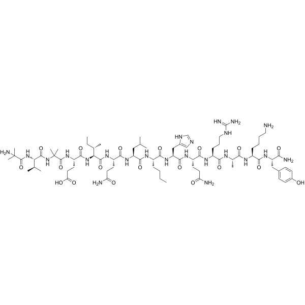
DPC-AJ1951, a 14 amino acid peptide that acts as a potent agonist of the parathyroid hormone (PTH)/PTH-related peptide receptor (PPR). And characterized the activity of DPC-AJ1951 in ex vivo and in vivo assays of bone resorption.
For research use only. We do not sell to patients.
- CAS No.: 943519-33-3
- Formula: C76H127N23O19
- Molecular Weight:1666.96
-
Storage:
Please store the product under the recommended conditions in the Certificate of Analysis.

To place orders, for customer services and technical support, please contact: MedChemExpress USA
Tel: 609-228-6898 E-mail: [email protected] [email protected]


-
 Biological Activity
-
Chemical Information
-
Solvent & Solubility
- Protocols
- Purity & Documentation
- References
-
 Help & FAQs





Biological Activity
Description
Cellular Effect
|
Cell Line

|
Type | Value | Description | References |
|---|---|---|---|---|
| HEK293 | EC50 |
0.15 nM
Compound: 6, DPC-AJ1951
|
Activity at human PTH/PTH-related peptide receptor expressed in HEK293 cells assessed as induction of luciferase activity by CRE-luciferase reporter assay
Activity at human PTH/PTH-related peptide receptor expressed in HEK293 cells assessed as induction of luciferase activity by CRE-luciferase reporter assay
|
[PMID: 17428923] |
| HEK293 | EC50 |
26 nM
Compound: 6, DPC-AJ1951
|
Activity at human PTH/PTH-related peptide receptor expressed in HEK293 cells assessed as induction of intracellular calcium mobilization
Activity at human PTH/PTH-related peptide receptor expressed in HEK293 cells assessed as induction of intracellular calcium mobilization
|
[PMID: 17428923] |
| SAOS-2 | EC50 |
2.2 nM
Compound: 6, DPC-AJ1951
|
Activity at human PTH/PTH-related peptide receptor expressed in SAOS2 cells assessed as cAMP formation
Activity at human PTH/PTH-related peptide receptor expressed in SAOS2 cells assessed as cAMP formation
|
[PMID: 17428923] |
In Vitro


Chemical Information
-
CAS No. 943519-33-3
-
Molecular Weight 1666.96
-
Formula C76H127N23O19
-
Sequence Shortening
{Aib}V{Aib}EIQL{Nle}HQRAKY-NH2
-
Shipping
Room temperature in continental US; may vary elsewhere.
-
Storage
Please store the product under the recommended conditions in the Certificate of Analysis.


Solvent & Solubility
In Vitro
H2O
Peptide Solubility and Storage Guidelines:
1. Calculate the length of the peptide.
2. Calculate the overall charge of the entire peptide according to the following table:
| Contents | Assign value | |
|---|---|---|
| Acidic amino acid | Asp (D), Glu (E), and the C-terminal -COOH. | -1 |
| Basic amino acid | Arg (R), Lys (K), His (H), and the N-terminal -NH2 | +1 |
| Neutral amino acid | Gly (G), Ala (A), Leu (L), Ile (I), Val (V), Cys (C), Met (M), Thr (T), Ser (S), Phe (F), Tyr (Y), Trp (W), Pro (P), Asn (N), Gln (Q) | 0 |
3. Recommended solution:
| Overall charge of peptide | Details |
|---|---|
| Negative (<0) |
1. Try to dissolve the peptide in water first. 2. If water fails, add NH4OH (<50 μL). 3. If the peptide still does not dissolve, add DMSO (50-100 μL) to solubilize the peptide. |
| Positive (>0) |
1. Try to dissolve the peptide in water first. 2. If water fails, try dissolving the peptide in a 10%-30% acetic acid solution. 3. If the peptide still does not dissolve, try dissolving the peptide in a small amount of DMSO. |
| Zero (=0) |
1. Try to dissolve the peptide in organic solvent (acetonitrile, methanol, etc.) first. 2. For very hydrophobic peptides, try dissolving the peptide in a small amount of DMSO, and then dilute the solution with water to the desired concentration. |

Protocols
-
Research Protocol for Endocrine Diseases
Endocrine diseases often arise from disrupted hormone production, hormone signaling, or target-tissue responsiveness; for diabetes-focused endocrine disease models, insulin signaling regulates glucose uptake, hepatic glucose output, lipid metabolism, and β-cell compensation. Type 2 diabetes develops through interacting defects in insulin resistance, β-cell dysfunction, adipose inflammation, hepatic glucose overproduction, altered incretin signaling, and ectopic lipid metabolism. A major unresolved question is whether endocrine dysfunction is driven primarily by target-tissue insulin resistance, intrinsic β-cell failure, immune/inflammatory stress, or combined multi-organ failure that differs by disease stage.
-
Osteoclast differentiation from monocyte/macrophage precursors
Osteoclast differentiation is an in vitro induction assay in which monocyte/macrophage-lineage precursors are exposed to macrophage colony-stimulating factor (M-CSF) and receptor activator of NF-κB ligand (RANKL), generating multinucleated osteoclasts that are commonly identified by tartrate-resistant acid phosphatase (TRAP) staining and functionally confirmed by resorption pits on dentin, bone, or mineralized substrates. M-CSF supports survival and expansion of osteoclast precursors, while RANKL binding to RANK drives osteoclast commitment, fusion, maturation, and resorptive function; osteoprotegerin inhibits this pathway by binding RANKL and preventing RANK activation. The main readouts are the number of TRAP-positive multinucleated cells, formation of F-actin rings, and resorbed surface area; TRAP-positive multinucleated cells indicate osteoclast differentiation, whereas pit formation on dentin, bone, or mineralized coating indicates functional bone-resorbing activity.


Purity & Documentation
References

Calculators
Concentration (start) × Volume (start) = Concentration (final) × Volume (final)


DPC-AJ1951 Related Classifications
HY-P1418 Related Classifications
 Powered by Bioz