



EGFR-IN-1
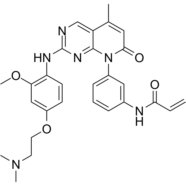
EGFR-IN-1 (compound 24) is an orally active and irreversible L858R/T790M mutant selective EGFR inhibitor. EGFR-IN-1 potently inhibits Gefitinib-resistant EGFR L858R, T790M with 100-fold selectivity over wild-type EGFR. EGFR-IN-1 displays strong antiproliferative activity against the H1975 cells and the first line mutant HCC827 cells. Antitumor activity.
For research use only. We do not sell to patients.
- CAS No.: 1625677-63-5
- Formula: C28H30N6O4
- Molecular Weight:514.58
-
Storage:
Please store the product under the recommended conditions in the Certificate of Analysis.

To place orders, for customer services and technical support, please contact: MedChemExpress USA
Tel: 609-228-6898 E-mail: [email protected] [email protected]


-
 Biological Activity
-
Chemical Information
- Protocols
- Purity & Documentation
- References
-
 Help & FAQs


All EGFR Isoforms
More

Biological Activity
Description
IC50 & Target
[1]|
EGFRL858R/T790M |
Cellular Effect
|
Cell Line

|
Type | Value | Description | References |
|---|---|---|---|---|
| A-431 | IC50 |
1054 nM
Compound: 24
|
Inhibition of wild type EGFR phosphorylation in human A431 cells after 60 mins by mesoscale multiplex assay
Inhibition of wild type EGFR phosphorylation in human A431 cells after 60 mins by mesoscale multiplex assay
|
[PMID: 26396685] |
| A-431 | IC50 |
2400 nM
Compound: 24
|
Cytotoxicity against human A431 cells expressing wild-type EGFR assessed as inhibition of cell proliferation after 72 hrs by CellTiter-Glo assay
Cytotoxicity against human A431 cells expressing wild-type EGFR assessed as inhibition of cell proliferation after 72 hrs by CellTiter-Glo assay
|
[PMID: 26396685] |
| HCC827 | IC50 |
28 nM
Compound: 24
|
Cytotoxicity against human HCC827 cells harboring EGFR 746 to 750 deletion mutant assessed as inhibition of cell proliferation after 72 hrs by CellTiter-Glo assay
Cytotoxicity against human HCC827 cells harboring EGFR 746 to 750 deletion mutant assessed as inhibition of cell proliferation after 72 hrs by CellTiter-Glo assay
|
[PMID: 26396685] |
| NCI-H1975 | IC50 |
4 nM
Compound: 24
|
Cytotoxicity against human NCI-H1975 cells harboring EGFR L858R/T790M double mutant assessed as inhibition of cell proliferation after 72 hrs by CellTiter-Glo assay
Cytotoxicity against human NCI-H1975 cells harboring EGFR L858R/T790M double mutant assessed as inhibition of cell proliferation after 72 hrs by CellTiter-Glo assay
|
[PMID: 26396685] |
| NCI-H1975 | IC50 |
4 nM
Compound: 24
|
Inhibition of EGFR L858R/T790M double mutant phosphorylation in human NCI-H1975 cells after 60 mins by mesoscale multiplex assay
Inhibition of EGFR L858R/T790M double mutant phosphorylation in human NCI-H1975 cells after 60 mins by mesoscale multiplex assay
|
[PMID: 26396685] |
In Vitro
EGFR-IN-1 (10 μM; 72 hours) displays strong antiproliferative activity against the H1975 and HCC827 cells with IC50s of 4 and 28 nM, respectively[1].
EGFR-IN-1 inhibits p-EGFR in H1975 and HCC827 cells with IC50s of 4 and 9 nM, respectively. EGFR-IN-1 highly selective against a panel of 100 kinases[1].
MedChemExpress (MCE) has not independently confirmed the accuracy of these methods. They are for reference only.
-
Cell Line:NSCLC cell lines H1975 (T790M/L858R), HCC827 (∆746-750)
-
Concentration:10 μM
-
Incubation Time:72 hours
-
Result:Inhibited H1975 nonsmall cell lung cancer cell line and the first line mutant HCC827 cell line with IC50s of 4 and 28 nM, respectively.
In Vivo
EGFR-IN-1 evaluates in a time course PD experiment upon oral dosing at 30 mg/kg. EGFR-IN-1 shows a >50% inhibition of phosphorylation of EGFR for >12 h. EGFR-IN-1 reaches maximal concentration of 0.10 μM at 2 h and systemic exposure (AUC0-inf.) is 0.33 μM. h[1].
MedChemExpress (MCE) has not independently confirmed the accuracy of these methods. They are for reference only.
-
Animal Model:Female athymic nude mice (H1975 Tumor Xenograft)[1]
-
Dosage:30 mg/kg
-
Administration:p.o.; daily for 2 weeks
-
Result:Led to significant tumor growth inhibition with no observed loss in body weight.


Chemical Information
-
CAS No. 1625677-63-5
-
Molecular Weight 514.58
-
Formula C28H30N6O4
-
SMILES
COC(C=C(OCCN(C)C)C=C1)=C1NC2=NC=C3C(N(C4=CC=CC(NC(C=C)=O)=C4)C(C=C3C)=O)=N2
-
Shipping
Room temperature in continental US; may vary elsewhere.
-
Storage
Please store the product under the recommended conditions in the Certificate of Analysis.


Purity & Documentation
References

Calculators
Concentration (start) × Volume (start) = Concentration (final) × Volume (final)


EGFR-IN-1 Related Classifications
HY-19617 Related Classifications
 Powered by Bioz