



ER 50891

Based on 1 Customer Validation
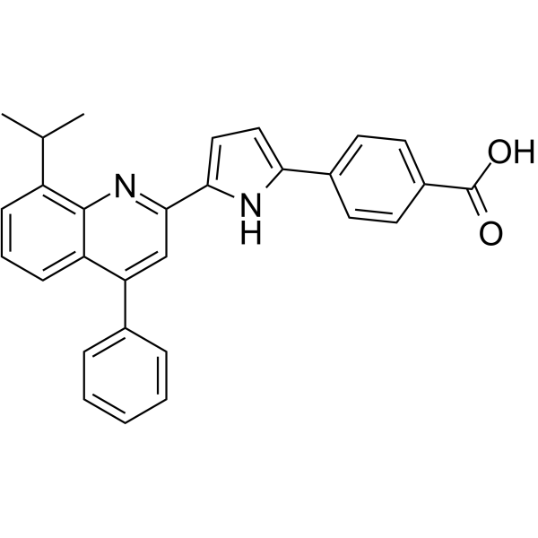
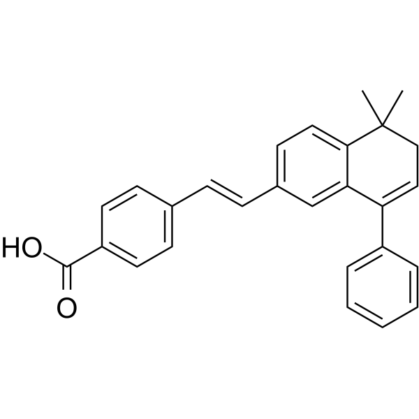
ER 50891 is a RARα antagonist. ER 50891 can significantly antagonize the inhibitory effect of all-trans retinoic acid (ATRA) on the total cell metabolic activity and proliferation of MC3T3-E1 preosteoblasts. ER 50891 rescues ATRA-inhibited osteocalcin (OCN) expression and extracellular matrix mineralization, and suppresses alkaline phosphatase (ALP) activity synergistically. ER 50891 can be used for the study of osteoporosis related to ATRA-induced inhibition of osteoblastogenesis.
For research use only. We do not sell to patients.
- Purity : 99.86%
- CAS No.: 187400-85-7
- Formula: C29H24N2O2
- Molecular Weight:432.51
-
Storage:
-20°C, sealed storage, away from moisture
* In solvent : -80°C, 6 months; -20°C, 1 month (sealed storage, away from moisture)

To place orders, for customer services and technical support, please contact: MedChemExpress USA
Tel: 609-228-6898 E-mail: [email protected] [email protected]


-
 Biological Activity
-
Chemical Information
- Protocols
- Purity & Documentation
- References
-
 Help & FAQs


-
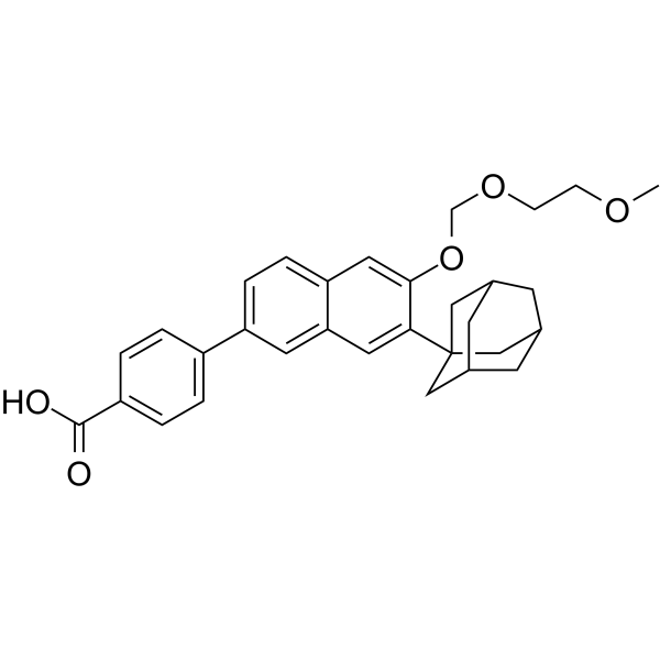
HY-107437
CD2665RAR-beta/gamma Antagonist -
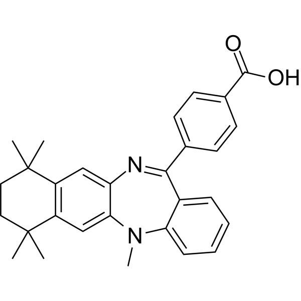
HY-107436
LE135RARβ/RARα Antagonist -
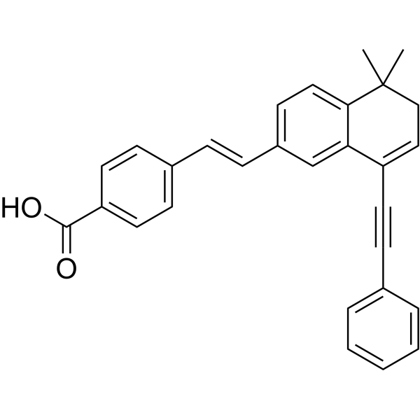
HY-108529
BMS493Inverse RAR Inhibitor -
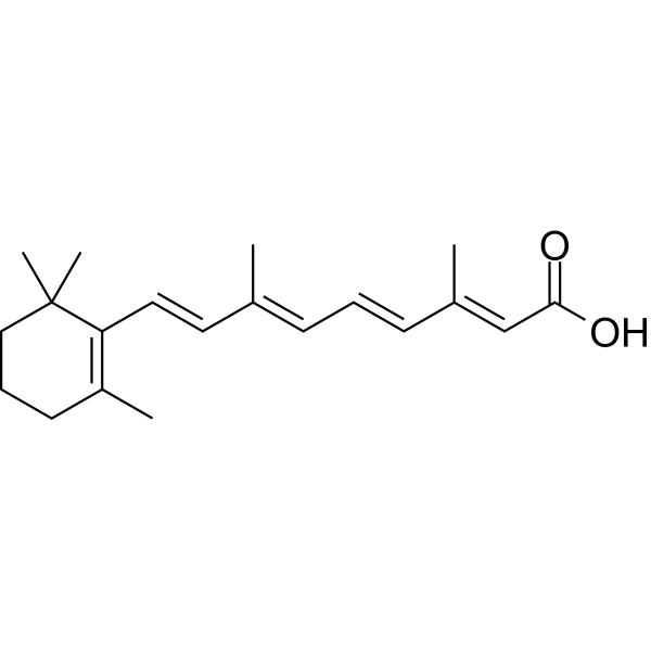
HY-14649
Retinoic acidRAR Agonist -
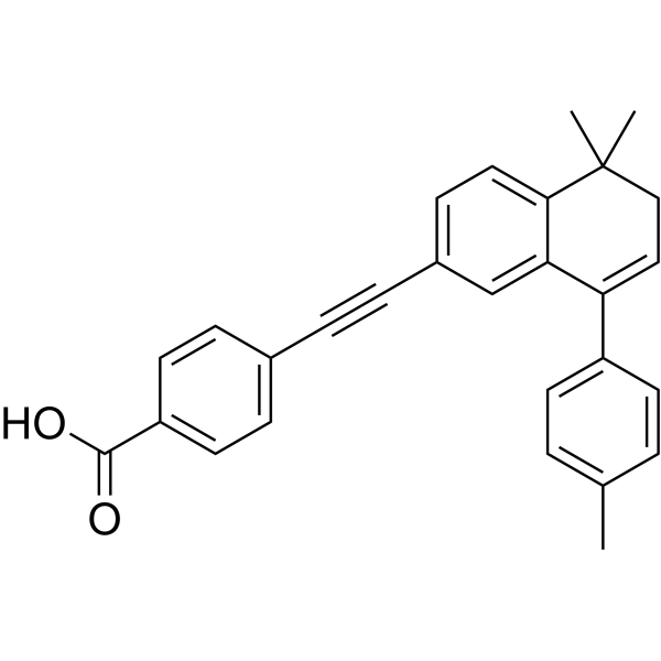
HY-U00449
AGN 193109RAR Antagonist -
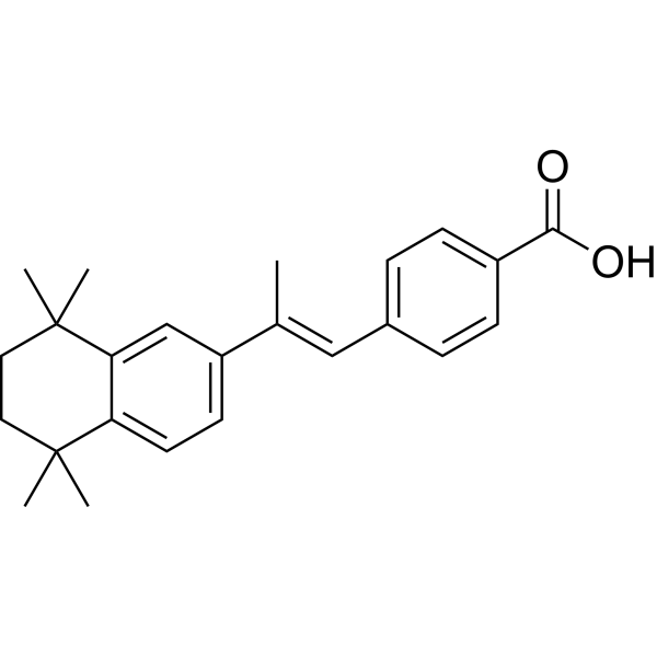
HY-15682
TTNPBApoptosis Inducer -
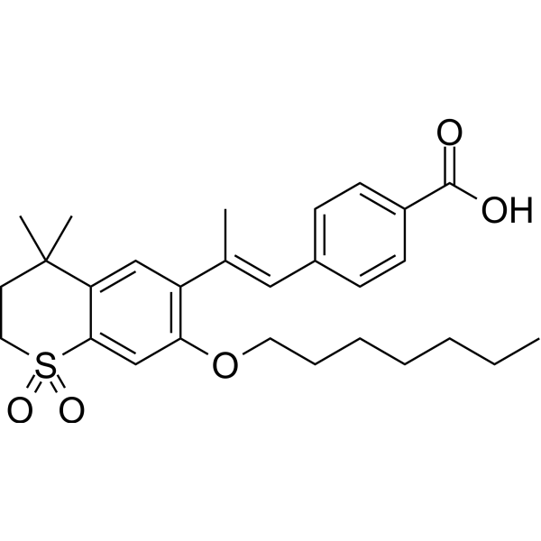
HY-116248
Ro 41-5253RARα Antagonist -
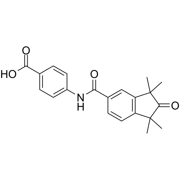
HY-107395
BMS 753RARα Agonist -
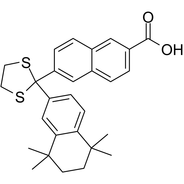
HY-108530
MM11253RARγ Antagonist -
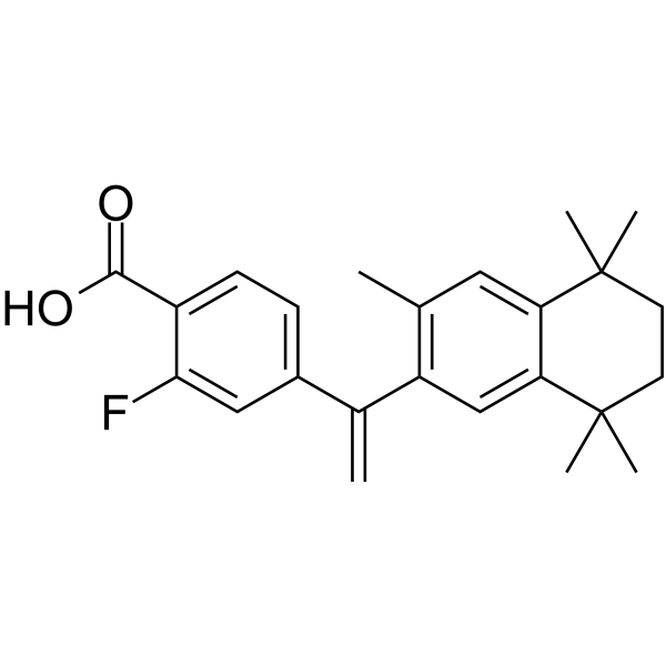
HY-108525
Fluorobexarotene -
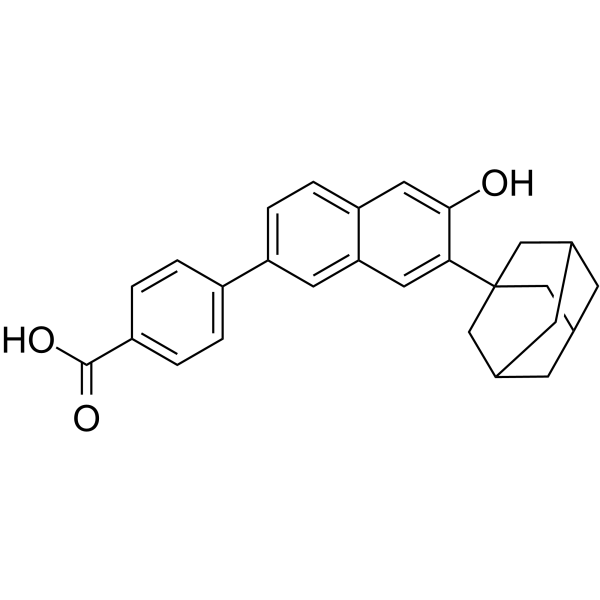
HY-108527
CD1530RARγ Agonist -
HY-100608
BMS453RARβ Agonist


Biological Activity
Description
IC50 & Target
[1]|
RXR α |
Cellular Effect
|
Cell Line

|
Type | Value | Description | References |
|---|---|---|---|---|
| HL-60 | IC50 |
6.4 nM
Compound: 12f
|
Antagonistic activity was evaluated in terms of inhibition of HL-60 cell differentiation activity by ATRA (10 nM)
Antagonistic activity was evaluated in terms of inhibition of HL-60 cell differentiation activity by ATRA (10 nM)
|
[PMID: 11354380] |
In Vitro
ER 50891 (1-3 μM, 3 days) significantly promotes total cell metabolic activity of MC3T3-E1 preosteoblasts alone and antagonizes the inhibitory effect of ATRA on total cell metabolic activity[1].
ER 50891 (0.25-3 μM, 3 days) dose-dependently relieves the inhibition of 1 μM ATRA on OCN expression in MC3T3-E1 preosteoblasts[1].
ER 50891 (1.5-3 μM, 19 days) rescues the extracellular matrix mineralization of MC3T3-E1 preosteoblasts inhibited by ATRA[1].
MedChemExpress (MCE) has not independently confirmed the accuracy of these methods. They are for reference only. Further protocols information, click here.


Chemical Information
-
CAS No. 187400-85-7
-
Appearance Solid
-
Molecular Weight 432.51
-
Formula C29H24N2O2
-
Color Light yellow to yellow
-
SMILES
O=C(O)C1=CC=C(C2=CC=C(C3=NC4=C(C(C)C)C=CC=C4C(C5=CC=CC=C5)=C3)N2)C=C1
-
Shipping
Room temperature in continental US; may vary elsewhere.
-
Storage
-20°C, sealed storage, away from moisture
* In solvent : -80°C, 6 months; -20°C, 1 month (sealed storage, away from moisture)

Protocols
-
Fibrosis/Collagen Morphometry
Fibrosis and collagen morphometry is based on the quantitative visualization of fibrillar collagen deposition in tissue sections using histochemical stains such as Sirius Red (Picrosirius Red) or Masson's trichrome, followed by image-based or polarization-enhanced analysis to estimate collagen proportional area as a surrogate of extracellular matrix accumulation during fibrotic remodeling. Sirius Red combined with polarized light microscopy enhances detection of collagen fibers due to birefringence properties, enabling more specific visualization of collagen type I and III fibrils compared to conventional bright-field histology, while whole-section or region-restricted digital morphometry reduces field-selection bias in fibrosis assessment. Alternative quantitative approaches include second harmonic generation (SHG) and two-photon excited fluorescence microscopy, which enable label-free detection of fibrillar collagen and have been validated against histological staining and biochemica
-
RNA extraction experimental
By lysing cells, releasing RNA, and removing impurities such as proteins and DNA, high-purity RNA products are finally obtained. The commonly used traditional method is the guanidine isothiocyanate/phenol/chloroform method (Trizol), which is suitable for a variety of animal materials including animal tissues, microorganisms, cultured cells, etc., and most plant materials.
-
Collagen: Sirius Red Staining
Sirius Red or picrosirius red staining is a histochemical method for visualizing collagen-rich extracellular matrix in tissue sections, and collagen fibers are detected as red-stained structures under bright-field microscopy with enhanced birefringence under polarized light. Picrosirius red is useful for assessing total collagen organization, distribution, and fibrosis burden, but polarized color should not be interpreted as a definitive collagen type I versus type III readout because color is affected by fiber orientation, thickness, and packing.
-
Mesenchymal stromal/stem cell osteogenic differentiation
Mesenchymal stromal/stem cells can be induced toward an osteoblast-like lineage in vitro by culture in osteogenic medium containing dexamethasone, ascorbic acid or ascorbate-2-phosphate, and β-glycerophosphate; the differentiation process is commonly evaluated by alkaline phosphatase activity, osteogenic marker expression, collagenous matrix formation, and calcium-rich matrix mineralization. The main readouts are alkaline phosphatase activity as an early osteogenic marker and Alizarin Red S staining as a calcium-deposit readout for mineralized extracellular matrix; Alizarin Red S can be inspected microscopically or extracted and measured colorimetrically at 405 nm.
-
ECM-Embedded Organoid (Matrigel/Dome) Culture
ECM-embedded organoid dome culture embeds epithelial stem cells, crypts, organoid fragments, or tumor-derived epithelial cells in a basement-membrane-like hydrogel such as Matrigel, allowing 3D growth, self-organization, lumen formation, budding or cystic morphogenesis, and lineage maintenance under defined niche-factor-containing medium. The primary readouts are organoid establishment efficiency, growth, morphology, passaging capacity, lineage-marker expression, and, when fluorescently labeled lines are used, microscopy- or flow-cytometry-based quantification of population behavior in 3D culture.
-
Matrigel/ECM Transwell Invasion Assay
The Matrigel/ECM Transwell invasion assay measures the ability of cells to move toward a chemoattractant while crossing an extracellular-matrix barrier placed on a porous membrane; therefore, the readout reflects both chemotactic motility and matrix invasion rather than migration alone. Matrigel is a basement-membrane-rich matrix derived from Engelbreth-Holm-Swarm mouse sarcoma and has been used as a reconstituted basement membrane barrier in chemoinvasion assays. The assay readout is generated by quantifying cells that reach the underside of the insert membrane or lower compartment after incubation, commonly by staining and counting invaded cells or by fluorescence-based quantification.
-
MTT Cell Proliferation Assay
The MTT assay is a colorimetric endpoint assay for estimating viable cell number, cell growth, cytotoxicity, or cell activation in cultured mammalian cells. Living cells reduce the yellow tetrazolium salt MTT into purple/blue formazan, while dead cells do not generate the same signal; the resulting color can be quantified with a multiwell spectrophotometer. MTT reduction is commonly interpreted as a readout of metabolic activity that often correlates with viable cell number, but it should not be treated as a direct cell-counting method unless the assay is optimized for the cell type and experimental condition. Studies show that MTT reduction can involve mitochondrial and non-mitochondrial reducing systems, and formazan may accumulate in intracellular lipid droplets rather than simply marking mitochondria.
-
Cell Viability Determination by MTT Colorimetric Assay
The following protocol uses the MTT colorimetric assay as a classic literature-established method for assessing cell viability/metabolic activity in cultured mammalian cells. MTT[3-(4,5-dimethylthiazol-2-yl)-2,5-diphenyltetrazolium bromide] is reduced by metabolically active cells to a colored formazan product; the amount of formazan is quantified spectrophotometrically and provides an indirect measure of metabolically active viable cells. Importantly, MTT reduction reflects cellular oxidoreductase/metabolic activity rather than an absolute direct count of living cells, so changes in cellular metabolism can alter the signal independently of cell number.
-
Matrigel Transwell/Boyden Chamber Invasion Assay
Matrigel Transwell/Boyden chamber invasion assay measures the ability of cells to degrade or traverse an extracellular matrix-coated porous membrane and move from an upper chamber toward a chemoattractant in a lower chamber. Invasion is distinguished from migration by coating the membrane with Matrigel or basement membrane matrix; uncoated inserts measure migration, while coated inserts require cells to cross an ECM barrier before reaching the underside of the membrane.


Purity & Documentation
-
Data Sheet (271 KB)
-
SDS (252 KB)
- English - EN (252 KB)
- Français - FR (252 KB)
- Deutsch - DE (252 KB)
- Norwegian - NO (252 KB)
- Español - ES (252 KB)
- Swedish - SV (252 KB)
- Italian - IT (252 KB)
- Korean - KR (252 KB)
- Portuguese - PT (252 KB)
-
Handling Instructions (2659 KB)
References

Calculators
Concentration (start) × Volume (start) = Concentration (final) × Volume (final)


ER 50891 Related Classifications
HY-108531 Related Classifications
 Powered by Bioz