



Olacaftor

Based on 1 Customer Validation
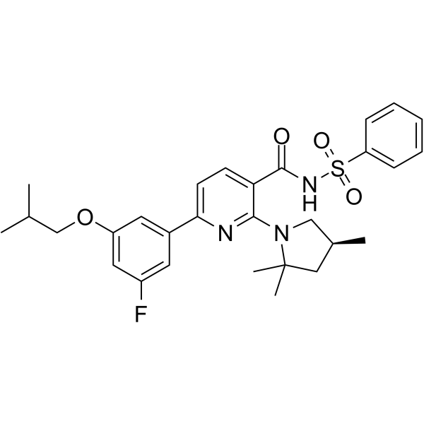
Olacaftor (VX-440) is a cystic fibrosis transmembrane conductance regulator (CFTR) modulator extracted from patent US9782408.
For research use only. We do not sell to patients.
- Purity : 99.60%
- CAS No.: 1897384-89-2
- Formula: C29H34FN3O4S
- Molecular Weight:539.66
-
Storage:Powder -20°C, 3 years ; In solvent -80°C, 6 months , -20°C, 1 month

To place orders, for customer services and technical support, please contact: MedChemExpress USA
Tel: 609-228-6898 E-mail: [email protected] [email protected]


-
 Biological Activity
-
Chemical Information
-
Solvent & Solubility
- Protocols
- Purity & Documentation
- References
-
 Help & FAQs


-
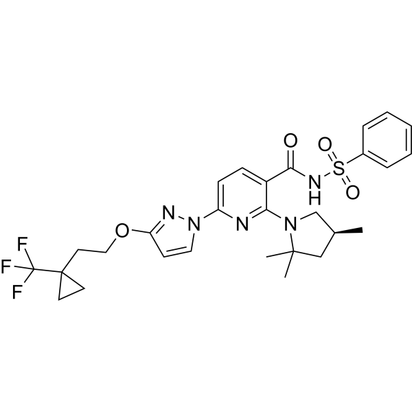
HY-126394
BamocaftorCFTR Corrector -
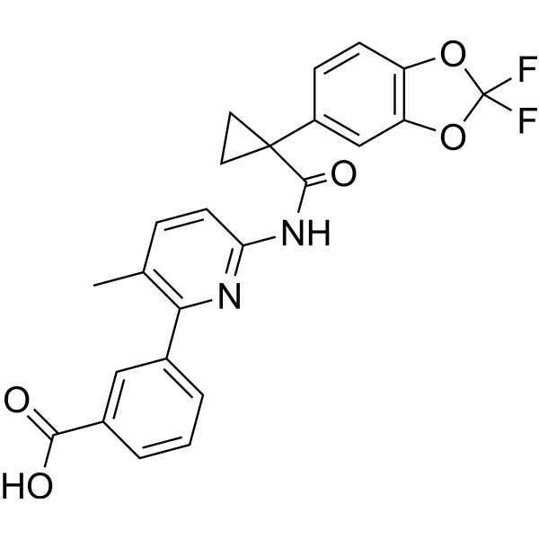
HY-13262
LumacaftorCFTR Modulator -
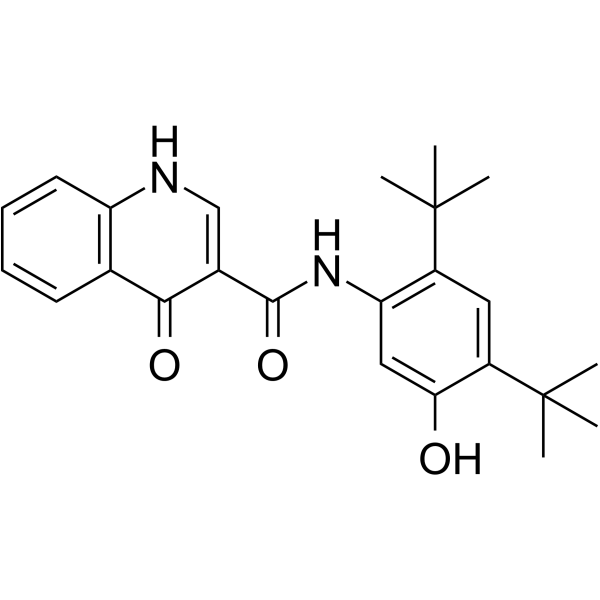
HY-13017
IvacaftorCFTR Potentiator -
HY-16671
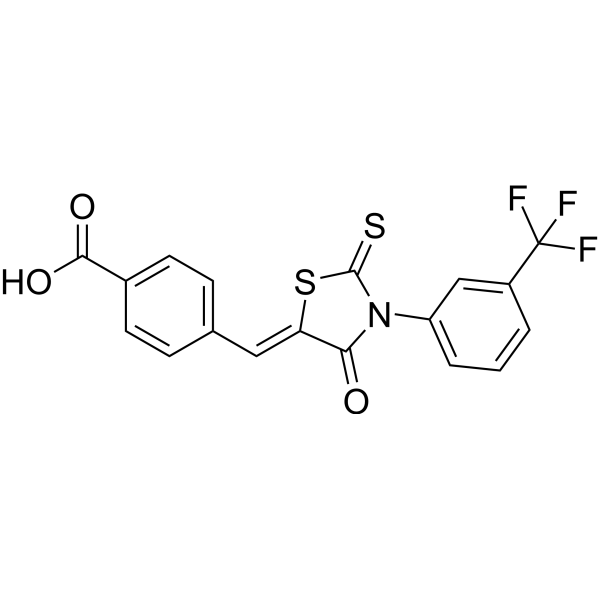
CFTR(inh)-172CFTR Inhibitor -
HY-14832
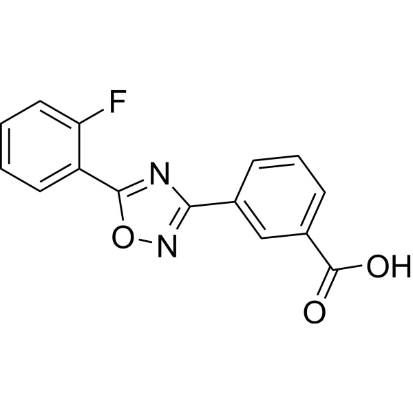
AtalurenCFTR Inhibitor -
HY-15448
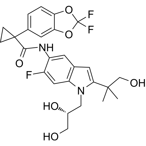
TezacaftorCFTR Corrector -
HY-111772
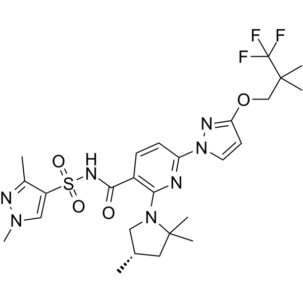
ElexacaftorCFTR Corrector -
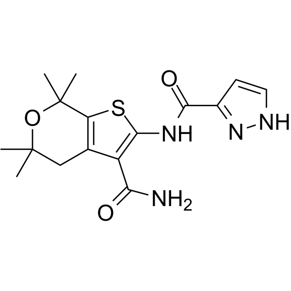
HY-111099
GLPG1837 -
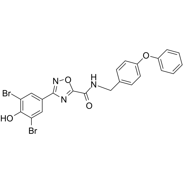
HY-18337
IOWH-032CFTR Inhibitor -
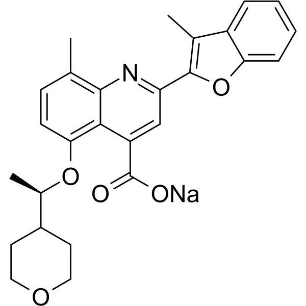
HY-109187A
Posenacaftor sodiumCFTR Modulator -
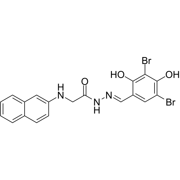
HY-18336
GlyH-101CFTR Inhibitor -
HY-19778A
BPO-27 racemateCFTR Inhibitor


Biological Activity
Description
IC50 & Target
CFTR[1]
Clinical Trial
| NCT Number | Sponsor | Condition | Start Date |
Phase

|
|---|---|---|---|---|
| NCT01329991 | 2011-05 | PHASE1 |


Chemical Information
-
CAS No. 1897384-89-2
-
Appearance Solid
-
Molecular Weight 539.66
-
Formula C29H34FN3O4S
-
Color White to yellow
-
SMILES
O=C(C1=CC=C(C2=CC(OCC(C)C)=CC(F)=C2)N=C1N3C(C)(C)C[C@H](C)C3)NS(=O)(C4=CC=CC=C4)=O
-
Synonyms
VX-440
-
Shipping
Room temperature in continental US; may vary elsewhere.
-
Storage
Powder -20°C 3 years In solvent -80°C 6 months -20°C 1 month


Solvent & Solubility
In Vitro:
DMSO : 50 mg/mL (92.65 mM; ultrasonic and warming and heat to 60°C; Hygroscopic DMSO has a significant impact on the solubility of product, please use newly opened DMSO)
Please refer to the solubility information to select the appropriate solvent. Once prepared, please aliquot and store the solution to prevent product inactivation from repeated freeze-thaw cycles.
Storage method and period of stock solution: -80°C, 6 months; -20°C, 1 month. When stored at -80°C, please use it within 6 months. When stored at -20°C, please use it within 1 month.
Please refer to the solubility information to select the appropriate solvent. Once prepared, please aliquot and store the solution to prevent product inactivation from repeated freeze-thaw cycles.
Storage method and period of stock solution: -80°C, 6 months; -20°C, 1 month. When stored at -80°C, please use it within 6 months. When stored at -20°C, please use it within 1 month.
Concentration (start) × Volume (start) = Concentration (final) × Volume (final)
In Vivo:
Select the appropriate dissolution method based on your experimental animal and administration route.
- For the following dissolution methods, please ensure to first prepare a clear stock solution using an In Vitro approach and then sequentially add co-solvents:
- To ensure reliable experimental results, the clarified stock solution can be appropriately stored based on storage conditions. As for the working solution for In Vivo experiments, it is recommended to prepare freshly and use it on the same day.
- The percentages shown for the solvents indicate their volumetric ratio in the final prepared solution. If precipitation or phase separation occurs during preparation, heat and/or sonication can be used to aid dissolution.
Add each solvent one by one: 10% DMSO 40% PEG300 5% Tween-80 45% Saline
Solubility: ≥ 0.62 mg/mL (1.15 mM); Clear solution
This protocol yields a clear solution of ≥ 0.62 mg/mL (saturation unknown).
Taking 1 mL working solution as an example, add 100 μL DMSO stock solution (6.2 mg/mL) to 400 μL PEG300, and mix evenly; then add 50 μL Tween-80 and mix evenly; then add 450 μL Saline to adjust the volume to 1 mL.
Preparation of Saline: Dissolve 0.9 g sodium chloride in ddH₂O and dilute to 100 mL to obtain a clear Saline solution.
In Vivo Dissolution Calculator
Please enter the basic information of animal experiments:
-
-
-
-
Recommended: Prepare an additional quantity of animals to account for potential losses during experiments.
Please enter your animal formula composition:
-
%DMSO +
Recommended: Keep the proportion of DMSO in working solution below 2% if your animal is weak.
-
%+
-
+%Tween-80 + +
-
%Saline +
The co-solvents required include: DMSO, . All of co-solvents are available by MedChemExpress (MCE). , Tween 80. All of co-solvents are available by MedChemExpress (MCE).
Working solution concentration: 0.22 mg/mL
Method for preparing stock solution: mg drug dissolved in μL DMSO. Stock solution concentration: mg/mL.
1. Take μL DMSO stock solution;
2. Add μL .
μL , mix evenly;
3. Then add μL Tween 80, mix evenly;
4. Then add μL
Please ensure that the stock solution in the first step is dissolved to a clear state, and add co-solvents in sequence. You can use ultrasonic heating (ultrasonic cleaner, recommended frequency 20-40 kHz), vortexing, etc. to assist dissolution.


Purity & Documentation
-
Data Sheet (271 KB)
-
SDS (393 KB)
- English - EN (393 KB)
- Français - FR (393 KB)
- Deutsch - DE (393 KB)
- Norwegian - NO (393 KB)
- Español - ES (393 KB)
- Swedish - SV (393 KB)
- Italian - IT (393 KB)
- Korean - KR (393 KB)
- Portuguese - PT (393 KB)
-
Handling Instructions (2659 KB)
References


Complete Stock Solution Preparation Table
Please refer to the solubility information to select the appropriate solvent. Once prepared, please aliquot and store the solution to prevent product inactivation from repeated freeze-thaw cycles.
Storage method and period of stock solution: -80°C, 6 months; -20°C, 1 month. When stored at -80°C, please use it within 6 months. When stored at -20°C, please use it within 1 month.
| Optional Solvent | Concentration Solvent Mass | 1 mg | 5 mg | 10 mg | 25 mg |
|---|---|---|---|---|---|
| DMSO | 1 mM | 1.8530 mL | 9.2651 mL | 18.5302 mL | 46.3255 mL |
| 5 mM | 0.3706 mL | 1.8530 mL | 3.7060 mL | 9.2651 mL | |
| 10 mM | 0.1853 mL | 0.9265 mL | 1.8530 mL | 4.6325 mL | |
| 15 mM | 0.1235 mL | 0.6177 mL | 1.2353 mL | 3.0884 mL | |
| 20 mM | 0.0927 mL | 0.4633 mL | 0.9265 mL | 2.3163 mL | |
| 25 mM | 0.0741 mL | 0.3706 mL | 0.7412 mL | 1.8530 mL | |
| 30 mM | 0.0618 mL | 0.3088 mL | 0.6177 mL | 1.5442 mL | |
| 40 mM | 0.0463 mL | 0.2316 mL | 0.4633 mL | 1.1581 mL | |
| 50 mM | 0.0371 mL | 0.1853 mL | 0.3706 mL | 0.9265 mL | |
| 60 mM | 0.0309 mL | 0.1544 mL | 0.3088 mL | 0.7721 mL | |
| 80 mM | 0.0232 mL | 0.1158 mL | 0.2316 mL | 0.5791 mL |


Olacaftor Related Classifications
HY-112267 Related Classifications
 Powered by Bioz