



Colchiceine

Based on 1 Customer Validation
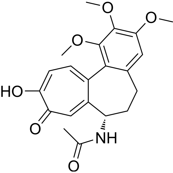
Colchiceine is one of several metabolites of the anti-gout medication Colchicine (HY-16569). Colchicine is a tubulin inhibitor and a microtubule disrupting agent, and may protect rats from developing liver injury and fibrosis.
For research use only. We do not sell to patients.
- CAS No.: 477-27-0
- Formula: C21H23NO6
- Molecular Weight:385.41
-
Storage:
4°C, stored under nitrogen
* In solvent : -80°C, 6 months; -20°C, 1 month (stored under nitrogen)

To place orders, for customer services and technical support, please contact: MedChemExpress USA
Tel: 609-228-6898 E-mail: [email protected] [email protected]


-
 Biological Activity
-
Chemical Information
- Protocols
- Purity & Documentation
- References
-
 Help & FAQs




Biological Activity
Description
Cellular Effect
|
Cell Line

|
Type | Value | Description | References |
|---|---|---|---|---|
| A549 | IC50 |
0.002 μg/mL
Compound: 1
|
In vitro cytotoxic activity against human lung carcinoma (A549) tumor cell line
In vitro cytotoxic activity against human lung carcinoma (A549) tumor cell line
|
[PMID: 15582446] |


Chemical Information
-
CAS No. 477-27-0
-
Appearance Solid
-
Molecular Weight 385.41
-
Formula C21H23NO6
-
Color Off-white to light yellow
-
SMILES
CC(N[C@H](C1=C2)CCC3=CC(OC)=C(OC)C(OC)=C3C1=CC=C(O)C2=O)=O
-
Synonyms
O10-Demethylcolchicine
-
Structure Classification
-
Initial Source
-
Shipping
Room temperature in continental US; may vary elsewhere.
-
Storage
4°C, stored under nitrogen
* In solvent : -80°C, 6 months; -20°C, 1 month (stored under nitrogen)


Purity & Documentation
-
Data Sheet (276 KB)
-
SDS (477 KB)
- English - EN (477 KB)
- Français - FR (477 KB)
- Deutsch - DE (477 KB)
- Norwegian - NO (477 KB)
- Español - ES (477 KB)
- Swedish - SV (477 KB)
- Italian - IT (477 KB)
- Korean - KR (477 KB)
- Portuguese - PT (477 KB)
-
Handling Instructions (2659 KB)
References
[1]. Rodríguez L, et al. Effects of colchicine and colchiceine in a biochemical model of liver injury and fibrosis. Arch Med Res. 1998;29(2):109-116. [Content Brief]
[2]. Nava-Ocampo AA, et al. Effect of colchiceine and ursodeoxycholic acid on hepatocyte and erythrocyte membranes and liver histology in experimentally induced carbon tetrachloride cirrhosis in rats. Eur J Clin Invest. 1997;27(1):77-84. [Content Brief]
[3]. Edström A, et al. Inhibition of fast axonal transport and microtubule polymerization in vitro by colchicine and colchiceine. Acta Physiol Scand. 1979;107(3):233-237. [Content Brief]

Calculators
Concentration (start) × Volume (start) = Concentration (final) × Volume (final)


Colchiceine Related Classifications
HY-127166 Related Classifications
 Powered by Bioz