



Ferroptosis-IN-1
Based on 1 publication(s) in Google Scholar
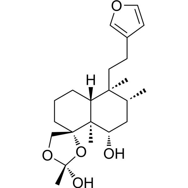
Ferroptosis-IN-1 is a A. campylantha diterpenes. Ferroptosis-IN-1 inhibits ferroptosis with an EC50 value of 10 μM. Ferroptosis-IN-1 can be used for neuroinflammation diseases research.
연구목적의 판매만을 진행합니다. 환자를 대상으로 한 판매는 하지 않습니다.
- 화학식: C22H34O5
- 분자량:378.50
-
보관:
Please store the product under the recommended conditions in the Certificate of Analysis.

To place orders, for customer services and technical support, please contact: MedChemExpress USA
Tel: 609-228-6898 E-mail: [email protected] [email protected]


-
 Biological Activity
-
Chemical Information
- Protocol
- 순도&문서
- References
-
 Help & FAQs


Publications Citing Use of MedChemExpress (MCE) Ferroptosis-IN-1
More

Biological Activity
제품 설명
Cellular Effect
|
Cell Line

|
Type | Value | Description | References |
|---|---|---|---|---|
| HT-22 | EC50 |
10 μM
Compound: 7
|
Inhibition of RSL3-induced ferroptosis in mouse HT-22 cells assessed as increase in cell viability incubated for 24 hrs by MTT assay
Inhibition of RSL3-induced ferroptosis in mouse HT-22 cells assessed as increase in cell viability incubated for 24 hrs by MTT assay
|
[PMID: 37566645] |


Chemical Information
-
분자량 378.50
-
화학식 C22H34O5
-
SMILES
C[C@@]1([C@@H](C[C@@H]([C@]2([C@@]3(CCC[C@@]21[H])O[C@@](C)(OC3)O)C)O)C)CCC4=COC=C4
-
Structure Classification
-
Initial Source
-
선적
Room temperature in continental US; may vary elsewhere.
-
보관
Please store the product under the recommended conditions in the Certificate of Analysis.


Publications (1)
-
Journal Impact Factor
-
Most Recent
-
Eur J Med Chem
Novel β-carboline derivatives show promise as dual-target inhibitors of DNA and TOP2A for the treatment of triple negative breast cancer. [Abstract]2025 Nov 15:298:118041. PMID: 40779814

Protocol
-
Ferroptosis Solutions
Ferroptosis is an iron-dependent, non-apoptotic form of regulated cell death characterized by lethal lipid peroxidation and sensitivity to suppression by iron chelators or lipophilic radical-trapping antioxidants. The core pathway links cystine uptake through system Xc−, glutathione availability, GPX4-dependent detoxification of phospholipid hydroperoxides, iron-dependent oxidative reactions, and polyunsaturated-phospholipid metabolism into a cell-death program that is biochemically and morphologically distinct from apoptosis, necrosis, and autophagy. The ferroptosis pathway is experimentally linked to phenotype through chemical and genetic perturbation. Erastin induces ferroptosis by inhibiting cystine uptake through system Xc− and weakening antioxidant defenses, while GPX4 inhibition or depletion causes lipid peroxide accumulation and ferroptotic cancer-cell death. ACSL4 and oxidizable arachidonoyl- or adrenoyl-containing phosphatidylethanolamines shape ferroptosis sensitivity by con
-
Research Protocol for Neurological Diseases
PINK1/Parkin-mediated mitophagy pathway is a mitochondrial quality-control signaling axis in which mitochondrial depolarization stabilizes PINK1 on damaged mitochondria, activates Parkin recruitment and E3 ubiquitin ligase activity, promotes ubiquitination of outer mitochondrial membrane proteins, recruits selective autophagy adaptors, and drives lysosomal degradation of damaged mitochondria. In neurological disease research, this pathway is experimentally important because neurons, especially dopaminergic neurons, are highly dependent on mitochondrial integrity, and defective mitochondrial turnover can lead to mitochondrial dysfunction, oxidative stress, impaired neuronal survival, α-synuclein accumulation, and neuroinflammatory damage-associated signals. The genetic disease link is strongest in Parkinson’s disease because mutations in PRKN/parkin cause autosomal recessive juvenile parkinsonism, mutations in PINK1 cause hereditary early-onset Parkinson’s disease, and Drosophila studie


순도&문서
References

Calculators
Concentration (start) × Volume (start) = Concentration (final) × Volume (final)


Ferroptosis-IN-1 Related Classifications
HY-N11885 Related Classifications
 Powered by Bioz