



Galloflavin potassium
Based on 19 publication(s) in Google Scholar
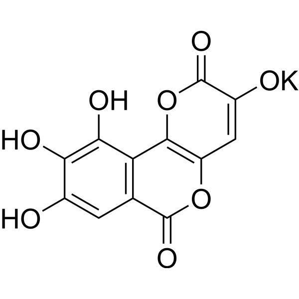
Galloflavin potassium is a novel lactate dehydrogenase inhibitor that can be used for the research of cancer. Galloflavin inhibits aerobic glycolysis in PLC/PRF/5 cells and triggers cell death via apoptosis.
For research use only. We do not sell to patients.
- CAS No.: 1780260-20-9
- Formula: C12H5KO8
- Molecular Weight:316.26
-
Storage:
Please store the product under the recommended conditions in the Certificate of Analysis.

To place orders, for customer services and technical support, please contact: MedChemExpress USA
Tel: 609-228-6898 E-mail: [email protected] [email protected]


-
 Biological Activity
-
Chemical Information
- Protocols
- Purity & Documentation
- References
-
 Help & FAQs


Publications Citing Use of MedChemExpress (MCE) Galloflavin potassium
More- Cell Res. 2023 Sep;33(9):679-698. [Abstract]
- Drug Resist Updat. 2024 Mar:73:101060. [Abstract]
- Redox Biol. 2024 May:71:103112. [Abstract]
- Mol Cell. 2024 Feb 15;84(4):744-759.e6. [Abstract]
- J Immunother Cancer. 2025 Dec 3;13(12):e013733. [Abstract]
- Cell Commun Signal. 2025 Feb 19;23(1):99. [Abstract]
- Cell Rep. 2024 Dec 12;43(12):115065. [Abstract]
- Sci Signal. 2026 Mar 3;19(927):eadz6443. [Abstract]
- J Dent. 2026 Jun:169:106664. [Abstract]
- Commun Biol. 2025 Nov 26;8(1):1761. [Abstract]
- Commun Biol. 2025 Jul 8;8(1):1020. [Abstract]
- Int Immunopharmacol. 2025 Mar 26:150:114265. [Abstract]
- J Immunol. 2025 Nov 6:vkaf218. [Abstract]
- Toxicol Appl Pharmacol. 2025 Nov:504:117508. [Abstract]
- Mol Genet Genomics. 2026 Aug 7;301(1):166.
- bioRxiv. 2026 Jul 10.
- SSRN. 2026 Apr 11.
- SSRN. 2025 Dec 2.
- bioRxiv. 2025 Jul 26:2025.07.22.666143. [Abstract]
 Customer Validation & Images

-
Bio/Physico-chemical Assay
-
Cell Proliferation/Viability Assay
-
Apoptosis Analysis
-
Cell Imaging/Staining
-
IP


Biological Activity
Description


Chemical Information
-
CAS No. 1780260-20-9
-
Molecular Weight 316.26
-
Formula C12H5KO8
-
SMILES
O=C1OC2=C(C=C1O[K])OC(C3=C2C(O)=C(O)C(O)=C3)=O
-
Shipping
Room temperature in continental US; may vary elsewhere.
-
Storage
Please store the product under the recommended conditions in the Certificate of Analysis.


Publications (19)
-
Journal Impact Factor
-
Most Recent
-
Cell Res
α-myosin heavy chain lactylation maintains sarcomeric structure and function and alleviates the development of heart failure. [Abstract]2023 Sep;33(9):679-698. PMID: 37443257
Galloflavin potassium purchased from MedChemExpress. Usage Cited in: Cell Res. 2023 Sep;33(9):679-698. [Abstract]
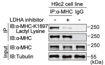
H9c2 cells stimulated with or without Galloflavin (LDHA inhibitor; 10 μM; 24 h) were lysed and immunoprecipitated using anti-α-MHC antibody or control IgG, followed by detection of αMHC K1897 Lactyl Lysine.
-
Drug Resist Updat
Polyphenol nanocomplex modulates lactate metabolic reprogramming and elicits immune responses to enhance cancer therapeutic effect. [Abstract]2024 Mar:73:101060. PMID: 38309140
Galloflavin potassium purchased from MedChemExpress. Usage Cited in: Drug Resist Updat. 2024 Mar:73:101060. [Abstract]
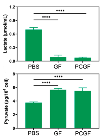
Inhibition of extracellular lactate secretion after different treatments (top). The concentration of pyruvate after various treatments (bottom). PBS, Galloflavin (GF;8 μM), and PCGF (containing GF 8 μM) were used to coculture with 4T1 cells. After incubation for 48 h, the tumor cell supernatant was collected for lactate and pyruvate measurement.
Galloflavin potassium purchased from MedChemExpress. Usage Cited in: Drug Resist Updat. 2024 Mar:73:101060. [Abstract]
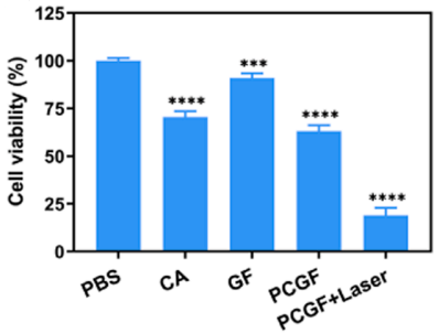
Cell viability after various treatments. The MTT assay was conducted after various treatments (PBS, CA 10 µM, Galloflavin (GF) 8 µM, PCGF (equivalent to CA 10 µM, GF 8 µM), and PCGF+Laser) for 48 h in 4T1 cells.
Galloflavin potassium purchased from MedChemExpress. Usage Cited in: Drug Resist Updat. 2024 Mar:73:101060. [Abstract]
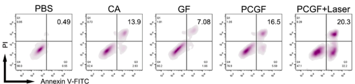
Characteristic flow cytometry profiles depicting 4T1 cell apoptosis post different treatment interventions. PBS, CA 10 µM, Galloflavin (GF) 8 µM, PCGF (equivalent to CA 10 µM, GF 8 µM), and PCGF+Laser) for 24 h.
Galloflavin potassium purchased from MedChemExpress. Usage Cited in: Drug Resist Updat. 2024 Mar:73:101060. [Abstract]
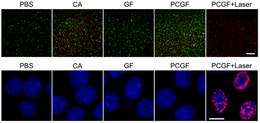
Results of the live/dead co-staining assay after different treatments (top). Levels of DNA double-strand breaks in 4T1 cells following treatments with PBS, CA, Galloflavin (GF), PCGF, and PCGF+Laser (bottom). PBS, CA 10 µM, Galloflavin (GF) 8 µM, PCGF (equivalent to CA 10 µM, GF 8 µM), and PCGF+Laser) for 24 h.
-
Redox Biol
Enteric coronavirus PDCoV evokes a non-Warburg effect by hijacking pyruvic acid as a metabolic hub. [Abstract]2024 May:71:103112. PMID: 38461791 -
Mol Cell
Serine synthesis sustains macrophage IL-1β production via NAD+-dependent protein acetylation. [Abstract]2024 Feb 15;84(4):744-759.e6. PMID: 38266638 -
J Immunother Cancer
2025 Dec 3;13(12):e013733. PMID: 41339107 -
Cell Commun Signal
Intrinsic STING of CD8 + T cells regulates self-metabolic reprogramming and memory to exert anti-tumor effects. [Abstract]2025 Feb 19;23(1):99. PMID: 39972350 -
Cell Rep
Mitochondrial transplantation rescues Ca2+ homeostasis imbalance and myocardial hypertrophy in SLC25A3-related hypertrophic cardiomyopathy. [Abstract]2024 Dec 12;43(12):115065. PMID: 39671292 -
Sci Signal
Glucose metabolism sustains aberrant STAT3 signaling in colorectal cancer through glycosylated local signaling factors. [Abstract]2026 Mar 3;19(927):eadz6443. PMID: 41774818 -
J Dent
2026 Jun:169:106664. PMID: 41921651 -
Commun Biol
Nicotinamide mononucleotide treatment improves spermatogenesis in obese mice by reducing lysine acetylation of lactate dehydrogenase C. [Abstract]2025 Nov 26;8(1):1761. PMID: 41298813 -
Commun Biol
CAF-derived GLUT1 and its role in modulating ovarian cancer progression: a multi-dimensional analysis of the tumor microenvironment. [Abstract]2025 Jul 8;8(1):1020. PMID: 40629043 -
Int Immunopharmacol
Galloflavin mitigates acute kidney injury by suppressing LDHA-dependent macrophage glycolysis. [Abstract]2025 Mar 26:150:114265. PMID: 39955920 -
J Immunol
The involvement of CLec-HTM/IgR-Syk-BCL10-mTOR/ERK signaling pathway in immune priming of oysters. [Abstract]2025 Nov 6:vkaf218. PMID: 41206721 -
Toxicol Appl Pharmacol
Myocardial ischemia/reperfusion-induced glycolysis enhances damage through TRPM7 histone lactylation. [Abstract]2025 Nov:504:117508. PMID: 40796076 -
-
-
-
-
bioRxiv
Glucose Metabolism Sustains Aberrant STAT3 Signaling in Colorectal Cancer via Glycosylated Paracrine Factors. [Abstract]2025 Jul 26:2025.07.22.666143. PMID: 40777356

Protocols
-
Apoptosis
Apoptosis, also called programmed cell death, is generally characterized by distinct morphological characteristics.
-
TUNEL staining for apoptotic DNA fragmentation
TUNEL staining detects DNA strand breaks by using terminal deoxynucleotidyl transferase to add labeled nucleotides to exposed 3′-OH DNA termini, generating either microscopic staining in fixed cells or tissue sections, or fluorescence/cytometric signal in cell suspensions. TUNEL positivity reflects DNA fragmentation but should not be interpreted alone as definitive apoptosis, because TUNEL can also label necrotic, autolytic, mechanically damaged, or DNA-repair-associated DNA breaks.
-
Cell Cytotoxicity Assay
Cytotoxicity assays are usually based on the assessment of cell membrane damage, which can also be indirectly detected by measuring cell viability. Detection methods include MTT assay, CKK-8 assay, LDH assay and ATP assay, etc.
-
Mammalian live/dead viability and cytotoxicity staining
Live/dead viability and cytotoxicity staining assays are based on the simultaneous detection of intracellular esterase activity in metabolically active (viable) cells and membrane integrity loss in non-viable cells. In commonly used dual-staining approaches, membrane-permeant fluorogenic substrates are converted by intracellular esterases into fluorescent products in live cells, while impermeant DNA-binding dyes selectively enter cells with compromised plasma membranes and label nucleic acids in dead or dying cells, enabling discrimination between viable and non-viable populations by fluorescence microscopy or flow cytometry.
-
Annexin V plus membrane-impermeant dye apoptosis staining
Annexin V-based apoptosis assays rely on the detection of phosphatidylserine (PS) externalization from the inner leaflet of the plasma membrane to the outer leaflet, an early biochemical hallmark of apoptosis. Fluorescently labeled Annexin V binds PS in a calcium-dependent manner, enabling identification of early apoptotic cells by flow cytometry or fluorescence microscopy. When combined with a membrane-impermeant DNA-binding dye (e. g. , propidium iodide), this approach allows discrimination between viable (Annexin V−/dye−), early apoptotic (Annexin V+/dye−), and late apoptotic or necrotic (Annexin V+/dye+) cell populations by assessing membrane integrity and PS exposure.
-
Apoptosis Solutions
Apoptosis is a regulated, generally non-lytic cell-death pathway that removes unwanted, damaged, infected, or abnormal cells through coordinated morphological changes, caspase activation, DNA fragmentation, and membrane remodeling. The intrinsic apoptosis pathway is controlled mainly by mitochondrial outer membrane permeabilization, BCL-2 family proteins, cytochrome c release, apoptosome formation, caspase-9 activation, and downstream executioner caspase-3/7 activation. The extrinsic apoptosis pathway is initiated by death receptors such as Fas, TNFR, and TRAIL receptors, which recruit adaptor proteins and activate caspase-8 before engaging executioner caspases or mitochondrial amplification through BID cleavage. Apoptosis is linked to many phenotypes, including cancer cell killing, tissue homeostasis, immune regulation, neurodegeneration, infection response, and treatment-induced cytotoxicity; unresolved questions include how apoptosis interacts with necroptosis, pyroptosis, ferroptos


Purity & Documentation
References

Calculators
Concentration (start) × Volume (start) = Concentration (final) × Volume (final)


Galloflavin potassium Related Classifications
HY-110119 Related Classifications
 Powered by Bioz