



EGFR-IN-71
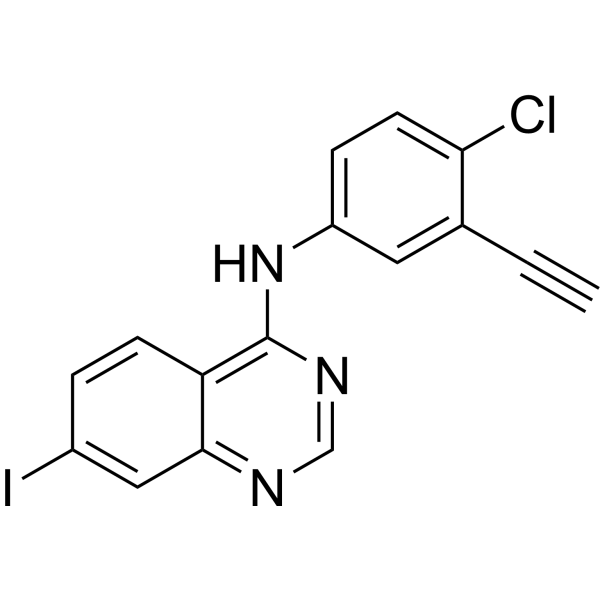
EGFR-IN-71 is a potent narrow spectrum epidermal growth factor receptor (EGFR) inhibitor with IC50 values of 3.7 μM. EGFR-IN-71 can be used for researching chordoma. EGFR-IN-71 is a click chemistry reagent, it contains an Alkyne group and can undergo copper-catalyzed azide-alkyne cycloaddition (CuAAc) with molecules containing Azide groups.
Para uso exclusivo en investigación. No vendemos a pacientes.
- No. CAS: 2676155-98-7
- Fòrmula: C16H9ClIN3
- Peso molecular:405.62
-
Almacenamiento:
Please store the product under the recommended conditions in the Certificate of Analysis.

To place orders, for customer services and technical support, please contact: MedChemExpress USA
Tel: 609-228-6898 E-mail: [email protected] [email protected]


-
 Actividad biológica
-
Chemical Information
- Protocolo
- Pureza y Documentación
- Referencias
-
 Help & FAQs


Ver todos los productos específicos de isoformas EGFR
More

Actividad biológica
Descripciòn
IC50 & Target
IC50: 3.7 μM (EGFR)[1]
In Vitro
EGFR-IN-71 (compound 41) (0-100 μM; 72 h) has inhibitory activity against U-CH1, U-CH2, CH22, UM-Chor1, U-CH12 and U-CH7 chordoma cell lines[1].
MedChemExpress (MCE) has not independently confirmed the accuracy of these methods. They are for reference only. Further protocols information, click here.
-
Cell Line:U-CH1, U-CH2, CH22, UM-Chor1, U-CH12 and U-CH7
-
Concentration:0-100 μM
-
Incubation Time:72 h
-
Result:Exhibited inhibitory activity against U-CH1, U-CH2, CH22, UM-Chor1, U-CH12 and U-CH7 chordoma cell lines with IC50s of 9.1 μM, 16 μM, 0.48 μM, 25 μM, 0.96 μM and 8.0 μM, respectively.


Chemical Information
-
No. CAS 2676155-98-7
-
Peso molecular 405.62
-
Fòrmula C16H9ClIN3
-
SMILES
IC1=CC2=NC=NC(NC3=CC=C(Cl)C(C#C)=C3)=C2C=C1
-
Envío
Room temperature in continental US; may vary elsewhere.
-
Almacenamiento
Please store the product under the recommended conditions in the Certificate of Analysis.

Protocolo
-
EdU Incorporation Assay (Click Chemistry-Based DNA Synthesis Measurement)
The EdU incorporation assay measures DNA synthesis by adding the thymidine analog 5-ethynyl-2′-deoxyuridine to cells or tissues, where it is incorporated into newly synthesized DNA during S phase. Incorporated EdU is detected by copper-catalyzed azide-alkyne cycloaddition, in which a fluorescent azide covalently reacts with the ethynyl group on EdU, allowing S-phase cells to be detected by fluorescence microscopy, flow cytometry, or high-content imaging. EdU detection does not require DNA denaturation or anti-BrdU antibody access, which preserves sample structure and improves compatibility with immunostaining and multiparameter cytometry compared with BrdU-based detection. EdU can be cytotoxic in a cell-type- and exposure-dependent manner, so pulse duration, concentration, and continuous-labeling designs should be validated for each cell type.
-
Cell Cytotoxicity Assay
Cytotoxicity assays are usually based on the assessment of cell membrane damage, which can also be indirectly detected by measuring cell viability. Detection methods include MTT assay, CKK-8 assay, LDH assay and ATP assay, etc.


Pureza y Documentación
Referencias

Calculators
Concentration (start) × Volume (start) = Concentration (final) × Volume (final)


EGFR-IN-71 Related Classifications
HY-150781 Related Classifications
 Powered by Bioz