



MKC-1

Based on 1 Customer Validation
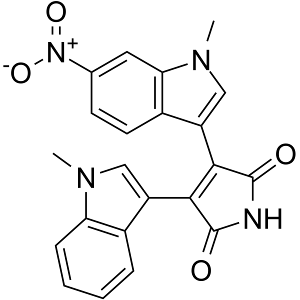
MKC-1 (Ro-31-7453) is an orally active and potent cell cycle inhibitor with broad antitumor activity. MKC-1 inhibits the Akt/mTOR pathway. MKC-1 arrests cellular mitosis and induces cell apoptosis by binding to a number of different cellular proteins including tubulin and members of the importin β family.
Para uso exclusivo en investigación. No vendemos a pacientes.
- Pureza : 99.78%
- No. CAS: 125313-92-0
- Fòrmula: C22H16N4O4
- Peso molecular:400.39
-
Almacenamiento:Powder -20°C, 3 years , 4°C, 2 years ; In solvent -80°C, 6 months , -20°C, 1 month

To place orders, for customer services and technical support, please contact: MedChemExpress USA
Tel: 609-228-6898 E-mail: [email protected] [email protected]


-
 Actividad biológica
-
Chemical Information
-
Solvente y solubilidad
- Protocolo
- Pureza y Documentación
- Referencias
-
 Help & FAQs


-

HY-13691R
MKC-1 (Standard)Akt/mTOR Inhibitor -
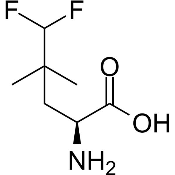
HY-114384
MefluleucinemTORC1 Activator -
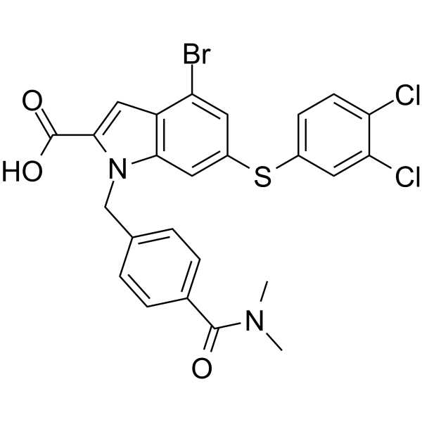
HY-124798
Rheb inhibitor NR1Rheb/mTORC1 Inhibitor -
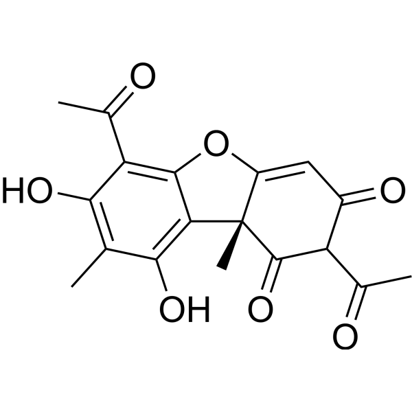
HY-N0656A
(+)-Usnic acidmTORC1/2 Inhibitor -
HY-100026
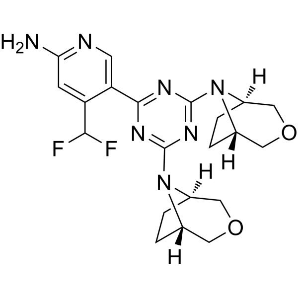
PQR620mTORC1/2 Inhibitor -
HY-10218S
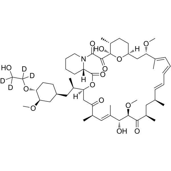
Everolimus-d4Stable Isotope -
HY-137175
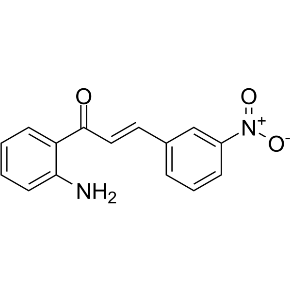
TMBIM6 antagonist-1TMBIM6 Antagonist -
HY-111508
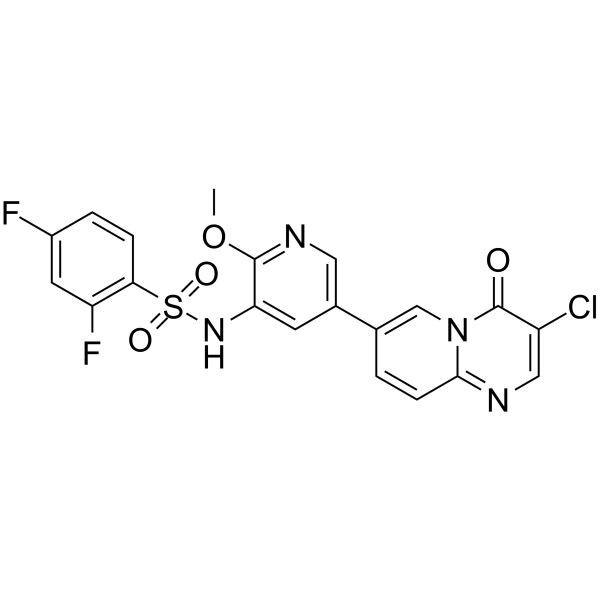
PI3K/mTOR Inhibitor-2 -
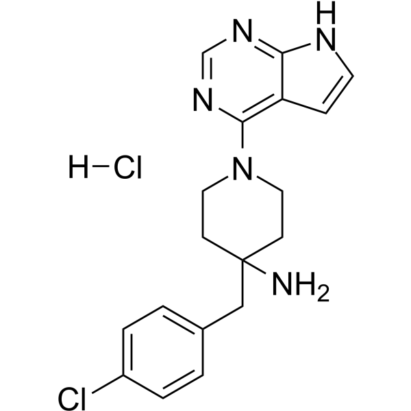
HY-13260A
CCT128930 hydrochlorideAKT Inhibitor -
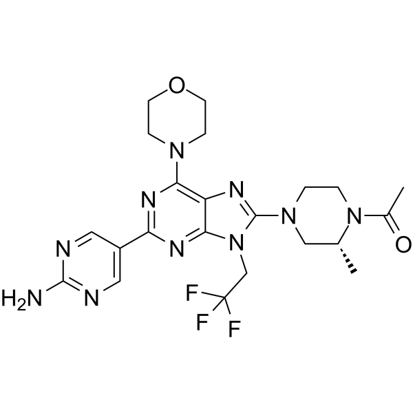
HY-124036
DS-7423PI3K/mTOR Inhibitor -
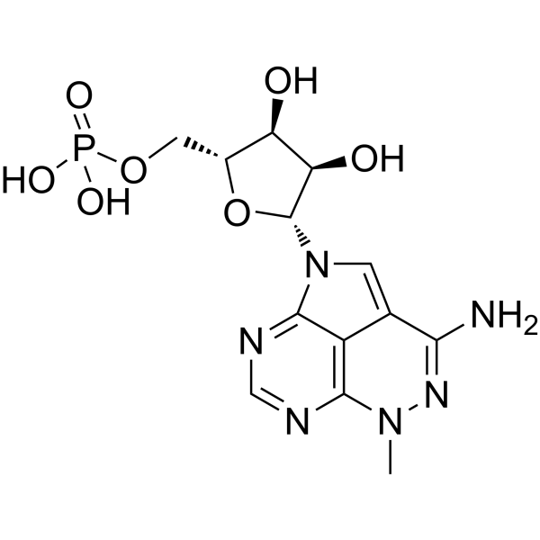
HY-10250
Triciribine phosphate -

HY-N0656AR
(+)-Usnic acid (Standard)mTORC1/2 Inhibitor


Actividad biológica
Descripciòn
In Vivo
Ensayo clínico
| NCT Number | Sponsor | Condition | Start Date |
Phase

|
|---|---|---|---|---|
| NCT01329991 | Plexxikon| | 2011-05 | PHASE1 |


Chemical Information
-
No. CAS 125313-92-0
-
Appearance Solid
-
Peso molecular 400.39
-
Fòrmula C22H16N4O4
-
Color Orange to red
-
SMILES
O=C(C(C1=CN(C)C2=C1C=CC=C2)=C3C4=CN(C)C5=C4C=CC([N+]([O-])=O)=C5)NC3=O
-
Synonyms
Ro-31-7453
-
Envío
Room temperature in continental US; may vary elsewhere.
-
Almacenamiento
Powder -20°C 3 years 4°C 2 years In solvent -80°C 6 months -20°C 1 month


Solvente y solubilidad
In Vitro:
DMSO : ≥ 50 mg/mL (124.88 mM; Hygroscopic DMSO has a significant impact on the solubility of product, please use newly opened DMSO)
* "≥" means soluble, but saturation unknown.
Please refer to the solubility information to select the appropriate solvent. Once prepared, please aliquot and store the solution to prevent product inactivation from repeated freeze-thaw cycles.
Storage method and period of stock solution: -80°C, 6 months; -20°C, 1 month. When stored at -80°C, please use it within 6 months. When stored at -20°C, please use it within 1 month.
Please refer to the solubility information to select the appropriate solvent. Once prepared, please aliquot and store the solution to prevent product inactivation from repeated freeze-thaw cycles.
Storage method and period of stock solution: -80°C, 6 months; -20°C, 1 month. When stored at -80°C, please use it within 6 months. When stored at -20°C, please use it within 1 month.
Concentration (start) × Volume (start) = Concentration (final) × Volume (final)

Protocolo
-
Apoptosis
Apoptosis, also called programmed cell death, is generally characterized by distinct morphological characteristics.
-
TUNEL staining for apoptotic DNA fragmentation
TUNEL staining detects DNA strand breaks by using terminal deoxynucleotidyl transferase to add labeled nucleotides to exposed 3′-OH DNA termini, generating either microscopic staining in fixed cells or tissue sections, or fluorescence/cytometric signal in cell suspensions. TUNEL positivity reflects DNA fragmentation but should not be interpreted alone as definitive apoptosis, because TUNEL can also label necrotic, autolytic, mechanically damaged, or DNA-repair-associated DNA breaks.
-
Flow cytometric DNA-content cell-cycle staining
Flow cytometric DNA-content cell-cycle staining measures the fluorescence intensity of DNA-bound fluorochromes in single cells or nuclei to estimate DNA content distributions, allowing assignment of populations to G0/G1, S, and G2/M phases by DNA histogram deconvolution. Propidium iodide (PI) intercalates into DNA, and PI fluorescence is proportional to cellular DNA content when staining is performed under conditions that make DNA accessible and minimize non-DNA signal. Cells with G2/M DNA content are expected to show approximately twice the fluorescence intensity of G0/G1 cells, while S-phase cells occupy intermediate fluorescence values. PI-based DNA-content analysis can also detect cells with fractional DNA content, often reported as sub-G1, when DNA fragmentation and extraction during staining reduce retained DNA signal in apoptotic cells. DAPI is an alternative DNA fluorochrome for univariate DNA-content analysis, while bivariate approaches combining DNA content with proliferation
-
Annexin V plus membrane-impermeant dye apoptosis staining
Annexin V-based apoptosis assays rely on the detection of phosphatidylserine (PS) externalization from the inner leaflet of the plasma membrane to the outer leaflet, an early biochemical hallmark of apoptosis. Fluorescently labeled Annexin V binds PS in a calcium-dependent manner, enabling identification of early apoptotic cells by flow cytometry or fluorescence microscopy. When combined with a membrane-impermeant DNA-binding dye (e. g. , propidium iodide), this approach allows discrimination between viable (Annexin V−/dye−), early apoptotic (Annexin V+/dye−), and late apoptotic or necrotic (Annexin V+/dye+) cell populations by assessing membrane integrity and PS exposure.
-
BrdU Incorporation Assay
Bromodeoxyuridine (BrdU) incorporation assay is based on the principle that BrdU, a thymidine analog, is incorporated into newly synthesized DNA during the S phase of the cell cycle, thereby serving as a marker of DNA replication and cellular proliferation. Incorporated BrdU can be detected using anti-BrdU antibodies following DNA denaturation, enabling visualization or quantification of proliferating cells through immunochemical detection methods such as immunofluorescence or immunohistochemistry.
-
Apoptosis Solutions
Apoptosis is a regulated, generally non-lytic cell-death pathway that removes unwanted, damaged, infected, or abnormal cells through coordinated morphological changes, caspase activation, DNA fragmentation, and membrane remodeling. The intrinsic apoptosis pathway is controlled mainly by mitochondrial outer membrane permeabilization, BCL-2 family proteins, cytochrome c release, apoptosome formation, caspase-9 activation, and downstream executioner caspase-3/7 activation. The extrinsic apoptosis pathway is initiated by death receptors such as Fas, TNFR, and TRAIL receptors, which recruit adaptor proteins and activate caspase-8 before engaging executioner caspases or mitochondrial amplification through BID cleavage. Apoptosis is linked to many phenotypes, including cancer cell killing, tissue homeostasis, immune regulation, neurodegeneration, infection response, and treatment-induced cytotoxicity; unresolved questions include how apoptosis interacts with necroptosis, pyroptosis, ferroptos
-
Protocol for Cell Cycle
Cell-cycle analysis by flow cytometry measures DNA content in single cells to estimate the fraction of cells in G0/G1, S, and G2/M phases. Propidium iodide intercalates into DNA, and after RNA removal with RNase, fluorescence intensity reflects cellular DNA content: 2N cells are assigned to G0/G1, cells between 2N and 4N to S phase, and 4N cells to G2/M. DNA-content analysis alone cannot reliably separate G0 from G1 or G2 from M. Ki-67 can distinguish quiescent G0 cells from cycling cells, EdU or BrdU incorporation marks active DNA synthesis in S phase, and phospho-histone H3 staining identifies mitotic cells within the 4N population.
-
Subcutaneous Cell-Line-Derived Xenograft
Subcutaneous cell-line-derived xenograft (CDX) models are established by implanting cultured human cancer cell lines into immunodeficient mice, where the injected cells form localized tumors that can be monitored in vivo as a measure of tumorigenic potential, growth kinetics, and treatment response. These models are widely used in oncology research because they allow reproducible tumor formation and enable comparative assessment of tumor growth between different cell lines or genetic manipulations in a controlled in vivo microenvironment. Subcutaneous implantation of cancer cells in immunodeficient mice is a standard approach for evaluating tumor growth behavior and therapeutic response across multiple cancer types, including prostate, esophageal, pancreatic, and colon cancer models.


Pureza y Documentación
-
Ficha de datos (283 KB)
-
SDS (252 KB)
- English - EN (252 KB)
- Français - FR (252 KB)
- Deutsch - DE (252 KB)
- Norwegian - NO (252 KB)
- Español - ES (252 KB)
- Swedish - SV (252 KB)
- Italian - IT (252 KB)
- Korean - KR (252 KB)
- Portuguese - PT (252 KB)
-
Instrucciones de manejo (2659 KB)
Referencias
[1]. Wang M, et al. The first design and synthesis of [11C]MKC-1 ([11C]Ro 31-7453), a new potential PET cancer imaging agent. Nucl Med Biol. 2010 Oct;37(7):763-75. [Content Brief]
[2]. Faris JE, et al. A phase 2 study of oral MKC-1, an inhibitor of importin-β, tubulin, and the mTOR pathway in patients with unresectable or metastatic pancreatic cancer. Invest New Drugs. 2012 Aug;30(4):1614-20. [Content Brief]


Complete Stock Solution Preparation Table
Please refer to the solubility information to select the appropriate solvent. Once prepared, please aliquot and store the solution to prevent product inactivation from repeated freeze-thaw cycles.
Storage method and period of stock solution: -80°C, 6 months; -20°C, 1 month. When stored at -80°C, please use it within 6 months. When stored at -20°C, please use it within 1 month.
| Optional Solvent | Concentration Solvent Mass | 1 mg | 5 mg | 10 mg | 25 mg |
|---|---|---|---|---|---|
| DMSO | 1 mM | 2.4976 mL | 12.4878 mL | 24.9756 mL | 62.4391 mL |
| 5 mM | 0.4995 mL | 2.4976 mL | 4.9951 mL | 12.4878 mL | |
| 10 mM | 0.2498 mL | 1.2488 mL | 2.4976 mL | 6.2439 mL | |
| 15 mM | 0.1665 mL | 0.8325 mL | 1.6650 mL | 4.1626 mL | |
| 20 mM | 0.1249 mL | 0.6244 mL | 1.2488 mL | 3.1220 mL | |
| 25 mM | 0.0999 mL | 0.4995 mL | 0.9990 mL | 2.4976 mL | |
| 30 mM | 0.0833 mL | 0.4163 mL | 0.8325 mL | 2.0813 mL | |
| 40 mM | 0.0624 mL | 0.3122 mL | 0.6244 mL | 1.5610 mL | |
| 50 mM | 0.0500 mL | 0.2498 mL | 0.4995 mL | 1.2488 mL | |
| 60 mM | 0.0416 mL | 0.2081 mL | 0.4163 mL | 1.0407 mL | |
| 80 mM | 0.0312 mL | 0.1561 mL | 0.3122 mL | 0.7805 mL | |
| 100 mM | 0.0250 mL | 0.1249 mL | 0.2498 mL | 0.6244 mL |


MKC-1 Related Classifications
HY-13691 Related Classifications
 Powered by Bioz