



NXPZ-2

Based on 1 Customer Validation
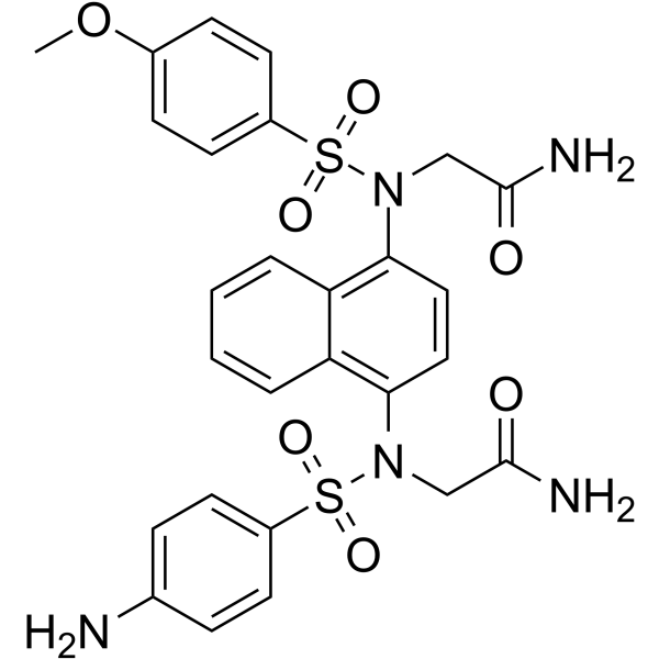
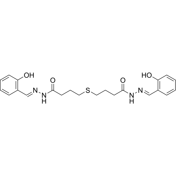
NXPZ-2 is an orally active Keap1-Nrf2 protein–protein interaction (PPI) inhibitor with a Ki value of 95 nM, EC50 value of 120 and 170 nM. NXPZ-2 can dose-dependently ameliorate Aβ[1-42]-Induced cognitive dysfunction, improve brain tissue pathological changes in Alzheimer’s disease (AD) mouse by increasing neuron quantity and function. NXPZ-2 can inhibit oxidative stress by increasing Nrf2 expression levels and promoting its cytoplasm to nuclear translocation, which is helpful for Keap1-Nrf2 PPI inhibitors and AD associated disease research.
Para uso exclusivo en investigación. No vendemos a pacientes.
- Pureza : 98.55%
- No. CAS: 2254492-08-3
- Fòrmula: C27H27N5O7S2
- Peso molecular:597.66
-
Almacenamiento:Powder -20°C, 3 years , 4°C, 2 years ; In solvent -80°C, 6 months , -20°C, 1 month

To place orders, for customer services and technical support, please contact: MedChemExpress USA
Tel: 609-228-6898 E-mail: [email protected] [email protected]


-
 Actividad biológica
-
Chemical Information
-
Solvente y solubilidad
- Protocolo
- Pureza y Documentación
- Referencias
-
 Help & FAQs


-
HY-112675
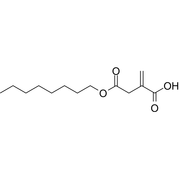
4-Octyl itaconateNrf2 Activator -
HY-110258
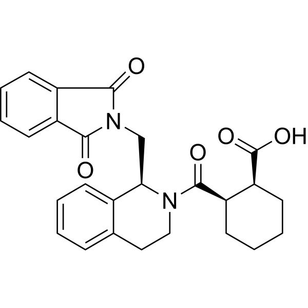
ML334NRF2 Activator -
HY-101140
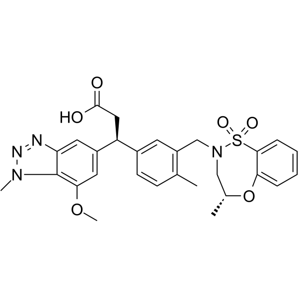
KI696KEAP1/NRF2 Protein-protein Interaction Inhibitor -
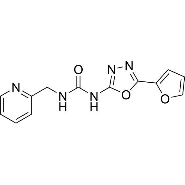
HY-19734
NK-252Nrf2 Activator -
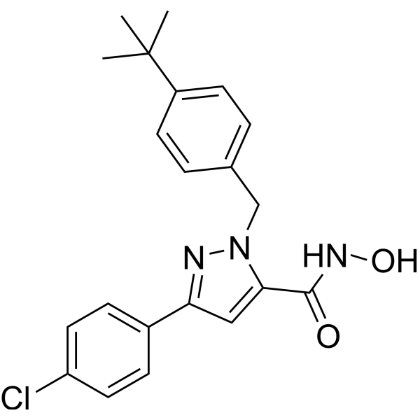
HY-101025
Nrf2-IN-1Nrf2 Inhibitor -
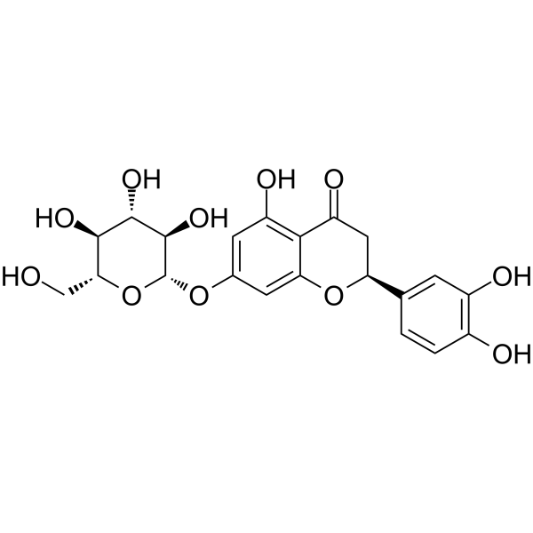
HY-N3847
Eriodictyol-7-O-glucosideNrf2 Activator -
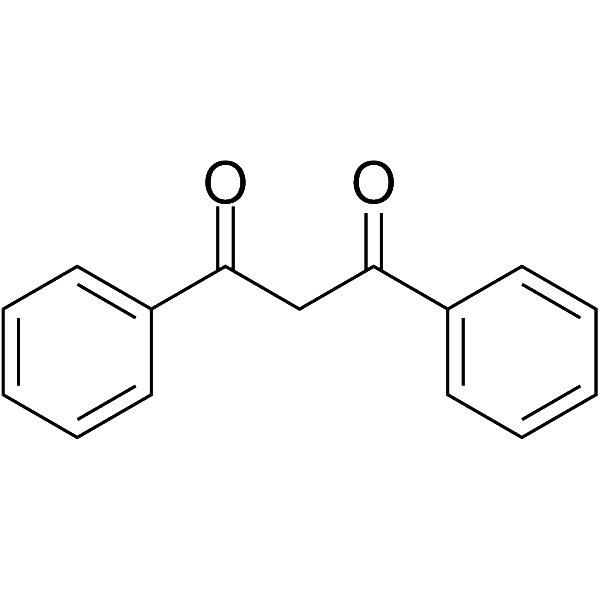
HY-W009731
Dibenzoylmethane -
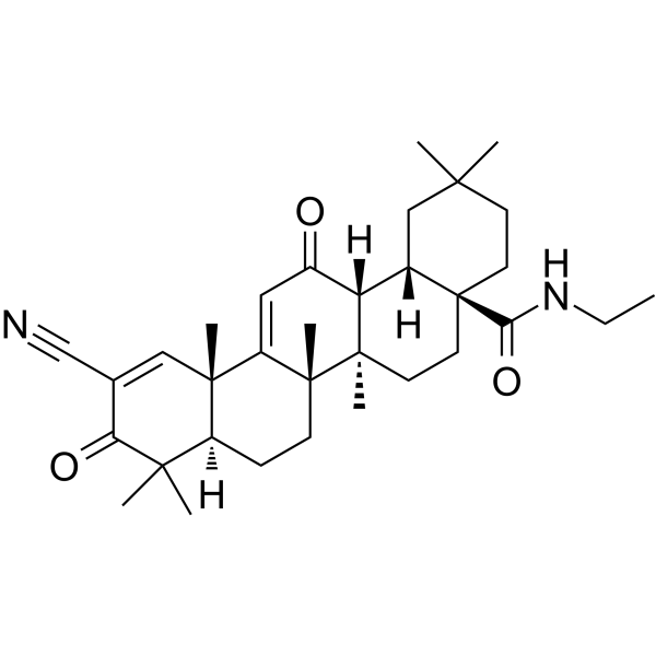
HY-12213
CDDO-EAKeap1-Nrf2 Activator -
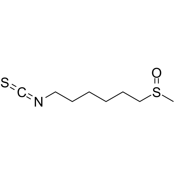
HY-101371
HesperinNrf2 Activator -
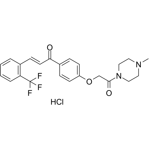
HY-146086
Nrf2 activator-4Nrf2 Activator -
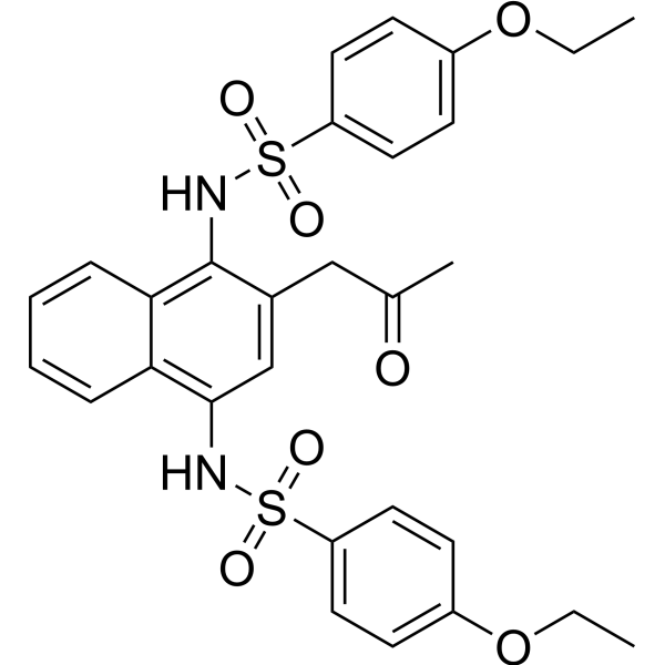
HY-111126
K67Keap1/S349-p-p62 Interaction Inhibitor -
HY-149508
Nrf2-IN-3Nrf2 Inhibitor


Actividad biológica
Descripciòn
IC50 & Target
Ki: 95 nM (Keap 1); EC50: 120 and 170 nM (Keap 1) [1]
Cellular Effect
|
Cell Line

|
Type | Value | Description | References |
|---|---|---|---|---|
| Peritoneal macrophage | CC50 |
>10 μM
Compound: NXPZ-2
|
Cytotoxicity against mouse peritoneal macrophages assessed as reduction in cell viability measured after 24 hrs by CCK8 assay
Cytotoxicity against mouse peritoneal macrophages assessed as reduction in cell viability measured after 24 hrs by CCK8 assay
|
[PMID: 34119834] |
| Peritoneal macrophage | CC50 |
>100 μM
Compound: NXPZ-2
|
Cytotoxicity against mouse peritoneal macrophages assessed as reduction in cell viability measured after 24 hrs by Cell Titer-Blue assay
Cytotoxicity against mouse peritoneal macrophages assessed as reduction in cell viability measured after 24 hrs by Cell Titer-Blue assay
|
[PMID: 35687391] |
| Peritoneal macrophage | CC50 |
>100 μM
Compound: 1; NXPZ-2
|
Cytotoxicity against mouse peritoneal macrophages assessed as reduction in cell viability incubated for 24 hrs by Cell titer-blue assay
Cytotoxicity against mouse peritoneal macrophages assessed as reduction in cell viability incubated for 24 hrs by Cell titer-blue assay
|
[PMID: 37257073] |
| Peritoneal macrophage | EC50 |
1.12 μM
Compound: NXPZ-2
|
Antiinflammatory activity against LPS-induced mouse peritoneal macrophage assessed as suppression of NO level pretreated with LPS followed by compound addition and measured after 24 hrs by Griess reagent based assay
Antiinflammatory activity against LPS-induced mouse peritoneal macrophage assessed as suppression of NO level pretreated with LPS followed by compound addition and measured after 24 hrs by Griess reagent based assay
|
[PMID: 35687391] |
| Peritoneal macrophage | EC50 |
1.4 μM
Compound: NXPZ-2
|
Antiinflammatory activity against LPS-induced mouse peritoneal macrophage assessed as inhibition of TNF alpha level after 4 hrs by ELISA method
Antiinflammatory activity against LPS-induced mouse peritoneal macrophage assessed as inhibition of TNF alpha level after 4 hrs by ELISA method
|
[PMID: 35687391] |
In Vitro
NXPZ-2 (0-200 μM, 7 days) has no obvious toxicity on primary cortical neuron[1].
MedChemExpress (MCE) has not independently confirmed the accuracy of these methods. They are for reference only. Further protocols information, click here.
-
Cell Line:primary cortical neuron
-
Concentration:0, 1.6, 8, 40 and 200 μM
-
Incubation Time:7 days
-
Result:Had no obvious toxicity on primary cortical neuron [1].
In Vivo
MedChemExpress (MCE) has not independently confirmed the accuracy of these methods. They are for reference only.
-
Animal Model:Male ICR mice, AD model[1].
-
Dosage:52.5 mg/kg, 105 mg/kg, 210 mg/kg.
-
Administration:P.o., once daily for 7 days.
-
Result:Showed statistically increased spontaneous alternation and no influence on basal motivation, displayed an increased number of active avoidance times, which improved the learning and memory ability of AD mice. Cell number and morphology in NPZX-2-treated groups were restored, dead neuron numbers of AD mice was lowered.
Increased serum Nrf2 level, displays more Nrf2 in the hippocampal and cortical nucleus and less expression level in the hippocampal and cortical cytoplasm. Increased Nrf2-ARE binding in both hippocampus and frontal cortex, dose-dependently restored SOD, GSH, and MDA levels, and decreased AD marker protein (p-Tau)[1].


Chemical Information
-
No. CAS 2254492-08-3
-
Appearance Solid
-
Peso molecular 597.66
-
Fòrmula C27H27N5O7S2
-
Color White to off-white
-
SMILES
O=C(N)CN(C1=C2C=CC=CC2=C(N(CC(N)=O)S(=O)(C3=CC=C(N)C=C3)=O)C=C1)S(=O)(C4=CC=C(OC)C=C4)=O
-
Envío
Room temperature in continental US; may vary elsewhere.
-
Almacenamiento
Powder -20°C 3 years 4°C 2 years In solvent -80°C 6 months -20°C 1 month


Solvente y solubilidad
In Vitro:
DMSO : 100 mg/mL (167.32 mM; ultrasonic and warming and heat to 60°C; Hygroscopic DMSO has a significant impact on the solubility of product, please use newly opened DMSO)
Please refer to the solubility information to select the appropriate solvent. Once prepared, please aliquot and store the solution to prevent product inactivation from repeated freeze-thaw cycles.
Storage method and period of stock solution: -80°C, 6 months; -20°C, 1 month. When stored at -80°C, please use it within 6 months. When stored at -20°C, please use it within 1 month.
Please refer to the solubility information to select the appropriate solvent. Once prepared, please aliquot and store the solution to prevent product inactivation from repeated freeze-thaw cycles.
Storage method and period of stock solution: -80°C, 6 months; -20°C, 1 month. When stored at -80°C, please use it within 6 months. When stored at -20°C, please use it within 1 month.
Concentration (start) × Volume (start) = Concentration (final) × Volume (final)
In Vivo:
Select the appropriate dissolution method based on your experimental animal and administration route.
- For the following dissolution methods, please ensure to first prepare a clear stock solution using an In Vitro approach and then sequentially add co-solvents:
- To ensure reliable experimental results, the clarified stock solution can be appropriately stored based on storage conditions. As for the working solution for In Vivo experiments, it is recommended to prepare freshly and use it on the same day.
- The percentages shown for the solvents indicate their volumetric ratio in the final prepared solution. If precipitation or phase separation occurs during preparation, heat and/or sonication can be used to aid dissolution.
Add each solvent one by one: 10% DMSO 40% PEG300 5% Tween-80 45% Saline
Solubility: 7.14 mg/mL (11.95 mM); Clear solution; Need ultrasonic
This protocol yields a clear solution of 7.14 mg/mL.
Taking 1 mL working solution as an example, add 100 μL DMSO stock solution (71.4 mg/mL) to 400 μL PEG300, and mix evenly; then add 50 μL Tween-80 and mix evenly; then add 450 μL Saline to adjust the volume to 1 mL.
Preparation of Saline: Dissolve 0.9 g sodium chloride in ddH₂O and dilute to 100 mL to obtain a clear Saline solution.
In Vivo Dissolution Calculator
Please enter the basic information of animal experiments:
-
-
-
-
Recommended: Prepare an additional quantity of animals to account for potential losses during experiments.
Please enter your animal formula composition:
-
%DMSO +
Recommended: Keep the proportion of DMSO in working solution below 2% if your animal is weak.
-
%+
-
+%Tween-80 + +
-
%Saline +
The co-solvents required include: DMSO, . All of co-solvents are available by MedChemExpress (MCE). , Tween 80. All of co-solvents are available by MedChemExpress (MCE).
Working solution concentration: 0.22 mg/mL
Method for preparing stock solution: mg drug dissolved in μL DMSO. Stock solution concentration: mg/mL.
1. Take μL DMSO stock solution;
2. Add μL .
μL , mix evenly;
3. Then add μL Tween 80, mix evenly;
4. Then add μL
Please ensure that the stock solution in the first step is dissolved to a clear state, and add co-solvents in sequence. You can use ultrasonic heating (ultrasonic cleaner, recommended frequency 20-40 kHz), vortexing, etc. to assist dissolution.

Protocolo
-
RNA extraction experimental
By lysing cells, releasing RNA, and removing impurities such as proteins and DNA, high-purity RNA products are finally obtained. The commonly used traditional method is the guanidine isothiocyanate/phenol/chloroform method (Trizol), which is suitable for a variety of animal materials including animal tissues, microorganisms, cultured cells, etc., and most plant materials.
-
Cytoplasmic-Nuclear Fractionated Protein Extraction
Cytoplasmic-nuclear fractionated protein extraction separates soluble cytoplasmic proteins from nuclear-enriched proteins by mild plasma-membrane permeabilization, differential centrifugation, washing of nuclei, and extraction of nuclear proteins for downstream immunoblotting or related molecular analysis. The readout is the relative abundance of a protein in cytoplasmic and nuclear fractions, commonly assessed by western blotting together with compartment markers such as tubulin or pyruvate kinase for cytoplasm and lamin, nucleoporin, hnRNP, H2AX, or Lamin B for nuclear fractions.
-
Pull-down
The pull-down assay is an in vitro technique used to detect physical interactions between two or more proteins and an invaluable tool for confirming a predicted protein-protein interaction or identifying novel interacting partners. This method typically involves the use of affinity purification with various wash and elution steps.
-
Immunoprecipitation
Immunoprecipitation (IP) is an experimental method that uses the principle of antibody specific binding to purify and enrich target proteins.
-
ROS/oxidative-stress fluorescent staining
ROS/oxidative-stress fluorescent staining uses cell-permeant fluorogenic probes that become fluorescent after oxidation inside cells or tissues; commonly used examples include DCFH-DA/DCFDA for broad cellular oxidant detection, DHE for superoxide-related signal detection, MitoSOX for mitochondrial superoxide-related signal detection, and CellROX probes for oxidative-stress-associated fluorescence readouts. The assay detects probe oxidation rather than a single ROS species unless the probe and analysis method have been chemically validated for that species. DCFH-DA enters cells, is deacetylated by intracellular esterases to DCFH, and produces fluorescent DCF after oxidation, so the readout is used as an operational measure of total cellular oxidative stress rather than a species-specific ROS measurement. DHE and MitoSOX can report superoxide-related oxidation, but red fluorescence alone can include non-specific ethidium-like oxidation products; HPLC or optimized spectral approaches are
-
Protocol for Bimolecular Fluorescence Complementation (BiFC) Assay
Bimolecular fluorescence complementation detects protein-protein proximity in living or fixed cells by fusing two candidate interaction partners to nonfluorescent N- and C-terminal fragments of a fluorescent protein; when the partners interact or remain close enough, the fluorescent fragments complement, mature, and generate a fluorescent signal at the site of the protein complex. The BiFC readout is fluorescence intensity and subcellular localization of the reconstituted fluorophore, which reflects formation or stabilization of a protein complex rather than direct biochemical binding kinetics; BiFC is therefore useful for mapping where interactions occur in cancer cells, neurons, macrophages, organoid-derived cells, or drug-screening systems, but results should be validated by independent assays such as co-IP or Western blot. BiFC signal formation is delayed by fluorophore maturation and can stabilize otherwise transient complexes, so it is not a real-time reversible interaction assay
-
Co-Immunoprecipitation
Co-immunoprecipitation technology can verify protein interaction based on the specific immune reaction between antibodies and antigens.
-
Protocol for Yeast Two-Hybrid (Y2H) Assay
The yeast two-hybrid assay detects binary protein-protein interactions by separating a transcription factor into a DNA-binding domain fused to a "bait" protein and a transcriptional activation domain fused to a "prey" protein; if bait and prey interact in yeast, the transcription factor is reconstituted and activates reporter genes such as HIS3, ADE2, lacZ, MEL1, or other selectable/readable reporters. The readout is yeast growth on selective medium and/or reporter activity, which reflects proximity-dependent transcriptional activation in the yeast nucleus rather than direct biochemical binding in the original mammalian, tumor, neuronal, macrophage, or organoid context. Because yeast two-hybrid can generate false positives and false negatives, interaction claims should be validated using independent assays such as co-immunoprecipitation, Western blot, immunofluorescence colocalization, BiFC, pull-down, or mammalian two-hybrid assays.
-
Alzheimer’s Disease Modeling
Alzheimer’s Disease (AD) is a neurodegenerative disorder characterized by a progressive decline in cognitive functions and loss of specific types of neurons and synapses. Alzheimer's symptoms can be simulated in mice by injecting drugs (such as Aβ) or genetically modified.


Pureza y Documentación
-
Ficha de datos (283 KB)
-
SDS (393 KB)
- English - EN (393 KB)
- Français - FR (393 KB)
- Deutsch - DE (393 KB)
- Norwegian - NO (393 KB)
- Español - ES (393 KB)
- Swedish - SV (393 KB)
- Italian - IT (393 KB)
- Korean - KR (393 KB)
- Portuguese - PT (393 KB)
-
Instrucciones de manejo (2659 KB)
Referencias


Complete Stock Solution Preparation Table
Please refer to the solubility information to select the appropriate solvent. Once prepared, please aliquot and store the solution to prevent product inactivation from repeated freeze-thaw cycles.
Storage method and period of stock solution: -80°C, 6 months; -20°C, 1 month. When stored at -80°C, please use it within 6 months. When stored at -20°C, please use it within 1 month.
| Optional Solvent | Concentration Solvent Mass | 1 mg | 5 mg | 10 mg | 25 mg |
|---|---|---|---|---|---|
| DMSO | 1 mM | 1.6732 mL | 8.3660 mL | 16.7319 mL | 41.8298 mL |
| 5 mM | 0.3346 mL | 1.6732 mL | 3.3464 mL | 8.3660 mL | |
| 10 mM | 0.1673 mL | 0.8366 mL | 1.6732 mL | 4.1830 mL | |
| 15 mM | 0.1115 mL | 0.5577 mL | 1.1155 mL | 2.7887 mL | |
| 20 mM | 0.0837 mL | 0.4183 mL | 0.8366 mL | 2.0915 mL | |
| 25 mM | 0.0669 mL | 0.3346 mL | 0.6693 mL | 1.6732 mL | |
| 30 mM | 0.0558 mL | 0.2789 mL | 0.5577 mL | 1.3943 mL | |
| 40 mM | 0.0418 mL | 0.2091 mL | 0.4183 mL | 1.0457 mL | |
| 50 mM | 0.0335 mL | 0.1673 mL | 0.3346 mL | 0.8366 mL | |
| 60 mM | 0.0279 mL | 0.1394 mL | 0.2789 mL | 0.6972 mL | |
| 80 mM | 0.0209 mL | 0.1046 mL | 0.2091 mL | 0.5229 mL | |
| 100 mM | 0.0167 mL | 0.0837 mL | 0.1673 mL | 0.4183 mL |


NXPZ-2 Related Classifications
HY-149010 Related Classifications
 Powered by Bioz