



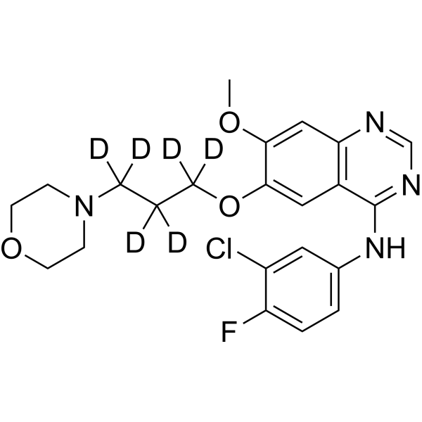
Gefitinib-d6

Based on 1 Customer Validation
Gefitinib-d6 is the deuterium labeled Gefitinib. Gefitinib (ZD1839) is a potent, selective and orally active EGFR tyrosine kinase inhibitor with an IC50 of 33 nM. Gefitinib selectively inhibits EGF-stimulated tumor cell growth (IC50 of 54 nM) and that blocks EGF-stimulated EGFR autophosphorylation in tumor cells. Gefitinib also induces autophagy. Gefitinib has antitumour activity.
For research use only. We do not sell to patients.
- Purity : 98.0%
- CAS No.: 1228664-49-0
- Formula: C22H18D6ClFN4O3
- Molecular Weight:452.94
-
Storage:
-20°C, sealed storage, away from moisture
* In solvent : -80°C, 6 months; -20°C, 1 month (sealed storage, away from moisture)

To place orders, for customer services and technical support, please contact: MedChemExpress USA
Tel: 609-228-6898 E-mail: [email protected] [email protected]


-
 Biological Activity
-
Chemical Information
-
Solvent & Solubility
- Protocols
- Purity & Documentation
- References
-
 Help & FAQs


All EGFR Isoforms
More

Biological Activity
Description
In Vitro
Stable heavy isotopes of hydrogen, carbon, and other elements have been incorporated into drug molecules, largely as tracers for quantitation during the drug development process. Deuteration has gained attention because of its potential to affect the pharmacokinetic and metabolic profiles of drugs[1].
MedChemExpress (MCE) has not independently confirmed the accuracy of these methods. They are for reference only.
- Protocol for Cell Counting and Cell Density Analysis
- Somatic Cell Culture
- Autophagy
- CCK-8/WST-8 Cell Proliferation Assay
- Cell Counting-Based Growth Curve Assay
- Lysosome and acidic-vesicle live-cell staining
- MTT Cell Proliferation Assay
- Macroautophagy Solutions
- Cell Viability Determination by MTT Colorimetric Assay
Application
1. This compound can be used as a tracer
2. This compound can be used as an internal standard for quantitative analysis by NMR, GC-MS, or LC-MS.


Chemical Information
-
CAS No. 1228664-49-0
-
Unlabeled CAS 184475-35-2
-
Appearance Solid
-
Molecular Weight 452.94
-
Formula C22H18D6ClFN4O3
-
Color White to off-white
-
SMILES
COC1=CC2=C(C=C1OC([2H])(C([2H])(C([2H])(N3CCOCC3)[2H])[2H])[2H])C(NC4=CC=C(C(Cl)=C4)F)=NC=N2
-
Synonyms
ZD1839-d6
-
Shipping
Room temperature in continental US; may vary elsewhere.
-
Storage
-20°C, sealed storage, away from moisture
* In solvent : -80°C, 6 months; -20°C, 1 month (sealed storage, away from moisture)


Solvent & Solubility
In Vitro:
DMF : ≥ 20 mg/mL (44.16 mM)
DMSO : ≥ 20 mg/mL (44.16 mM; Hygroscopic DMSO has a significant impact on the solubility of product, please use newly opened DMSO)
Ethanol : ≥ 0.3 mg/mL (0.66 mM)
* "≥" means soluble, but saturation unknown.
Please refer to the solubility information to select the appropriate solvent. Once prepared, please aliquot and store the solution to prevent product inactivation from repeated freeze-thaw cycles.
Storage method and period of stock solution: -80°C, 6 months; -20°C, 1 month (sealed storage, away from moisture). When stored at -80°C, please use it within 6 months. When stored at -20°C, please use it within 1 month.
Please refer to the solubility information to select the appropriate solvent. Once prepared, please aliquot and store the solution to prevent product inactivation from repeated freeze-thaw cycles.
Storage method and period of stock solution: -80°C, 6 months; -20°C, 1 month (sealed storage, away from moisture). When stored at -80°C, please use it within 6 months. When stored at -20°C, please use it within 1 month.
Concentration (start) × Volume (start) = Concentration (final) × Volume (final)


Purity & Documentation
-
Data Sheet (279 KB)
-
SDS (925 KB)
- English - EN (925 KB)
- Français - FR (925 KB)
- Deutsch - DE (925 KB)
- Norwegian - NO (925 KB)
- Español - ES (925 KB)
- Swedish - SV (925 KB)
- Italian - IT (925 KB)
- Korean - KR (925 KB)
- Portuguese - PT (925 KB)
-
Handling Instructions (2659 KB)
References
[1]. Russak EM, et al. Impact of Deuterium Substitution on the Pharmacokinetics of Pharmaceuticals. Ann Pharmacother. 2019;53(2):211-216. [Content Brief]
[2]. Wakeling AE, et al. ZD1839: an orally active inhibitor of epidermal growth factor signaling with potential for cancer therapy. Cancer Res. 2002 Oct 15;62(20):5749-54. [Content Brief]
[3]. Pedersen MW, et al. Differential response to gefitinib of cells expressing normal EGFR and the mutant EGFRvIII. Br J Cancer. 2005 Oct 17;93(8):915-23. [Content Brief]
[4]. Moasser MM, et al. The tyrosine kinase inhibitor ZD1839 inhibits HER2-driven signaling and suppresses the growth of HER2-overexpressing tumor cells. Cancer Res. 2001 Oct 1;61(19):7184-8. [Content Brief]
[5]. Morgillo F, et al. Synergistic effects of 1,1-Dimethylbiguanide treatment in combination with gefitinib, a selective EGFR tyrosine kinase inhibitor, in LKB1 wild-type NSCLC cell lines. Clin Cancer Res. 2013 Jul 1;19(13):3508-19. [Content Brief]
[6]. Miyake K, et al. Epidermal growth factor receptor-tyrosine kinase inhibitor (gefitinib) augments pneumonitis, but attenuates lung fibrosis in response to radiation injury in rats. J Med Invest. 2012;59(1-2):174-85. [Content Brief]
[7]. Noh CK, et al. Simultaneous quantification of volitinib and gefitinib in rat plasma by HPLC-MS/MS for application to a pharmacokinetic study in rats. J Sep Sci. 2017 Jul 27. [Content Brief]
[8]. Dhar D, et al. Liver Cancer Initiation Requires p53 Inhibition by CD44-Enhanced Growth Factor Signaling. Cancer Cell. 2018 Jun 11;33(6):1061-1077.e6. [Content Brief]


Complete Stock Solution Preparation Table
Please refer to the solubility information to select the appropriate solvent. Once prepared, please aliquot and store the solution to prevent product inactivation from repeated freeze-thaw cycles.
Storage method and period of stock solution: -80°C, 6 months; -20°C, 1 month (sealed storage, away from moisture). When stored at -80°C, please use it within 6 months. When stored at -20°C, please use it within 1 month.
| Optional Solvent | Concentration Solvent Mass | 1 mg | 5 mg | 10 mg | 25 mg |
|---|---|---|---|---|---|
| DMF / DMSO | 1 mM | 2.2078 mL | 11.0390 mL | 22.0780 mL | 55.1949 mL |
| 5 mM | 0.4416 mL | 2.2078 mL | 4.4156 mL | 11.0390 mL | |
| 10 mM | 0.2208 mL | 1.1039 mL | 2.2078 mL | 5.5195 mL | |
| 15 mM | 0.1472 mL | 0.7359 mL | 1.4719 mL | 3.6797 mL | |
| 20 mM | 0.1104 mL | 0.5519 mL | 1.1039 mL | 2.7597 mL | |
| 25 mM | 0.0883 mL | 0.4416 mL | 0.8831 mL | 2.2078 mL | |
| 30 mM | 0.0736 mL | 0.3680 mL | 0.7359 mL | 1.8398 mL | |
| 40 mM | 0.0552 mL | 0.2760 mL | 0.5519 mL | 1.3799 mL |


Gefitinib-d6 Related Classifications
HY-50895S1 Related Classifications
 Powered by Bioz