



Glyburide-d3

Based on 1 Customer Validation
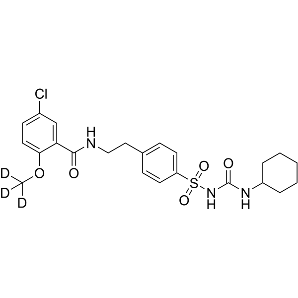
Glyburide-d3 is the deuterium labeled Glibenclamide. Glibenclamide (Glyburide) is an orally active ATP-sensitive K+ channel (KATP) inhibitor and can be used for the research of diabetes and obesity[1]. Glibenclamide inhibits P-glycoprotein. Glibenclamide directly binds and blocks the SUR1 subunits of KATP and inhibits the cystic fibrosis transmembrane conductance regulator protein (CFTR)[3]. Glibenclamide interferes with mitochondrial bioenergetics by inducing changes on membrane ion permeability[4]. Glibenclamide can induce autophagy[5].
For research use only. We do not sell to patients.
- Purity : 99.64%
- CAS No.: 1219803-02-7
- Formula: C23H25D3ClN3O5S
- Molecular Weight:497.02
-
Storage:Powder -20°C, 3 years , 4°C, 2 years ; In solvent -80°C, 6 months , -20°C, 1 month

To place orders, for customer services and technical support, please contact: MedChemExpress USA
Tel: 609-228-6898 E-mail: [email protected] [email protected]


-
 Biological Activity
-
Chemical Information
-
Solvent & Solubility
- Protocols
- Purity & Documentation
- References
-
 Help & FAQs


-
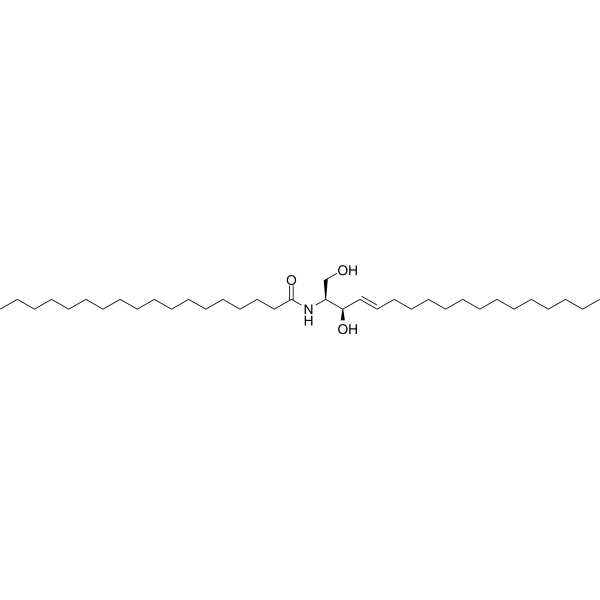
HY-100355
C18-Ceramide (d18:1/18:0)Ceramide -
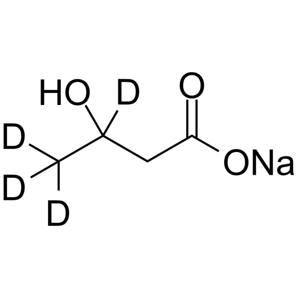
HY-113378S
3-Hydroxybutyric acid-d4 sodiumStable Isotope -
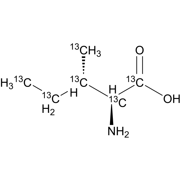
HY-N0771S
L-Isoleucine-13C6Stable Isotope -
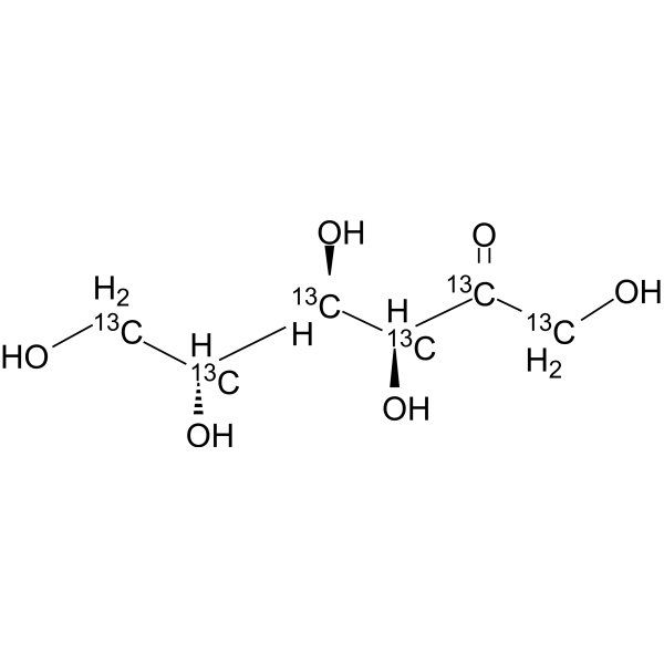
HY-N7092S
D-Fructose-13C6Stable Isotope -
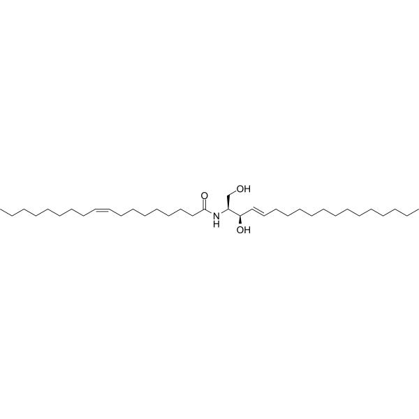
HY-141578
C18:1-CeramideCeramide -
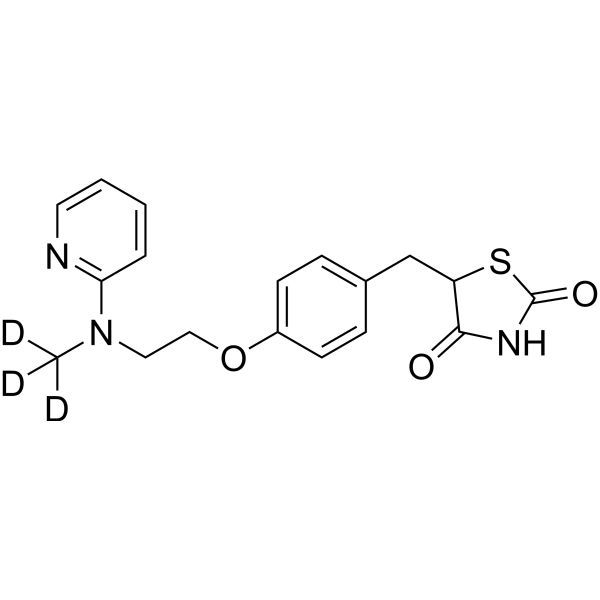
HY-17386S
Rosiglitazone-d3Stable Isotope -
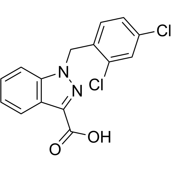
HY-B0486
LonidamineHexokinase Inhibitor -
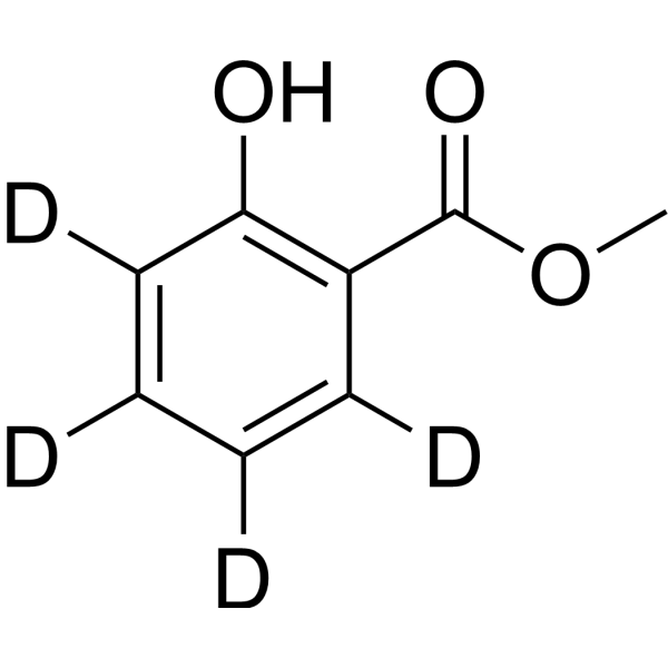
HY-Y0189S
Methyl Salicylate-d4Stable Isotope -
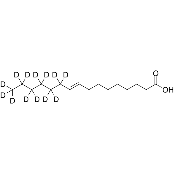
HY-N2341S
Palmitelaidic acid-d13Stable Isotope -
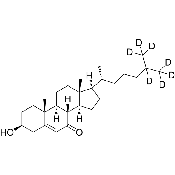
HY-113342S
7-Keto Cholesterol-d7Stable Isotope -

HY-100355R
C18-Ceramide (Standard)Ceramide -
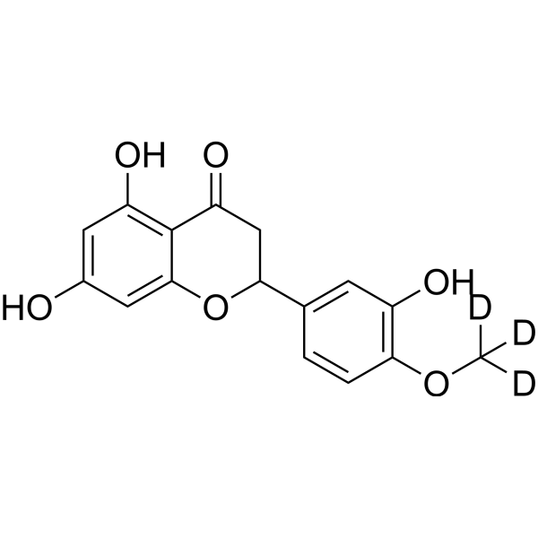
HY-N0168AS
(Rac)-Hesperetin-d3Stable Isotope


Biological Activity
Description
In Vitro
Stable heavy isotopes of hydrogen, carbon, and other elements have been incorporated into drug molecules, largely as tracers for quantitation during the drug development process. Deuteration has gained attention because of its potential to affect the pharmacokinetic and metabolic profiles of drugs[1].
MedChemExpress (MCE) has not independently confirmed the accuracy of these methods. They are for reference only. Further protocols information, click here.
Application
1. This compound can be used as a tracer
2. This compound can be used as an internal standard for quantitative analysis by NMR, GC-MS, or LC-MS.


Chemical Information
-
CAS No. 1219803-02-7
-
Unlabeled CAS 10238-21-8
-
Appearance Solid
-
Molecular Weight 497.02
-
Formula C23H25D3ClN3O5S
-
Color White to off-white
-
SMILES
O=C(NCCC1=CC=C(S(=O)(NC(NC2CCCCC2)=O)=O)C=C1)C3=CC(Cl)=CC=C3OC([2H])([2H])[2H]
-
Synonyms
Glyburide-d3
-
Shipping
Room temperature in continental US; may vary elsewhere.
-
Storage
Powder -20°C 3 years 4°C 2 years In solvent -80°C 6 months -20°C 1 month


Solvent & Solubility
In Vitro:
DMSO : 100 mg/mL (201.20 mM; Need ultrasonic; Hygroscopic DMSO has a significant impact on the solubility of product, please use newly opened DMSO)
H2O : 0.1 mg/mL (0.20 mM; Need ultrasonic and warming)
Please refer to the solubility information to select the appropriate solvent. Once prepared, please aliquot and store the solution to prevent product inactivation from repeated freeze-thaw cycles.
Storage method and period of stock solution: -80°C, 6 months; -20°C, 1 month. When stored at -80°C, please use it within 6 months. When stored at -20°C, please use it within 1 month.
* Note: If you choose water as the stock solution, please dilute it to the working solution, then filter and sterilize it with a 0.22 μm filter before use.
Please refer to the solubility information to select the appropriate solvent. Once prepared, please aliquot and store the solution to prevent product inactivation from repeated freeze-thaw cycles.
Storage method and period of stock solution: -80°C, 6 months; -20°C, 1 month. When stored at -80°C, please use it within 6 months. When stored at -20°C, please use it within 1 month.
* Note: If you choose water as the stock solution, please dilute it to the working solution, then filter and sterilize it with a 0.22 μm filter before use.
Concentration (start) × Volume (start) = Concentration (final) × Volume (final)

Protocols
-
Fibrosis/Collagen Morphometry
Fibrosis and collagen morphometry is based on the quantitative visualization of fibrillar collagen deposition in tissue sections using histochemical stains such as Sirius Red (Picrosirius Red) or Masson's trichrome, followed by image-based or polarization-enhanced analysis to estimate collagen proportional area as a surrogate of extracellular matrix accumulation during fibrotic remodeling. Sirius Red combined with polarized light microscopy enhances detection of collagen fibers due to birefringence properties, enabling more specific visualization of collagen type I and III fibrils compared to conventional bright-field histology, while whole-section or region-restricted digital morphometry reduces field-selection bias in fibrosis assessment. Alternative quantitative approaches include second harmonic generation (SHG) and two-photon excited fluorescence microscopy, which enable label-free detection of fibrillar collagen and have been validated against histological staining and biochemica
-
Collagen: Sirius Red Staining
Sirius Red or picrosirius red staining is a histochemical method for visualizing collagen-rich extracellular matrix in tissue sections, and collagen fibers are detected as red-stained structures under bright-field microscopy with enhanced birefringence under polarized light. Picrosirius red is useful for assessing total collagen organization, distribution, and fibrosis burden, but polarized color should not be interpreted as a definitive collagen type I versus type III readout because color is affected by fiber orientation, thickness, and packing.
-
Connective Tissue: Masson's Trichrome/Collagen Trichrome Staining
Masson’s Trichrome (collagen/trichrome staining) is a histological technique that differentially stains tissue compartments using sequential acidic dyes to distinguish collagen from muscle and cytoplasmic components based on dye affinity and tissue permeability differences, enabling visualization of fibrosis and connective tissue architecture in histological sections. The classical formulation typically uses Weigert's iron hematoxylin for nuclear staining, Biebrich scarlet-acid fuchsin for cytoplasm and muscle, and aniline blue (or light green variants) for collagen, producing a characteristic blue/green collagen signal contrasted against red cytoplasm and dark nuclei. The staining principle relies on selective displacement of smaller dye molecules by larger anionic dyes in collagen-rich regions under controlled acidified conditions, which enhances collagen-specific dye retention. This property makes the method widely used for fibrosis assessment in organs such as heart, liver, lung, a
-
Autophagy
Autophagy is a process in which eukaryotic cells use lysosomes to degrade their own cytoplasmic proteins and damaged organelles under the regulation of autophagy related gene (Atg). Microtubule-associated proteins light chain 3 (LC3) is recognized as autophagy marker, which transfers from cytoplasmic LC3 (LC3-I) to membrane type (LC3-II). LC3-II/I ratio could be detected by Western Blot and fluorescence microscopy.
-
Transepithelial/transendothelial electrical resistance assay
TEER measures electrical resistance across epithelial or endothelial monolayers cultured on permeable supports, and the readout reflects ionic conductance through the cell barrier, especially the paracellular pathway regulated by junctional integrity. TEER can be measured without destroying the monolayer and is commonly used before or during transport, permeability, barrier-disruption, and barrier-maturation experiments. TEER values are influenced by biological maturation and technical conditions; reported factors include temperature, medium formulation, passage number, electrode geometry, membrane properties, and junctional length during early monolayer maturation. Therefore, TEER should be interpreted with blank-insert subtraction, area normalization, repeated readings, and, when possible, orthogonal barrier readouts such as FITC-dextran flux or tight-junction staining.
-
Lysosome and acidic-vesicle live-cell staining
Lysosome and acidic-vesicle live-cell staining detects acidic intracellular compartments by using membrane-permeant acidotropic probes that accumulate in low-pH vesicles, including lysosomes, late endosomes, autolysosomes, and acidic phagosomes. LysoTracker staining is commonly used as an intensity-based readout of acidic lysosomal compartment abundance or enlargement, while acridine orange produces green fluorescence in less concentrated compartments and red fluorescence after concentration-dependent accumulation in acidic vesicular organelles. Loss or reduction of acridine-orange red signal can be used as a readout of lysosomal membrane permeabilization or reduced acidic-vesicle integrity. This protocol is designed for live cultured cells and can be adapted for fluorescence microscopy, high-content imaging, plate-reader readout, or flow cytometry when the selected literature supports the readout. Because these dyes report acidotropic accumulation rather than lysosome identity alone,
-
Research Protocol for Metabolic Diseases
AMP-activated protein kinase, AMPK, is a conserved cellular energy sensor that responds to reduced cellular energy status and coordinates metabolism by increasing ATP-generating catabolic pathways while suppressing ATP-consuming anabolic processes. In metabolic disease research, the AMPK pathway is experimentally relevant because it regulates hepatic lipid synthesis, fatty acid oxidation, glucose production, skeletal-muscle glucose disposal, mTORC1-linked biosynthesis, autophagy, mitochondrial homeostasis, and whole-body energy balance. The central pathway logic is that energy stress, metformin, exercise-like stimulation, or direct AMPK activators increase AMPKα Thr172 phosphorylation and downstream substrate phosphorylation, including ACC and RAPTOR. Phosphorylation of ACC suppresses lipogenesis and supports fatty acid oxidation, whereas phosphorylation of RAPTOR suppresses mTORC1 signaling and links cellular energy status to growth and protein synthesis control. The pathway is linked
-
Macroautophagy Solutions
Macroautophagy is a conserved lysosome-dependent degradation pathway in which cytoplasmic material is sequestered into double-membrane autophagosomes and delivered to lysosomes for degradation and recycling. The pathway supports cellular homeostasis during nutrient limitation, organelle stress, protein-aggregate accumulation, infection, differentiation, and tissue remodeling by coupling cargo sequestration, autophagosome maturation, lysosomal fusion, and degradation of cargo-derived macromolecules. The core molecular sequence includes initiation by nutrient- and stress-regulated autophagy machinery, autophagosome nucleation, LC3/ATG8-family conjugation to autophagosomal membranes, cargo selection through receptors such as SQSTM1/p62, autophagosome-lysosome fusion, and lysosomal degradation. LC3 was identified as a mammalian homolog of yeast Atg8 that localizes to autophagosomal membranes after processing, and p62/SQSTM1 was shown to connect ubiquitinated cargo with autophagic degradati


Purity & Documentation
-
Data Sheet (272 KB)
-
SDS (418 KB)
- English - EN (418 KB)
- Français - FR (418 KB)
- Deutsch - DE (418 KB)
- Norwegian - NO (418 KB)
- Español - ES (418 KB)
- Swedish - SV (418 KB)
- Italian - IT (418 KB)
- Korean - KR (418 KB)
- Portuguese - PT (418 KB)
-
Handling Instructions (2659 KB)
References
[1]. Russak EM, et al. Impact of Deuterium Substitution on the Pharmacokinetics of Pharmaceuticals. Ann Pharmacother. 2019;53(2):211-216. [Content Brief]
[2]. Heo R, et al. The anti-diabetic drug trelagliptin induces vasodilation via activation of Kv channels and SERCA pumps. Life Sci. 2021;283:119868. [Content Brief]
[3]. Qiu Y, et al. Glyburide Regulates UCP1 Expression in Adipocytes Independent of KATP Channel Blockade. iScience. 2020;23(9):101446. [Content Brief]
[4]. Golstein PE, et al. P-glycoprotein inhibition by glibenclamide and related compounds. Pflugers Arch. 1999;437(5):652-660. [Content Brief]
[5]. Fernandes MA, et al. Glibenclamide interferes with mitochondrial bioenergetics by inducing changes on membrane ion permeability. J Biochem Mol Toxicol. 2004;18(3):162-169. [Content Brief]
[6]. Zhou J, et al. Glibenclamide-Induced Autophagy Inhibits Its Insulin Secretion-Improving Function in β Cells. Int J Endocrinol. 2019;2019:1265175. [Content Brief]


Complete Stock Solution Preparation Table
Please refer to the solubility information to select the appropriate solvent. Once prepared, please aliquot and store the solution to prevent product inactivation from repeated freeze-thaw cycles.
Storage method and period of stock solution: -80°C, 6 months; -20°C, 1 month. When stored at -80°C, please use it within 6 months. When stored at -20°C, please use it within 1 month.
| Optional Solvent | Concentration Solvent Mass | 1 mg | 5 mg | 10 mg | 25 mg |
|---|---|---|---|---|---|
| DMSO | 1 mM | 2.0120 mL | 10.0600 mL | 20.1199 mL | 50.2998 mL |
| 5 mM | 0.4024 mL | 2.0120 mL | 4.0240 mL | 10.0600 mL | |
| 10 mM | 0.2012 mL | 1.0060 mL | 2.0120 mL | 5.0300 mL | |
| 15 mM | 0.1341 mL | 0.6707 mL | 1.3413 mL | 3.3533 mL | |
| 20 mM | 0.1006 mL | 0.5030 mL | 1.0060 mL | 2.5150 mL | |
| 25 mM | 0.0805 mL | 0.4024 mL | 0.8048 mL | 2.0120 mL | |
| 30 mM | 0.0671 mL | 0.3353 mL | 0.6707 mL | 1.6767 mL | |
| 40 mM | 0.0503 mL | 0.2515 mL | 0.5030 mL | 1.2575 mL | |
| 50 mM | 0.0402 mL | 0.2012 mL | 0.4024 mL | 1.0060 mL | |
| 60 mM | 0.0335 mL | 0.1677 mL | 0.3353 mL | 0.8383 mL | |
| 80 mM | 0.0251 mL | 0.1257 mL | 0.2515 mL | 0.6287 mL | |
| 100 mM | 0.0201 mL | 0.1006 mL | 0.2012 mL | 0.5030 mL |
* Note: If you choose water as the stock solution, please dilute it to the working solution, then filter and sterilize it with a 0.22 μm filter before use.


Glyburide-d3 Related Classifications
HY-15206S1 Related Classifications
 Powered by Bioz