



Oxfenicine

Based on 1 Customer Validation
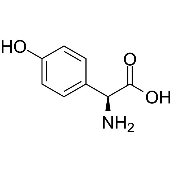
Oxfenicine (L-p-Hydroxyphenylglycine) is an orally active carnitine palmitoyltransferase-1 inhibitor. Oxfenicine inhibits the oxidation of fatty acids in the heart, protecting cardiac tissue from necrotic damage during ischemia, and also has an inhibitory effect on cardiac tissue apoptosis. In addition, Oxfenicine promotes lipolysis in a high-fat diet rat model. Oxfenicine can be used in the study of cardiovascular and metabolic diseases.
For research use only. We do not sell to patients.
- Purity : 99.98%
- CAS No.: 32462-30-9
- Formula: C8H9NO3
- Molecular Weight:167.16
-
Storage:Powder -20°C, 3 years , 4°C, 2 years ; In solvent -80°C, 2 years , -20°C, 1 year

To place orders, for customer services and technical support, please contact: MedChemExpress USA
Tel: 609-228-6898 E-mail: [email protected] [email protected]


-
 Biological Activity
-
Chemical Information
-
Solvent & Solubility
- Protocols
- Purity & Documentation
- References
-
 Help & FAQs


-
HY-50202
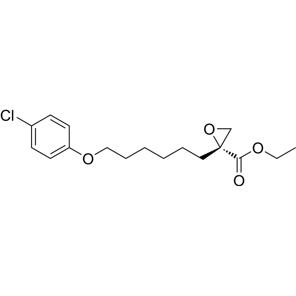
EtomoxirCPT1a Inhibitor -
HY-B0968A
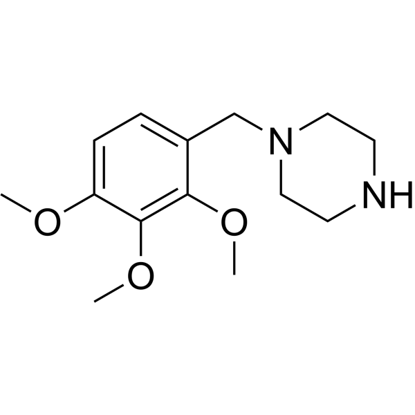
TrimetazidineAntianginal Agent -
HY-126015
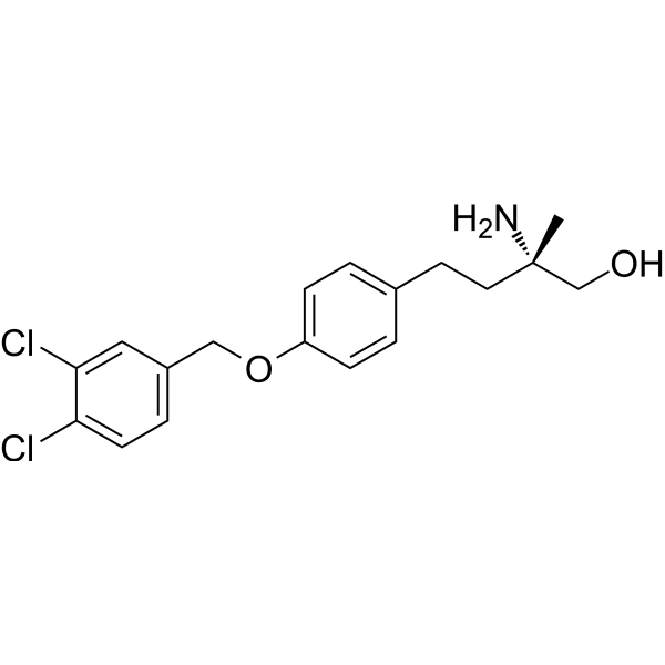
P053CerS1 Inhibitor -

HY-10219
RapamycinmTOR Inhibitor -
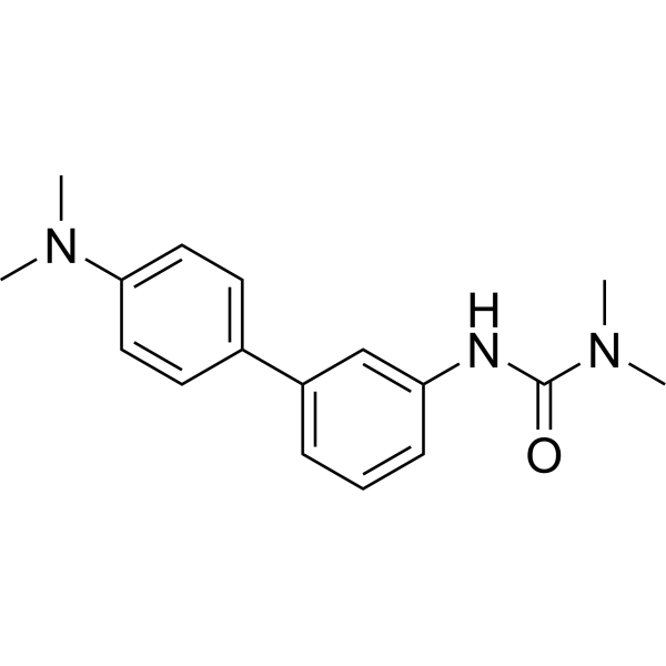
HY-15859
AtglistatinATGL Inhibitor -
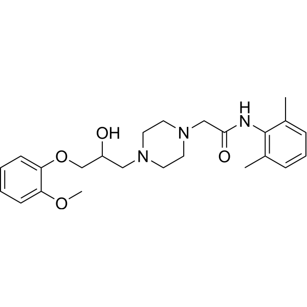
HY-B0280
RanolazineSodium Current/FAO Inhibitor -
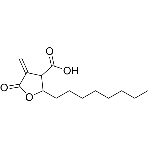
HY-12364
C75Fatty Acid Synthase Inhibitor -
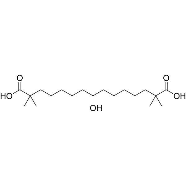
HY-12357
Bempedoic acidACL Inhibitor/AMPK Activator -
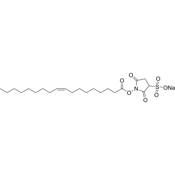
HY-112847A
Sulfosuccinimidyl oleate sodiumFatty Acid Transport Inhibitor -
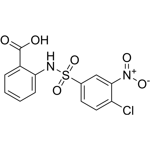
HY-123986
CTPI-2SLC25A1 Inhibitor -
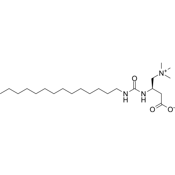
HY-16482
TeglicarL-CPT1 Inhibitor -
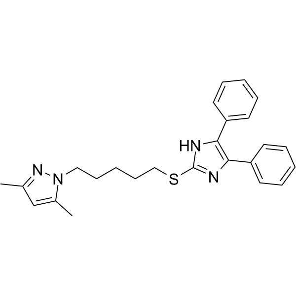
HY-101576
RP 70676ACAT Inhibitor


Biological Activity
Description
IC50 & Target
carnitine palmitoyltransferase-1[1]
Cellular Effect
|
Cell Line

|
Type | Value | Description | References |
|---|---|---|---|---|
| KB | IC50 |
>100 μM
Compound: 46
|
Cytotoxicity in human KB cells
Cytotoxicity in human KB cells
|
[PMID: 21504156] |
In Vitro
Oxfenicine (1 mM, 2 hours) reduces fatty acid oxidation and glucose incorporation into lipids in subcutaneous and epididymal adipocytes[4]. Oxfenicine (10 mM, 24 hours) blunts carnitine plus palmitate-induced formation of apoptotic nuclei in chick embryonic ventricular cells[5].
MedChemExpress (MCE) has not independently confirmed the accuracy of these methods. They are for reference only. Further protocols information, click here.
-
Cell Line:Carnitine and Palmitate-induced chick embryonic ventricular cells (7-day-old chick embryos from white Leghorn eggs)
-
Concentration:10 mM
-
Incubation Time:24 h
-
Result:Significantly reduced the formation of apoptotic cell nuclei.
In Vivo
MedChemExpress (MCE) has not independently confirmed the accuracy of these methods. They are for reference only.
-
Animal Model:Male Sprague-Dawley rats fed a high-fat (HF) diet[4]
-
Dosage:150 mg/kg
-
Administration:intraperitoneal injection (i.p.) once daily for 3 weeks
-
Result:Reduced whole-body fat oxidation, body weight, adiposity, and improved insulin sensitivity in HF-fed rats.


Chemical Information
-
CAS No. 32462-30-9
-
Appearance Solid
-
Molecular Weight 167.16
-
Formula C8H9NO3
-
Color White to off-white
-
SMILES
O=C([C@H](C1=CC=C(C=C1)O)N)O
-
Synonyms
L-p-Hydroxyphenylglycine; 4-Hydroxy-L-phenylglycine; UK 25842
-
Shipping
Room temperature in continental US; may vary elsewhere.
-
Storage
Powder -20°C 3 years 4°C 2 years In solvent -80°C 2 years -20°C 1 year


Solvent & Solubility
In Vitro:
H2O : 4.55 mg/mL (27.22 mM; Need ultrasonic)
DMSO : < 1 mg/mL (insoluble or slightly soluble)
Please refer to the solubility information to select the appropriate solvent. Once prepared, please aliquot and store the solution to prevent product inactivation from repeated freeze-thaw cycles.
Storage method and period of stock solution: -80°C, 2 years; -20°C, 1 year. When stored at -80°C, please use it within 2 years. When stored at -20°C, please use it within 1 year.
* Note: If you choose water as the stock solution, please dilute it to the working solution, then filter and sterilize it with a 0.22 μm filter before use.
Please refer to the solubility information to select the appropriate solvent. Once prepared, please aliquot and store the solution to prevent product inactivation from repeated freeze-thaw cycles.
Storage method and period of stock solution: -80°C, 2 years; -20°C, 1 year. When stored at -80°C, please use it within 2 years. When stored at -20°C, please use it within 1 year.
* Note: If you choose water as the stock solution, please dilute it to the working solution, then filter and sterilize it with a 0.22 μm filter before use.
Concentration (start) × Volume (start) = Concentration (final) × Volume (final)
In Vivo:
For the following dissolution methods, please prepare the working solution directly:
It is recommended to prepare fresh solutions and use them promptly within a short period of time.
The percentages shown for the solvents indicate their volumetric ratio in the final prepared solution. If precipitation or phase separation occurs during preparation, heat and/or sonication can be used to aid dissolution.
Add each solvent one by one: PBS
Solubility: 2 mg/mL (11.96 mM); Clear solution; Need ultrasonic and warming and heat to 60°C

Protocols
-
Apoptosis
Apoptosis, also called programmed cell death, is generally characterized by distinct morphological characteristics.
-
TUNEL staining for apoptotic DNA fragmentation
TUNEL staining detects DNA strand breaks by using terminal deoxynucleotidyl transferase to add labeled nucleotides to exposed 3′-OH DNA termini, generating either microscopic staining in fixed cells or tissue sections, or fluorescence/cytometric signal in cell suspensions. TUNEL positivity reflects DNA fragmentation but should not be interpreted alone as definitive apoptosis, because TUNEL can also label necrotic, autolytic, mechanically damaged, or DNA-repair-associated DNA breaks.
-
Annexin V plus membrane-impermeant dye apoptosis staining
Annexin V-based apoptosis assays rely on the detection of phosphatidylserine (PS) externalization from the inner leaflet of the plasma membrane to the outer leaflet, an early biochemical hallmark of apoptosis. Fluorescently labeled Annexin V binds PS in a calcium-dependent manner, enabling identification of early apoptotic cells by flow cytometry or fluorescence microscopy. When combined with a membrane-impermeant DNA-binding dye (e. g. , propidium iodide), this approach allows discrimination between viable (Annexin V−/dye−), early apoptotic (Annexin V+/dye−), and late apoptotic or necrotic (Annexin V+/dye+) cell populations by assessing membrane integrity and PS exposure.
-
How to Select a Suitable Non-Mouse Animal Model
Selecting a suitable non-mouse animal model is a structured decision based on the research question, required anatomy or physiology, disease mechanism, endpoint feasibility, translational relevance, and ethical justification. Non-mouse models are preferred when mice cannot reproduce key human-relevant features, such as organ size, surgical anatomy, cardiovascular physiology, neuroanatomy, immune features, pharmacology, toxicology, or long-term clinical procedures. Candidate species may include rats, rabbits, guinea pigs, ferrets, zebrafish, pigs, sheep, goats, dogs, cats, horses, and non-human primates, but each species must be justified by its specific scientific advantage rather than convenience or tradition. Unresolved questions include how to quantify translational superiority across species, how to balance increased biological relevance against higher ethical burden, and when human-derived systems or new approach methodologies should replace animal use.
-
Apoptosis Solutions
Apoptosis is a regulated, generally non-lytic cell-death pathway that removes unwanted, damaged, infected, or abnormal cells through coordinated morphological changes, caspase activation, DNA fragmentation, and membrane remodeling. The intrinsic apoptosis pathway is controlled mainly by mitochondrial outer membrane permeabilization, BCL-2 family proteins, cytochrome c release, apoptosome formation, caspase-9 activation, and downstream executioner caspase-3/7 activation. The extrinsic apoptosis pathway is initiated by death receptors such as Fas, TNFR, and TRAIL receptors, which recruit adaptor proteins and activate caspase-8 before engaging executioner caspases or mitochondrial amplification through BID cleavage. Apoptosis is linked to many phenotypes, including cancer cell killing, tissue homeostasis, immune regulation, neurodegeneration, infection response, and treatment-induced cytotoxicity; unresolved questions include how apoptosis interacts with necroptosis, pyroptosis, ferroptos
-
How to Choose the Right Model Animal
Choosing the right model animal is a validity-driven decision in which the species, strain, sex, age, genetic background, disease-induction method, outcome measures, and welfare burden must match the scientific question rather than laboratory tradition or convenience. A model should be selected by judging face validity, construct validity, and predictive validity: whether it resembles the human phenotype, whether it reproduces relevant mechanisms, and whether results are likely to predict human biology or treatment response. Animal studies often fail to translate because of species differences, weak disease resemblance, poor experimental design, inadequate reporting, publication bias, and underuse of randomization, blinding, and sample-size justification. Unresolved questions include how to rank competing models objectively, how much human-disease complexity must be reproduced for a given objective, and when non-animal systems such as organoids, ex vivo tissue, or computational models


Purity & Documentation
-
Data Sheet (274 KB)
-
SDS (394 KB)
- English - EN (394 KB)
- Français - FR (394 KB)
- Deutsch - DE (394 KB)
- Norwegian - NO (394 KB)
- Español - ES (394 KB)
- Swedish - SV (394 KB)
- Italian - IT (394 KB)
- Korean - KR (394 KB)
- Portuguese - PT (394 KB)
-
Handling Instructions (2659 KB)
References
[1]. Stephens TW, et, al. Two mechanisms produce tissue-specific inhibition of fatty acid oxidation by oxfenicine. Biochem J. 1985 Apr 15;227(2):651-60. [Content Brief]
[2]. Greaves P, et, al. Cardiac hypertrophy in the dog and rat induced by oxfenicine, an agent which modifies muscle metabolism. Arch Toxicol Suppl. 1984;7:488-93. [Content Brief]
[3]. Kennedy JA, et al. Effect of perhexiline and oxfenicine on myocardial function and metabolism during low-flow ischemia/reperfusion in the isolated rat heart. J Cardiovasc Pharmacol. 2000 Dec;36(6):794-801. [Content Brief]
[4]. Sepa-Kishi DM, et al. Antilipolytic and antilipogenic effects of the CPT-1b inhibitor oxfenicine in the white adipose tissue of rats. Am J Physiol Regul Integr Comp Physiol. 2016 Oct 1;311(4):R779-R787. [Content Brief]
[5]. Kong JY, et al. Palmitate-induced cardiac apoptosis is mediated through CPT-1 but not influenced by glucose and insulin. Am J Physiol Heart Circ Physiol. 2002 Feb;282(2):H717-25. [Content Brief]


Complete Stock Solution Preparation Table
Please refer to the solubility information to select the appropriate solvent. Once prepared, please aliquot and store the solution to prevent product inactivation from repeated freeze-thaw cycles.
Storage method and period of stock solution: -80°C, 2 years; -20°C, 1 year. When stored at -80°C, please use it within 2 years. When stored at -20°C, please use it within 1 year.
| Optional Solvent | Concentration Solvent Mass | 1 mg | 5 mg | 10 mg | 25 mg |
|---|---|---|---|---|---|
| H2O | 1 mM | 5.9823 mL | 29.9115 mL | 59.8229 mL | 149.5573 mL |
| 5 mM | 1.1965 mL | 5.9823 mL | 11.9646 mL | 29.9115 mL | |
| 10 mM | 0.5982 mL | 2.9911 mL | 5.9823 mL | 14.9557 mL | |
| 15 mM | 0.3988 mL | 1.9941 mL | 3.9882 mL | 9.9705 mL | |
| 20 mM | 0.2991 mL | 1.4956 mL | 2.9911 mL | 7.4779 mL | |
| 25 mM | 0.2393 mL | 1.1965 mL | 2.3929 mL | 5.9823 mL |
* Note: If you choose water as the stock solution, please dilute it to the working solution, then filter and sterilize it with a 0.22 μm filter before use.


Oxfenicine Related Classifications
HY-W018026 Related Classifications
 Powered by Bioz