



hAChE-IN-5
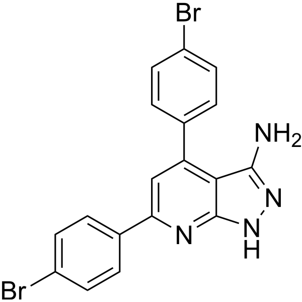
hAChE-IN-5 (compound 49) is a potent hAChE and hBuChE inhibitor with IC50 values of 0.17 μM and 0.17 μM, respectively. hAChE-IN-5 shows potent GSK3β inhibition with an IC50 value of 0.21 μM. hAChE-IN-5 is used as tau protein aggregation and Aβ1-42 self-aggregation inhibitor. hAChE-IN-5 can bind virtually with the PAS affecting Aβ aggregation, thus preventing Aβ-dependent neurotoxicity. hAChE-IN-5 can penetrate BBB and has the potential for multi-targeted anti-Alzheimer's agents research.
For research use only. We do not sell to patients.
- CAS No.: 3007667-15-1
- Formula: C18H12Br2N4
- Molecular Weight:444.12
-
Storage:
Please store the product under the recommended conditions in the Certificate of Analysis.

To place orders, for customer services and technical support, please contact: MedChemExpress USA
Tel: 609-228-6898 E-mail: [email protected] [email protected]


-
 Biological Activity
-
Chemical Information
- Protocols
- Purity & Documentation
- References
-
 Help & FAQs




Biological Activity
Description
Cellular Effect
|
Cell Line

|
Type | Value | Description | References |
|---|---|---|---|---|
| WI-38 | IC50 |
58.28 μM
Compound: 49
|
Cytotoxicity against human WI-38 cells assessed as inhibition of cell growth incubated for 72 hrs by MTT assay
Cytotoxicity against human WI-38 cells assessed as inhibition of cell growth incubated for 72 hrs by MTT assay
|
[PMID: 37871406] |


Chemical Information
-
CAS No. 3007667-15-1
-
Molecular Weight 444.12
-
Formula C18H12Br2N4
-
SMILES
BrC1=CC=C(C2=C3C(NN=C3N)=NC(C4=CC=C(Br)C=C4)=C2)C=C1
-
Shipping
Room temperature in continental US; may vary elsewhere.
-
Storage
Please store the product under the recommended conditions in the Certificate of Analysis.

Protocols
-
Neurotoxicity Study
This protocol assesses in vitro neurotoxicity by combining neuronal viability, mitochondrial/metabolic activity, neurite outgrowth, and optional neuronal network function readouts. Calcein-AM or resazurin/PrestoBlue readouts estimate viable or metabolically active cells; βIII-tubulin immunofluorescence detects neuronal morphology and neurite networks; TMRE detects mitochondrial membrane potential; and MEA recordings detect functional changes in neuronal network activity.
-
Cell Cytotoxicity Assay
Cytotoxicity assays are usually based on the assessment of cell membrane damage, which can also be indirectly detected by measuring cell viability. Detection methods include MTT assay, CKK-8 assay, LDH assay and ATP assay, etc.
-
Carbohydrates and Mucins: Periodic Acid-Schiff (PAS) Staining
Periodic acid-Schiff staining detects tissue carbohydrates and mucosubstances by oxidizing carbohydrate glycol groups with periodic acid to generate aldehydes, which then react with Schiff reagent to produce a magenta reaction product; classic reports established the method for mucin and polysaccharide-containing structures in fixed tissue sections. PAS staining can demonstrate neutral mucins and goblet-cell mucin, but it is not specific for mucin because glycogen and other PAS-positive tissue components can also stain; diastase/PAS-D is used when glycogen removal is needed to distinguish glycogen-dependent PAS signal from non-glycogen PAS-positive mucosubstances.
-
Alzheimer’s Disease Modeling
Alzheimer’s Disease (AD) is a neurodegenerative disorder characterized by a progressive decline in cognitive functions and loss of specific types of neurons and synapses. Alzheimer's symptoms can be simulated in mice by injecting drugs (such as Aβ) or genetically modified.


Purity & Documentation
References

Calculators
Concentration (start) × Volume (start) = Concentration (final) × Volume (final)


hAChE-IN-5 Related Classifications
HY-155365 Related Classifications
 Powered by Bioz