



Hirudin
Based on 4 publication(s) in Google Scholar
Hirudin is a thrombin inhibitor with blood anticoagulant property. Hirudin has potent anti-thrombotic, wound repair, anti-fibrosis, anti-tumor and anti-hyperuricemia effects. Hirudin also affects diabetic complications, cerebral hemorrhage, and others.
For research use only. We do not sell to patients.
- Purity : 95.0%
- CAS No.: 8001-27-2
-
Storage:
-80°C


To place orders, for customer services and technical support, please contact: MedChemExpress USA
Tel: 609-228-6898 E-mail: [email protected] [email protected]


-
 Biological Activity
-
Chemical Information
- Protocols
- Purity & Documentation
- References
-
 Help & FAQs


-
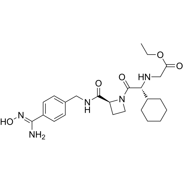
HY-10787
XimelagatranThrombin Inhibitor -
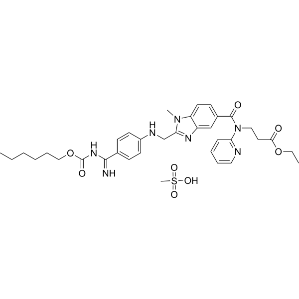
HY-10274A
Dabigatran etexilate mesylateAntithrombotic Agent -

HY-10787R
Ximelagatran (Standard)Reference Standard -
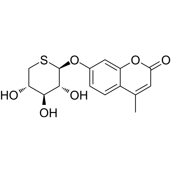
HY-10277
Odiparcilβ-D-xyloside Derivative -

HY-10274AR
Dabigatran etexilate mesylate (Standard)Antithrombotic Agent -
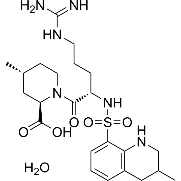
HY-B0375A
Argatroban monohydrateThrombin Inhibitor -
HY-15664
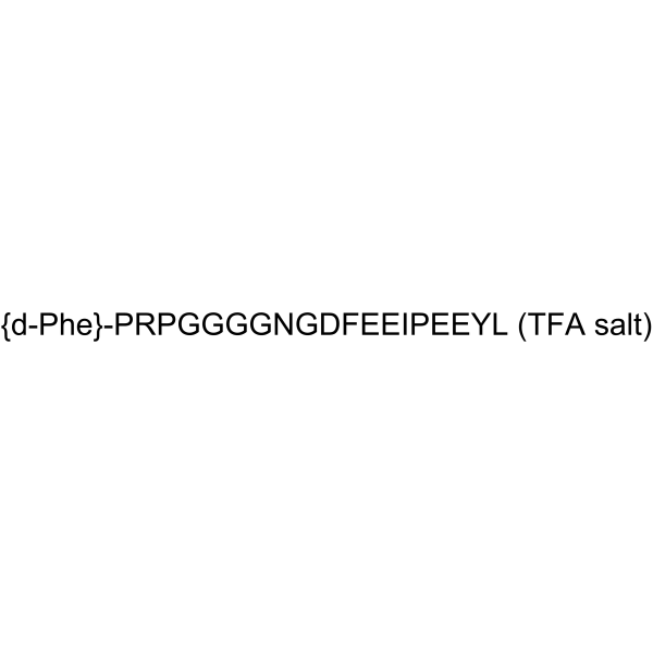
Bivalirudin TFAThrombin Inhibitor -
HY-122542
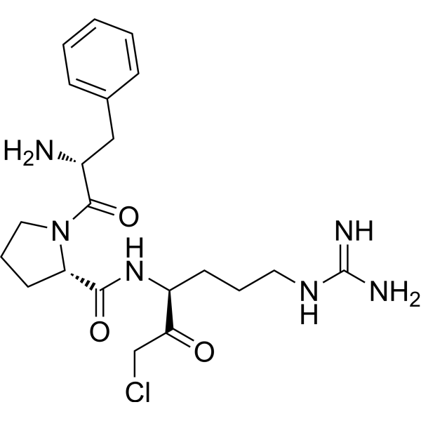
PPACKThrombin/GZMK Inhibitor -

HY-10277R
Odiparcil (Standard)Reference Standard -

HY-B0375AR
Argatroban monohydrate (Standard)Thrombin Inhibitor
Publications Citing Use of MedChemExpress (MCE) Hirudin
More

Biological Activity
Description
In Vitro
Hirudin inhibits the activity of thrombin, deprives the ability of thrombin to cleave fibrinogen, prevents the formation of fibrin and the cross-linking polymerization process of fibrin monomer in internal and external coagulation pathway[1]. Hirudin reduces cell apoptosis of human microvascular endothelial cells (HMVECs) and suppresses the expression of p-JAK2 via antagonizing thrombin[1]. Hirudin inhibits VEGF-Notch pathway and cell proliferation of HMVECs at high doses[1]. Hirudin (3-10 mg/mL) reverses the abnormal proliferation and fibrosis in HK-2 cells caused by TGF-β1[1]. Hirudin depresses the myocardial fibroblasts induced by angiotensin II by dose-dependently inhibits oxidative stress, regulates fibrosis-related factors, and represses the ERK1/2 pathway[1].
MedChemExpress (MCE) has not independently confirmed the accuracy of these methods. They are for reference only. Further protocols information, click here.
In Vivo
MedChemExpress (MCE) has not independently confirmed the accuracy of these methods. They are for reference only.
-
Animal Model:Male balb/c mice with underwent unilateral ureteral ligation (UUO)[2]
-
Dosage:10 and 15 mg/kg
-
Administration:Oral gavage; 10 and 15 mg/kg, once daily for 21 days
-
Result:Reduced renal damages and suppressed the upregulation of α-SMA, collagen deposition in UUO mice. Increased the level of fibrosis (collagen-I, FN, α-SMA), N-cad, slug and E-cad in UUO mice. Decreased the level of IL-1β, IL-6 and TNF-α, apoptosis of renal tubular cells in UUO mice. Decreased the expression of inflammatory factors, the occurrence of EMT, the incidence of fibrosis and the apoptosis of TGF-β-induced renal tubular epithelial cell.
Clinical Trial
| NCT Number | Sponsor | Condition | Start Date |
Phase

|
|---|---|---|---|---|
| NCT01329991 | Plexxikon| | 2011-05 | PHASE1 |


Chemical Information


Publications (4)
-
Journal Impact Factor
-
Most Recent
-
J Interferon Cytokine Res
2025 Oct;45(10):335-343. PMID: 40916805 -
Acta Cir Bras
Hirudin inhibits ferroptosis to improve renal fibrosis by targeting the STAT3/NLRP3 signaling pathway. [Abstract]2025 Apr 28:40:e403325. PMID: 40298655 -
Biosci Biotechnol Biochem
Hirudin Alleviates Renal Fibrosis by Inducing Autophagy to Suppress NLRP3 Inflammasome Activation. [Abstract]2025 Jul 29:zbaf114. PMID: 40728927 -

Protocols
-
Fibrosis/Collagen Morphometry
Fibrosis and collagen morphometry is based on the quantitative visualization of fibrillar collagen deposition in tissue sections using histochemical stains such as Sirius Red (Picrosirius Red) or Masson's trichrome, followed by image-based or polarization-enhanced analysis to estimate collagen proportional area as a surrogate of extracellular matrix accumulation during fibrotic remodeling. Sirius Red combined with polarized light microscopy enhances detection of collagen fibers due to birefringence properties, enabling more specific visualization of collagen type I and III fibrils compared to conventional bright-field histology, while whole-section or region-restricted digital morphometry reduces field-selection bias in fibrosis assessment. Alternative quantitative approaches include second harmonic generation (SHG) and two-photon excited fluorescence microscopy, which enable label-free detection of fibrillar collagen and have been validated against histological staining and biochemica
-
Collagen: Sirius Red Staining
Sirius Red or picrosirius red staining is a histochemical method for visualizing collagen-rich extracellular matrix in tissue sections, and collagen fibers are detected as red-stained structures under bright-field microscopy with enhanced birefringence under polarized light. Picrosirius red is useful for assessing total collagen organization, distribution, and fibrosis burden, but polarized color should not be interpreted as a definitive collagen type I versus type III readout because color is affected by fiber orientation, thickness, and packing.
-
Connective Tissue: Masson's Trichrome/Collagen Trichrome Staining
Masson’s Trichrome (collagen/trichrome staining) is a histological technique that differentially stains tissue compartments using sequential acidic dyes to distinguish collagen from muscle and cytoplasmic components based on dye affinity and tissue permeability differences, enabling visualization of fibrosis and connective tissue architecture in histological sections. The classical formulation typically uses Weigert's iron hematoxylin for nuclear staining, Biebrich scarlet-acid fuchsin for cytoplasm and muscle, and aniline blue (or light green variants) for collagen, producing a characteristic blue/green collagen signal contrasted against red cytoplasm and dark nuclei. The staining principle relies on selective displacement of smaller dye molecules by larger anionic dyes in collagen-rich regions under controlled acidified conditions, which enhances collagen-specific dye retention. This property makes the method widely used for fibrosis assessment in organs such as heart, liver, lung, a


Purity & Documentation
-
Data Sheet (274 KB)
-
SDS (251 KB)
- English - EN (251 KB)
- Français - FR (251 KB)
- Deutsch - DE (251 KB)
- Norwegian - NO (251 KB)
- Español - ES (251 KB)
- Swedish - SV (251 KB)
- Italian - IT (251 KB)
- Korean - KR (251 KB)
- Portuguese - PT (251 KB)
-
Handling Instructions (2659 KB)
References
[1]. Junren C,et al. Pharmacological Activities and Mechanisms of Hirudin and Its Derivatives - A Review. Front Pharmacol. 2021 Apr 16;12:660757. [Content Brief]
[2]. Xie Y, et al. Hirudin improves renal interstitial fibrosis by reducing renal tubule injury and inflammation in unilateral ureteral obstruction (UUO) mice. Int Immunopharmacol. 2020 Apr;81:106249. [Content Brief]

Calculators
Concentration (start) × Volume (start) = Concentration (final) × Volume (final)


Hirudin Related Classifications
HY-P2813 Related Classifications
 Powered by Bioz