



Hyaluronic acid (Mw:1000-2000Da)

Based on 1 Customer Validation
Hyaluronic acid (Mw: 1000-2000 Da) is a long-chain unbranched polysaccharide with a molecular weight of 1000-2000 Da. Hyaluronic acid (Mw: 2000 Da) inhibits IL-1β expression and increases LC3-II. Hyaluronic acid (Mw: 1000-2000Da) can regulate tissue homeostasis and stress resistance, promote angiogenesis and tissue remodeling. Hyaluronic acid (Mw: 1000-2000 Da) has good biocompatibility and biodegradability, and can be used in drug delivery systems and tissue engineering. Hyaluronic acid (Mw: 2000 Da) can reduce facial skin blemishes and sagging.
For research use only. We do not sell to patients.
- CAS No.: 9004-61-9
- Formula: (C14H21NO11)n
- Molecular Weight:1000-2000
-
Storage:
4°C, protect from light
* In solvent : -80°C, 6 months; -20°C, 1 month (protect from light)


To place orders, for customer services and technical support, please contact: MedChemExpress USA
Tel: 609-228-6898 E-mail: [email protected] [email protected]


-
 Biological Activity
-
Chemical Information
-
Solvent & Solubility
- Protocols
- Purity & Documentation
- References
-
 Help & FAQs


-

HY-B0633D
Hyaluronic acid sodium (MW 200-1560)Biopolymer Composed of Repeating Disaccharide Units -
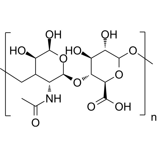
HY-B0633A
Hyaluronic acidGlycosaminoglycan -

HY-B0633B
Hyaluronic acid (MW 50000-100000)Biopolymer -

HY-B0633O
Hyaluronic acid sodium (MW 5000)Glycosaminoglycan -
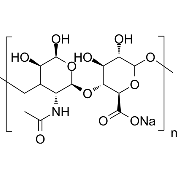
HY-B0633
Hyaluronic acid sodiumGlycosaminoglycan -

HY-B0633E
Hyaluronic acid, low endotoxinBiopolymer -

HY-B0633I
Hyaluronic acid sodium (MW 800kDa)Biopolymer -

HY-B0633P
Hyaluronic acid sodium (MW 0.9M-1.0Mda)Biopolymer -
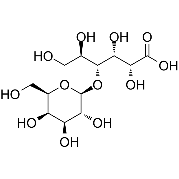
HY-N7059
Lactobionic acidBacterial Inhibitor -

HY-N7059R
Lactobionic acid (Standard)Bacterial Inhibitor


Biological Activity
Description
IC50 & Target
[2]|
IL-1β |
In Vitro
Hyaluronic acid (Mw:1000-2000Da) (2 kDa; 1-100 μg/mL; 48 h) inhibits the increase in inflammatory cytokine gene expression levels (TNF-α and IL-1β mRNA) and improves cell viability in H2O2-treated NHEK cells[2].
Hyaluronic acid (Mw:1000-2000Da) (2 kDa; 10-100 μg/mL; 48 h) significantly increases the cell viability of NHEK cells after UVB stimulation and increases LC3-II in NHEK cells irradiated with UVB[2].
MedChemExpress (MCE) has not independently confirmed the accuracy of these methods. They are for reference only. Further protocols information, click here.
-
Cell Line:Normal human epidermal keratinocytes (NHEK)
-
Concentration:0.1 μg/mL, 1 μg/mL, 10 μg/mL, 100 μg/mL
-
Incubation Time:24 h, 72 h (for cell viability evaluation in unstimulated cells); 48 h (for cell viability evaluation in stimulated cells)
-
Result:Showed no significant difference in cell viability after 24 h and 72 h (unstimulated NHEK cells).
Showed higher, and concentration-dependent cell viability than the control group after 1 μM H2O2 stimulation.
Showed higher cell viability than the control group after UVB stimulation (10 μg/mL and 100 μg/mL).


Chemical Information
-
CAS No. 9004-61-9
-
Appearance Solid
-
Molecular Weight 1000-2000
-
Formula (C14H21NO11)n
-
Color White to off-white
-
SMILES
CO[C@H]1[C@H](O)[C@@H](CO)O[C@@H](O[C@H]2[C@@H]([C@H]([C@@H](O[C@H]2C(O)=O)OC)O)O)[C@@H]1NC(C)=O.[n]
-
Shipping
Room temperature in continental US; may vary elsewhere.
-
Storage
4°C, protect from light
* In solvent : -80°C, 6 months; -20°C, 1 month (protect from light)


Solvent & Solubility
In Vitro:
H2O : 40 mg/mL (ultrasonic and warming and heat to 60°C)

Protocols
-
3D Hydrogel Synthetic Scaffold Culture
3D hydrogel synthetic scaffold culture embeds cells, spheroids, organoids, or tissue fragments inside a hydrated crosslinked polymer network so that cells receive matrix and cell-cell cues in three dimensions rather than from a flat plastic surface. A literature-supported model protocol is PEG-4MAL hydrogel culture, in which four-arm maleimide-terminated PEG is functionalized with cysteine-containing adhesive peptides such as RGD and crosslinked with protease-degradable peptides such as GPQ-W; this creates a defined, modular scaffold that supports human organoid generation and culture. The readouts are scaffold-supported growth, morphology, lumen formation, budding, viability, proliferation, lineage-marker expression, and matrix-dependent expansion or differentiation; reported assays include transmitted-light imaging, immunofluorescence, in situ hybridization, qRT-PCR, and rheological characterization.
-
RNA extraction experimental
By lysing cells, releasing RNA, and removing impurities such as proteins and DNA, high-purity RNA products are finally obtained. The commonly used traditional method is the guanidine isothiocyanate/phenol/chloroform method (Trizol), which is suitable for a variety of animal materials including animal tissues, microorganisms, cultured cells, etc., and most plant materials.
-
Human pluripotent stem cell endothelial-cell differentiation
Human pluripotent stem cell endothelial differentiation is based on stepwise developmental patterning: early activation of WNT/GSK3β inhibition promotes mesodermal or vascular progenitor entry, followed by endothelial specification using VEGF-related signaling, BMP4, FGF2, Notch modulation, or cAMP depending on the published protocol. Endothelial differentiation is read out by acquisition of CD31, CD34, VE-cadherin/CD144, KDR/VEGFR2, vWF, Tie2, NOS3, acetylated LDL uptake, tube/network formation, barrier function, and in vivo vessel-forming capacity where tested.
-
Vascular/Branching Fractal Analysis
Vascular/branching fractal analysis quantifies the geometric complexity of vessel trees or vascular networks from segmented 2D images, commonly by converting vessels into binary and/or skeletonized maps and estimating fractal dimension using box-counting or related approaches. Fractal dimension is interpreted as an image-derived readout of vascular branching complexity, space filling, or density, and has been applied to retinal photographs, fluorescein angiography, OCT angiography, capillary perfusion maps, and in vitro Matrigel angiogenesis networks. The assay readout is generated from vessel-positive pixels after image preprocessing, vessel segmentation, binarization, and optional skeletonization; reported outputs include fractal dimension, vessel density, branchpoint density, endpoint density, vessel length density, tortuosity, and generation-based branching metrics when VESGEN-style analysis is used. The biological interpretation is limited to quantitative vascular patterning and s
-
Autophagy
Autophagy is a process in which eukaryotic cells use lysosomes to degrade their own cytoplasmic proteins and damaged organelles under the regulation of autophagy related gene (Atg). Microtubule-associated proteins light chain 3 (LC3) is recognized as autophagy marker, which transfers from cytoplasmic LC3 (LC3-I) to membrane type (LC3-II). LC3-II/I ratio could be detected by Western Blot and fluorescence microscopy.
-
Endothelial Tube Formation Assay
Endothelial tube formation assay evaluates the ability of endothelial cells to attach, migrate, align, and organize into capillary-like networks when cultured on gelled basement membrane extract or Matrigel; the readout is the morphology and quantity of tube-like networks, which reflects an in vitro endothelial morphogenesis step related to angiogenesis. Basement membrane extract/Matrigel provides laminin-rich extracellular matrix cues that support endothelial differentiation into capillary-like structures, but it can contain biologically active growth factors, so growth-factor-reduced matrix is preferred when testing defined angiogenic stimulators or inhibitors.
-
Macroautophagy Solutions
Macroautophagy is a conserved lysosome-dependent degradation pathway in which cytoplasmic material is sequestered into double-membrane autophagosomes and delivered to lysosomes for degradation and recycling. The pathway supports cellular homeostasis during nutrient limitation, organelle stress, protein-aggregate accumulation, infection, differentiation, and tissue remodeling by coupling cargo sequestration, autophagosome maturation, lysosomal fusion, and degradation of cargo-derived macromolecules. The core molecular sequence includes initiation by nutrient- and stress-regulated autophagy machinery, autophagosome nucleation, LC3/ATG8-family conjugation to autophagosomal membranes, cargo selection through receptors such as SQSTM1/p62, autophagosome-lysosome fusion, and lysosomal degradation. LC3 was identified as a mammalian homolog of yeast Atg8 that localizes to autophagosomal membranes after processing, and p62/SQSTM1 was shown to connect ubiquitinated cargo with autophagic degradati
-
Endothelial Cell Migration/Angiogenic Sprouting Assay
Endothelial cell migration and angiogenic sprouting assays are in vitro (and partially ex vivo-adapted) functional models that quantify the ability of endothelial cells to undergo coordinated migration, extracellular matrix invasion, and multicellular organization into capillary-like sprouts in response to pro-angiogenic stimuli such as VEGF, bFGF, or conditioned microenvironments. These assays are used to model early angiogenic events including tip-cell formation, directional migration, and lumen-like sprout extension, which collectively reflect angiogenic activation and vascular morphogenesis processes observed in vivo.
-
Pyroptosis Solutions
Pyroptosis is a lytic inflammatory cell-death pathway executed by gasdermin pores, most classically through inflammasome-mediated activation of caspase-1, cleavage of gasdermin D, membrane pore formation, LDH release, and secretion of IL-1β and IL-18. The canonical pathway is commonly modeled by priming cells with an inflammatory signal such as LPS to induce pro-IL-1β and inflammasome components, followed by an activation signal such as ATP or nigericin to activate NLRP3, ASC speck formation, caspase-1 cleavage, GSDMD cleavage, cytokine release, and pyroptotic membrane rupture. The non-canonical pathway is triggered when cytosolic LPS activates mouse caspase-11 or human caspase-4/5, leading to GSDMD cleavage and pyroptosis, and this can secondarily activate NLRP3-dependent IL-1β release. Pyroptosis is linked to inflammatory injury, infection, cancer, liver disease, ocular disease, placental inflammation, and other disease phenotypes, but unresolved questions include which gasdermin fam


Purity & Documentation
-
Data Sheet (275 KB)
-
SDS (252 KB)
- English - EN (252 KB)
- Français - FR (252 KB)
- Deutsch - DE (252 KB)
- Norwegian - NO (252 KB)
- Español - ES (252 KB)
- Swedish - SV (252 KB)
- Italian - IT (252 KB)
- Korean - KR (252 KB)
- Portuguese - PT (252 KB)
-
Handling Instructions (2659 KB)
References

Calculators
Concentration (start) × Volume (start) = Concentration (final) × Volume (final)


Hyaluronic acid (Mw:1000-2000Da) Related Classifications
HY-B0633C Related Classifications
 Powered by Bioz