



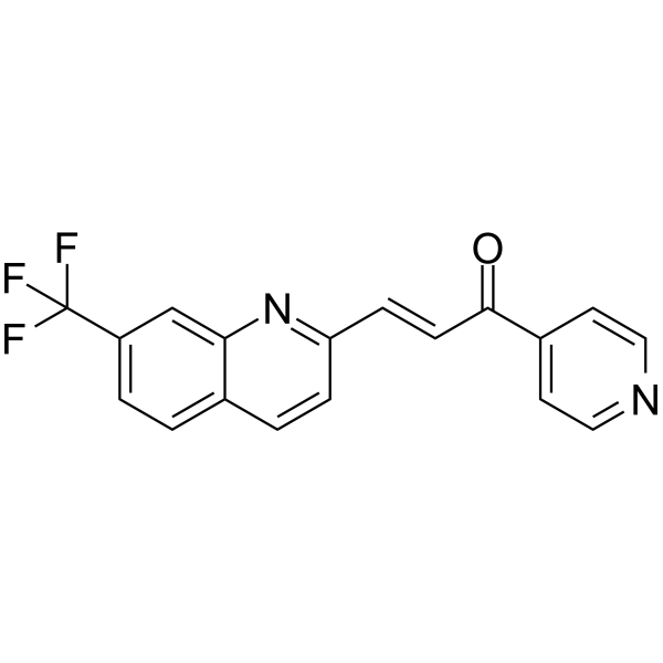
PFK-158
Based on 12 publication(s) in Google Scholar
PFK-158 is a potent and selective PFKFB3 inhibitor with an IC50 value 137 nM. PFK-158 reduces glucose uptake, ATP production, lactate release, and induces apoptosis and autophagy in cancer cells. PFK-158 has broad anti-tumor activity. PFK-158 can also enhance Colistin's resistance to bacteria.
For research use only. We do not sell to patients.
- Purity : 98.95%
- CAS No.: 1462249-75-7
- Formula: C18H11F3N2O
- Molecular Weight:328.29
-
Storage:Powder -20°C, 3 years , 4°C, 2 years ; In solvent -80°C, 6 months , -20°C, 1 month

To place orders, for customer services and technical support, please contact: MedChemExpress USA
Tel: 609-228-6898 E-mail: [email protected] [email protected]


-
 Biological Activity
-
Chemical Information
-
Solvent & Solubility
- Protocols
- Purity & Documentation
- References
-
 Help & FAQs


-
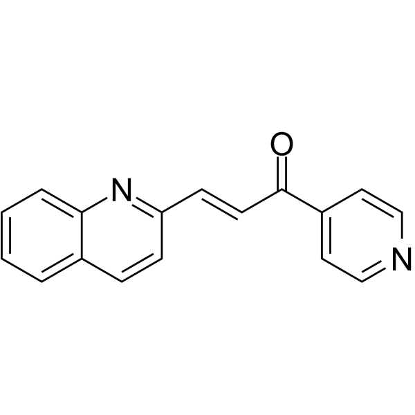
HY-12204
PFK-015PFKFB3 Inhibitor -
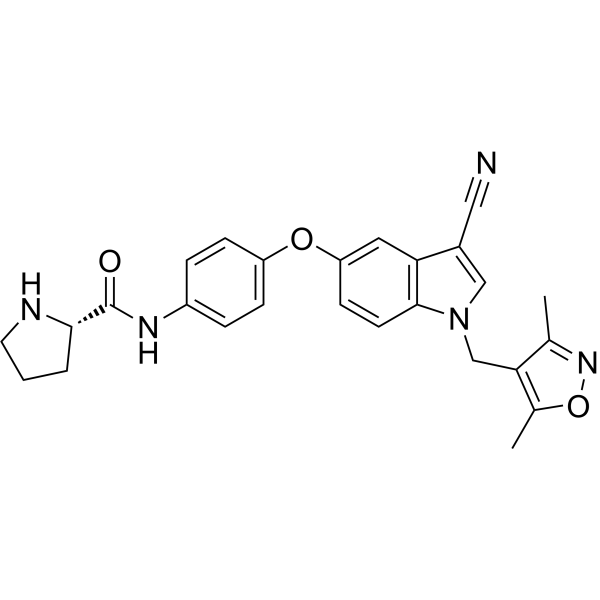
HY-101972
AZ-PFKFB3-67PFKFB3 Inhibitor -
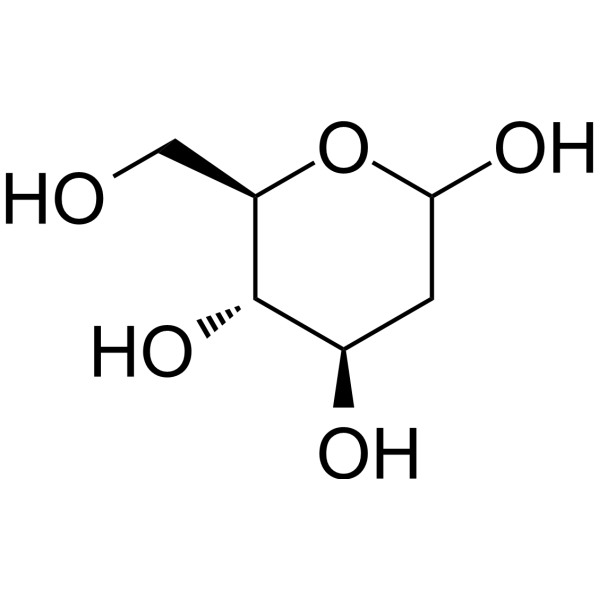
HY-13966
2-Deoxy-D-glucoseGlycolysis Inhibitor -
HY-50202
EtomoxirCPT1a Inhibitor -
HY-15452
SelisistatSirT1/Sir2 Inhibitor -
HY-W107464
G6PDi-1G6PD Inhibitor -

HY-13259
MG-132Proteasome Inhibitor -

HY-N6782
OligomycinH+-ATP-Synthase Inhibitor -

HY-10071
Y-27632ROCK Inhibitor -
HY-10295
SB 202190p38 MAPK Inhibitor -

HY-Y0320
Dimethyl sulfoxide, meets analytical specification of Ch.P.Aprotic Solvent -
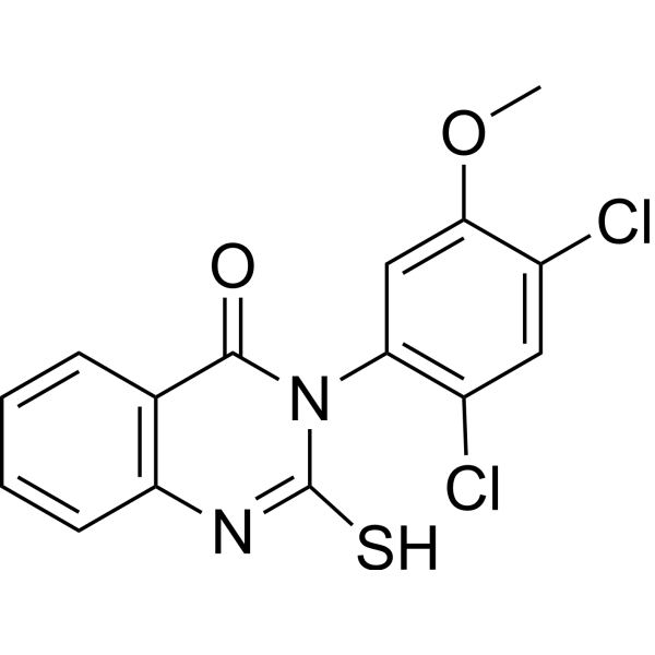
HY-15886
Mdivi-1Drp1 Inhibitor
Publications Citing Use of MedChemExpress (MCE) PFK-158
More- Cell. 2025 Aug 21;188(17):4567-4585.e32. [Abstract]
- Nat Commun. 2026 Oct 1.
- Adv Sci (Weinh). 2025 May;12(19):e2411980. [Abstract]
- Cell Rep Med. 2025 Feb 18;6(2):101966. [Abstract]
- Cell Rep. 2023 Dec 18;43(1):113557. [Abstract]
- Cells. 2021 Jul 3;10(7):1679. [Abstract]
- Cancer Metab. 2024 Dec 18;12(1):37. [Abstract]
- Exp Neurol. 2024 Jan:371:114590. [Abstract]
- J Biol Chem. 2019 Jul 5;294(27):10530-10543. [Abstract]
- Infect Drug Resist. 2021 Jun 9:14:2143-2154. [Abstract]
- bioRxiv. 2024 Mar 27.
- Research Square Preprint. 2021 Nov.
 Customer Validation & Images

-
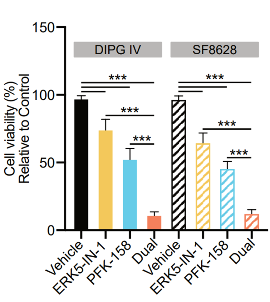
Cell Proliferation/Viability Assay
-
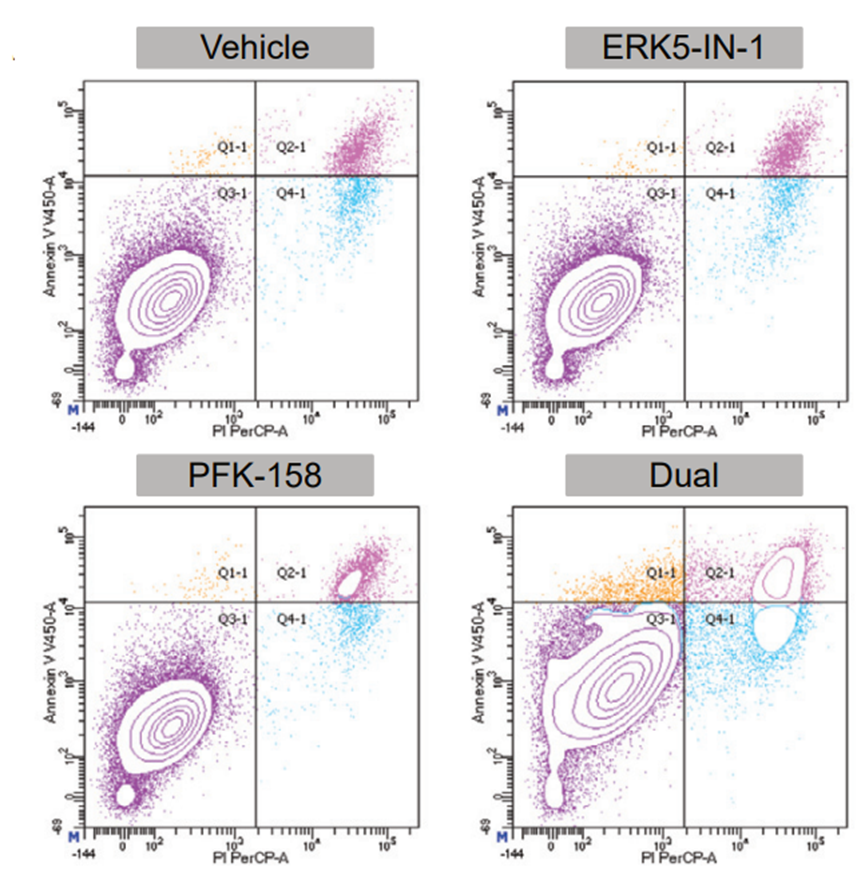
Flow Cytometry
-
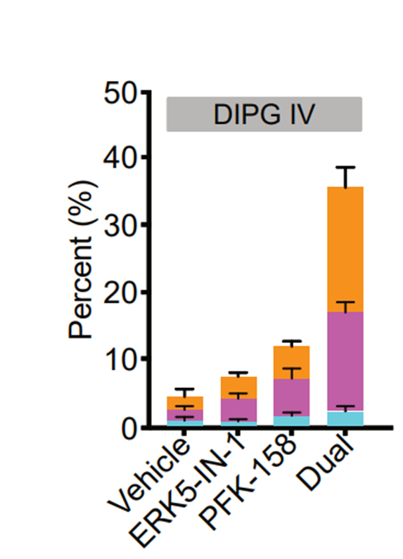
Apoptosis Analysis
-
WB
-
Cell Imaging/Staining


Biological Activity
Description
IC50 & Target
Cellular Effect
|
Cell Line

|
Type | Value | Description | References |
|---|---|---|---|---|
| A549 | IC50 |
15 μM
Compound: A13; PFK-158
|
Cytotoxicity against human A549 cells
Cytotoxicity against human A549 cells
|
[PMID: 33766773] |
In Vitro
PFK-158 (10 μM ; 24 hours; OV2008 and C13 cells) combined with Carboplatin (CBPt; 77-453 μM) results in significant increase in apoptosis in C13 (45%) and OV2008 cells (24.6%)[1].
PFK-158 (0-10 μM ; 24 hours; C13 and HeyA8MDR cells) treatment results in a dose-dependent decrease in p-PFKFB3, p-cPLA2 and lipid droplet (LD) levels[1].
PFK-158 (10 μM; 24 hours) has synergistic anti-proliferative effects in vitro when combined with Cisplatin in C13 and HeyA8MDR cells compared to OV2008 and HeyA8, respectively[1].
PFK-158 (0‐10 μM; 24 h) treatment shows a dose-dependent downregulation of p62/SQSTM1 and upregulation of LC3BII, two markers of autophagy induction, in both C13 and HeyA8MDR cells. PFK-158 treatment also reduces the numbers of LDs[1].
MedChemExpress (MCE) has not independently confirmed the accuracy of these methods. They are for reference only.
-
Cell Line:OV2008 and C13 cells
-
Concentration:10 µM
-
Incubation Time:24 hours
-
Result:Combined with Carboplatin (CBPt) treatment resulted in significant increase in apoptosis.
-
Cell Line:C13 and HeyA8MDR cells
-
Concentration:0 µM, 5 µM, 10 µM
-
Incubation Time:24 hours
-
Result:Demonstrated a dose-dependent decrease in p-PFKFB3, p-cPLA2 and lipid droplet (LD) levels.
In Vivo
MedChemExpress (MCE) has not independently confirmed the accuracy of these methods. They are for reference only.
-
Animal Model:Female athymic nude mice (nu/nu) (5-6 weeks old) injected with HeyA8MDR cells[1]
-
Dosage:15 mg/kg
-
Administration:Intraperitoneal injection; once a week; for 4 weeks
-
Result:A marked reduction of tumor growth was observed in the combination treatment.
Clinical Trial
| NCT Number | Sponsor | Condition | Start Date |
Phase
|
|---|---|---|---|---|
| NCT01329991 | Plexxikon| | 2011-05 | PHASE1 |


Chemical Information
-
CAS No. 1462249-75-7
-
Appearance Solid
-
Molecular Weight 328.29
-
Formula C18H11F3N2O
-
Color White to light yellow
-
SMILES
O=C(C1=CC=NC=C1)/C=C/C2=NC3=CC(C(F)(F)F)=CC=C3C=C2
-
Shipping
Room temperature in continental US; may vary elsewhere.
-
Storage
Powder -20°C 3 years 4°C 2 years In solvent -80°C 6 months -20°C 1 month


Publications (12)
-
Journal Impact Factor
-
Most Recent
-
Cell
The immunoproteasome disturbs neuronal metabolism and drives neurodegeneration in multiple sclerosis. [Abstract]2025 Aug 21;188(17):4567-4585.e32. PMID: 40532699 -
-
Adv Sci (Weinh)
The Novel Dual GIP and GLP-1 Receptor Agonist Tirzepatide Attenuates Colon Cancer Development by Regulating Glucose Metabolism. [Abstract]2025 May;12(19):e2411980. PMID: 40125821 -
Cell Rep Med
KRASG12D-driven pentose phosphate pathway remodeling imparts a targetable vulnerability synergizing with MRTX1133 for durable remissions in PDAC. [Abstract]2025 Feb 18;6(2):101966. PMID: 39970873 -
Cell Rep
An ERK5-PFKFB3 axis regulates glycolysis and represents a therapeutic vulnerability in pediatric diffuse midline glioma. [Abstract]2023 Dec 18;43(1):113557. PMID: 38113141
PFK-158 purchased from MedChemExpress. Usage Cited in: Cell Rep. 2023 Dec 18;43(1):113557. [Abstract]
Cell viability of DIPG IV and SF8628 cells treated with ERK5-IN-1, PFK-158, or combination ERK5-IN-1 and PFK-158 (dual) compared with vehicle control.
PFK-158 purchased from MedChemExpress. Usage Cited in: Cell Rep. 2023 Dec 18;43(1):113557. [Abstract]
Flow cytometry plots with annexin-propidium iodide (PI) assay in SF8268 cells treated with ERK5-IN-1, PFK-158, or combination ERK5-IN-1 and PFK-158 (dual) compared with vehicle control.
PFK-158 purchased from MedChemExpress. Usage Cited in: Cell Rep. 2023 Dec 18;43(1):113557. [Abstract]
Apoptosis of DIPG IV cells treated with ERK5-IN-1, PFK-158, or combination ERK5-IN-1 and PFK-158 (dual) compared with vehicle control.
-
Cells
2021 Jul 3;10(7):1679. PMID: 34359849
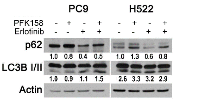
PFK-158 purchased from MedChemExpress. Usage Cited in: Cells. 2021 Jul 3;10(7):1679. [Abstract]
PC9 and H522 cells were transfected with a pEGFP-LC3 plasmid and treated with erlotinib (PC9: 0.5 µM; H522: 2 µM) and/or PFK158 (PC9: 5 µM; H522: 0.75 µM) for 36 h or 24 h respectively, Whole cell lysates were immunoblotted with the indicated antibodies.
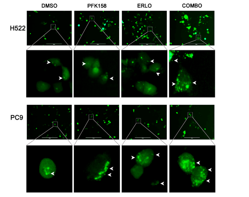
PFK-158 purchased from MedChemExpress. Usage Cited in: Cells. 2021 Jul 3;10(7):1679. [Abstract]
PC9 and H522 cells were transfected with a pEGFP-LC3 plasmid and treated with erlotinib (PC9: 0.5 µM; H522: 2 µM) and/or PFK158 (PC9: 5 µM; H522: 0.75 µM) for 36 h or 24 h, respectively. GFP-LC3 localization was evaluated by fluorescence microscopy.
-
Cancer Metab
PFKFB3-dependent redox homeostasis and DNA repair support cell survival under EGFR-TKIs in non-small cell lung carcinoma. [Abstract]2024 Dec 18;12(1):37. PMID: 39696407 -
Exp Neurol
2024 Jan:371:114590. PMID: 37907123 -
J Biol Chem
Increased 6-phosphofructo-2-kinase/fructose-2,6-bisphosphatase-3 activity in response to EGFR signaling contributes to non-small cell lung cancer cell survival. [Abstract]2019 Jul 5;294(27):10530-10543. PMID: 31126985 -
Infect Drug Resist
Synergistic Activity and Biofilm Formation Effect of Colistin Combined with PFK-158 Against Colistin-Resistant Gram-Negative Bacteria. [Abstract]2021 Jun 9:14:2143-2154. PMID: 34135604 -
-


Solvent & Solubility
In Vitro:
DMSO : 50 mg/mL (152.30 mM; Need ultrasonic; Hygroscopic DMSO has a significant impact on the solubility of product, please use newly opened DMSO)
Please refer to the solubility information to select the appropriate solvent. Once prepared, please aliquot and store the solution to prevent product inactivation from repeated freeze-thaw cycles.
Storage method and period of stock solution: -80°C, 6 months; -20°C, 1 month. When stored at -80°C, please use it within 6 months. When stored at -20°C, please use it within 1 month.
Please refer to the solubility information to select the appropriate solvent. Once prepared, please aliquot and store the solution to prevent product inactivation from repeated freeze-thaw cycles.
Storage method and period of stock solution: -80°C, 6 months; -20°C, 1 month. When stored at -80°C, please use it within 6 months. When stored at -20°C, please use it within 1 month.
Concentration (start) × Volume (start) = Concentration (final) × Volume (final)
In Vivo:
For the following dissolution methods, please prepare the working solution directly:
It is recommended to prepare fresh solutions and use them promptly within a short period of time.
The percentages shown for the solvents indicate their volumetric ratio in the final prepared solution. If precipitation or phase separation occurs during preparation, heat and/or sonication can be used to aid dissolution.


Purity & Documentation
-
Data Sheet (277 KB)
-
SDS (581 KB)
- English - EN (581 KB)
- Français - FR (581 KB)
- Deutsch - DE (581 KB)
- Norwegian - NO (581 KB)
- Español - ES (581 KB)
- Swedish - SV (581 KB)
- Italian - IT (581 KB)
- Korean - KR (581 KB)
- Portuguese - PT (581 KB)
-
Handling Instructions (2659 KB)
References
[1]. Mondal S, et al. Therapeutic targeting of PFKFB3 with a novel glycolytic inhibitor PFK158 promotes lipophagy and chemosensitivity in gynecologic cancers. Int J Cancer. 2019 Jan 1;144(1):178-189. [Content Brief]
[2]. Zhang Y, et al. Synergistic Effect of Colistin Combined with PFK-158 against Colistin-Resistant Enterobacteriaceae. Antimicrob Agents Chemother. 2019 Jun 24;63(7). pii: e00271-19. [Content Brief]


Complete Stock Solution Preparation Table
Please refer to the solubility information to select the appropriate solvent. Once prepared, please aliquot and store the solution to prevent product inactivation from repeated freeze-thaw cycles.
Storage method and period of stock solution: -80°C, 6 months; -20°C, 1 month. When stored at -80°C, please use it within 6 months. When stored at -20°C, please use it within 1 month.
| Optional Solvent | Concentration Solvent Mass | 1 mg | 5 mg | 10 mg | 25 mg |
|---|---|---|---|---|---|
| DMSO | 1 mM | 3.0461 mL | 15.2304 mL | 30.4609 mL | 76.1522 mL |
| 5 mM | 0.6092 mL | 3.0461 mL | 6.0922 mL | 15.2304 mL | |
| 10 mM | 0.3046 mL | 1.5230 mL | 3.0461 mL | 7.6152 mL | |
| 15 mM | 0.2031 mL | 1.0154 mL | 2.0307 mL | 5.0768 mL | |
| 20 mM | 0.1523 mL | 0.7615 mL | 1.5230 mL | 3.8076 mL | |
| 25 mM | 0.1218 mL | 0.6092 mL | 1.2184 mL | 3.0461 mL | |
| 30 mM | 0.1015 mL | 0.5077 mL | 1.0154 mL | 2.5384 mL | |
| 40 mM | 0.0762 mL | 0.3808 mL | 0.7615 mL | 1.9038 mL | |
| 50 mM | 0.0609 mL | 0.3046 mL | 0.6092 mL | 1.5230 mL | |
| 60 mM | 0.0508 mL | 0.2538 mL | 0.5077 mL | 1.2692 mL | |
| 80 mM | 0.0381 mL | 0.1904 mL | 0.3808 mL | 0.9519 mL | |
| 100 mM | 0.0305 mL | 0.1523 mL | 0.3046 mL | 0.7615 mL |


PFK-158 Related Classifications
HY-12203 Related Classifications
 Powered by Bioz