



Benoxacor

Based on 1 Customer Validation
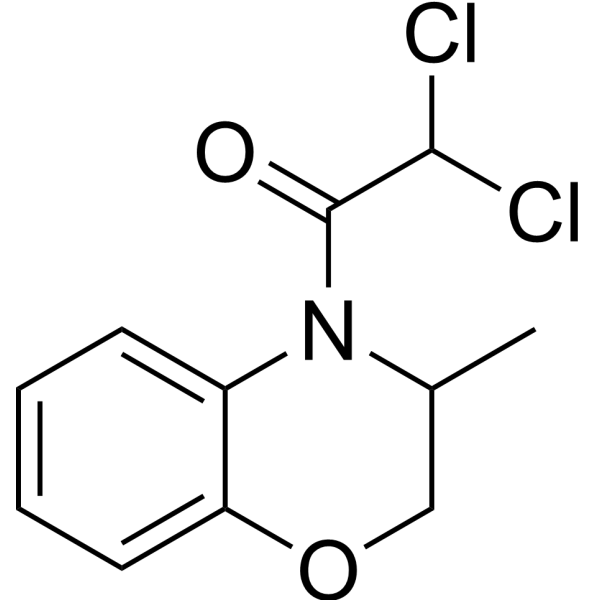
Benoxacor (CGA 154281) is a herbicide safener and xenobiotic metabolism regulator. Benoxacor protects maize from the toxicity of metolachlor mainly by inducing detoxifying enzymes such as Glutathione S-transferase. Benoxacor also activates FXR, PXR and ERRα, and inhibits aromatase (aromatase). However, Benoxacor exhibits potential subacute oral toxicity and a high risk of hepatotoxicity in animal models. Benoxacor induces reactive oxygen species accumulation, interferes with embryonic heart development, and causes increased liver and kidney weights as well as alterations in gut microbiota in mice. Benoxacor can be used in studies related to hepatic steatosis, infertility, breast cancer and developmental toxicity.
For research use only. We do not sell to patients.
- Purity : 99.39%
- CAS No.: 98730-04-2
- Formula: C11H11Cl2NO2
- Molecular Weight:260.12
-
Storage:
4°C, stored under nitrogen
* In solvent : -80°C, 6 months; -20°C, 1 month (stored under nitrogen)

To place orders, for customer services and technical support, please contact: MedChemExpress USA
Tel: 609-228-6898 E-mail: [email protected] [email protected]


-
 Biological Activity
-
Chemical Information
-
Solvent & Solubility
- Protocols
- Purity & Documentation
- References
-
 Help & FAQs


-
HY-W009861
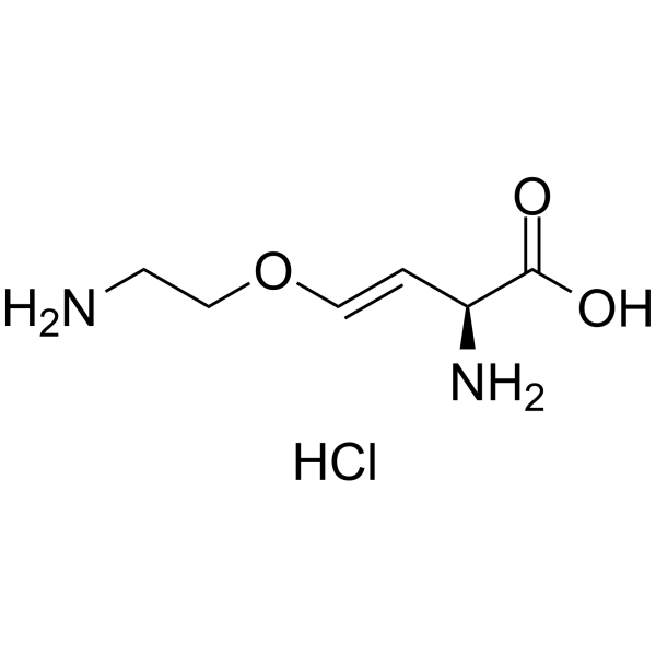
Aviglycine hydrochlorideEthylene Biosynthesis Inhibitor -
HY-N0548
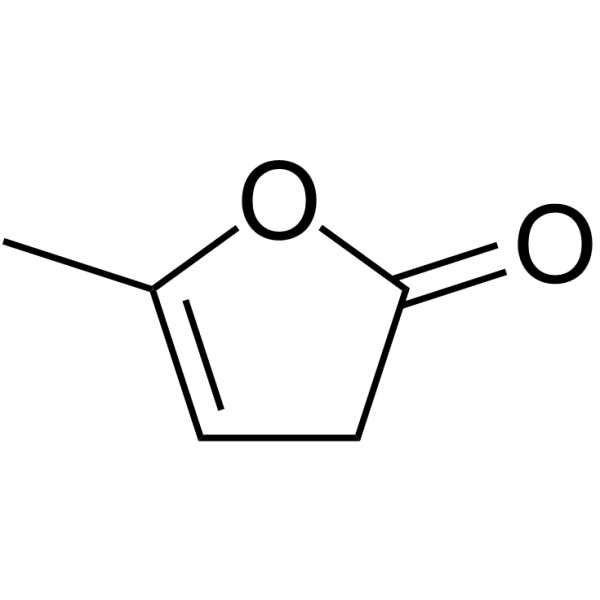
α-Angelica lactoneChemopreventative Agent -
HY-111530
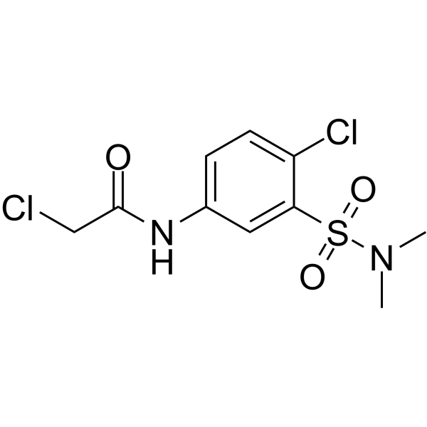
GSTO1-IN-1Gutathione S-transferase Inhibitor -
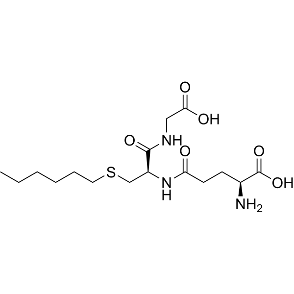
HY-W013754
S-HexylglutathioneGlutathione-S-transferase Inhibitor -
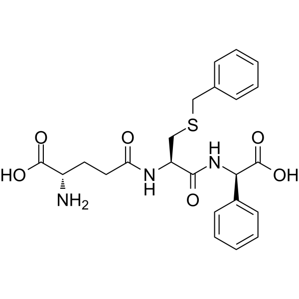
HY-13634B
TLK117GSTP1-1 Inhibitor -
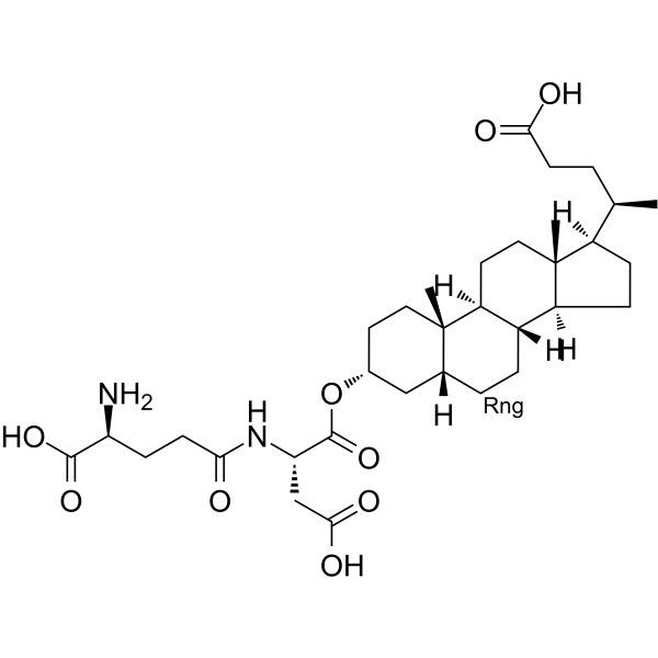
HY-112534
GSTO-IN-2Glutathione S-transferase Inhibitor -
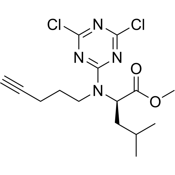
HY-115673
LAS17GSTP1 Inhibitor -
HY-137877
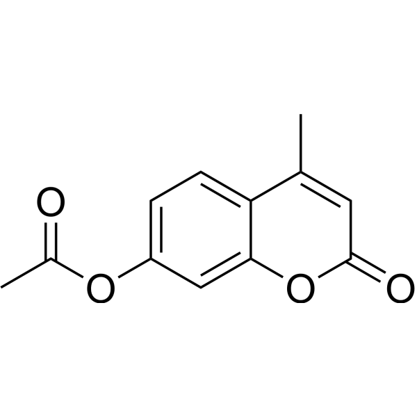
7-Acetoxy-4-methylcoumarinGST Inhibitor -
HY-117143
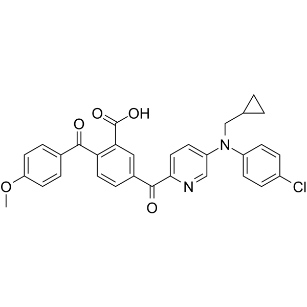
TK05LTC4S Inhibitor -
HY-126329
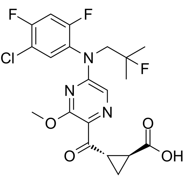
AZD9898Gutathione S-transferase Inhibitor -

HY-N0548R
α-Angelica lactone (Standard)Chemopreventative Agent -

HY-W020788R
Benoxacor (Standard)Herbicide Safety Agent


Biological Activity
Description
IC50 & Target
|
ERRα |
In Vitro
Benoxacor (5 μM; 0-30 min) is metabolized by liver microsomes from male C57BL/6 mice in a time-dependent and enantioselective manner. Cytochrome P450 enzymes primarily metabolize the E2-benoxacor enantiomer, while microsomal carboxylesterases mainly metabolize the E1-benoxacor enantiomer. The intrinsic clearance of microsomal CYP-mediated elimination is 1.05 mL/min/mg protein, with a normalized clearance of 40.22 mL/min/g liver, whereas the intrinsic clearance of microsomal CES-mediated elimination is 0.54 mL/min/mg protein, with a normalized clearance of 20.77 mL/min/g liver[1].
Benoxacor (5 μM; 0-30 min) is metabolized in a time-dependent and enantioselective manner by glutathione S-transferase in the liver cytosol of male C57BL/6 mice, with an intrinsic clearance rate of 1.46 mL/min/mg protein and a normalized clearance rate of 132.52 mL/min/g liver for GST-mediated elimination[1].
Benoxacor (10 μM; 0.25-48 h) is rapidly taken up by suspension-cultured cells of maize (cultivar: Black Mexican Sweet, same below), and the uptake amount increases continuously within 48 h after treatment with 10 μM[3].
Treatment of maize suspension-cultured cells with Benoxacor (10 μM; 0-48 h) doubles the activity of GST-M, a change detectable as early as 4 h post-treatment and peaking at 24 h[3].
Benoxacor (10 μM; 24 h) increases the activities of three distinct GSTs (a, c, d) isolated via anion exchange chromatography from treated maize suspension-cultured cells[3].
Isolation of GST fractions (b, c, d) from maize suspension-cultured cells treated with Benoxacor (10 μM; 24 h) reveals GST polypeptides (26,000-27,000 Mr) that cross-react with maize GST I and GST I/III antisera, a pattern consistent with that observed in benzofenap-treated maize seedlings[3].
Treatment of maize suspension-cultured cells with Benoxacor (10 μM; 24 h) increases GST-M activity by 3-fold, and this effect is partially blocked by pre-treatment with Cycloheximide (HY-12320) (10/100 μM; 2 h), indicating that this induction activity requires de novo protein synthesis[3].
Treatment of maize suspension-cultured cells with Benoxacor (10 μM; 24 h) increases GST-M activity by 3-fold, and this enhancing effect is significantly blocked by pretreatment with Cordycepin (HY-N0262) (10/100 μM; 2 h), indicating that this inductive activity requires de novo RNA synthesis[3].
MedChemExpress (MCE) has not independently confirmed the accuracy of these methods. They are for reference only. Further protocols information, click here.
-
Cell Line:Zea mays (cv. Black Mexican Sweet) suspension-cultured cells
-
Concentration:10 μM
-
Incubation Time:24 h
-
Result:Detected a 27,000 Mr polypeptide in fractions corresponding to activities b and c with maize GST I antisera.
Detected a 27,000 Mr polypeptide in fraction corresponding to activity b, and a 27,000 Mr plus a slightly reactive 26,000 Mr polypeptide in fraction corresponding to activity c with maize GST I/III antisera.
Detected a slightly reactive 26,000 Mr polypeptide in fraction corresponding to activity d with maize GST I/III antisera.
In Vivo
Benoxacor (0.5-2.0 mg/L; waterborne exposure; continuous exposure; 5.5 to 72 hpf) induces concentration-dependent developmental toxicity, cardiac morphological and functional abnormalities, oxidative stress, as well as apoptosis and dysregulated expression of cardiac development-related genes in zebrafish embryos, with the most severe effects observed at the 2.0 mg/L dose[2].
MedChemExpress (MCE) has not independently confirmed the accuracy of these methods. They are for reference only.
-
Animal Model:C57BL/6J mice (male, 8-week-old, acclimated for 4 weeks from 4-week-old)[1]
-
Dosage:0.5 mg/kg b.w.; 5 mg/kg b.w.; 50 mg/kg b.w.
-
Administration:p.o.; daily; 3 consecutive days
-
Result:Increased bodyweight-adjusted liver/testes weight significantly at 50 mg/kg.
Caused no significant changes in body weight or bodyweight-adjusted spleen, kidney, or brain weights at any dose.
Induced no overt pathological changes in liver or intestinal tissues at any dose.
Caused no significant effects on cecal microbiome α-diversity (Shannon, Chao-1 indices) or β-diversity (Weighted Unifrac distance) at any dose.
Identified 163 hepatic differentially expressed genes (DEGs) across all doses, including xenobiotic processing genes Cyp7a1, Cyp3a13, and Cyp26a1; the gene regulation profile matched that of dichloroacetic acid and sedaxane, with 15/16 and 15/21 consistent DE targets respectively, though this was not significant after multiple comparison correction.
Altered 9 serum and 15 liver metabolites before multiple comparison correction; in liver, creatinine and trigonelline were significantly altered, but no changes remained significant after false discovery rate correction.
Caused no significant changes in hepatic catalase, glutathione peroxidase (GPx), superoxide dismutase (SOD), or glutathione S-transferase (GST) activity at any dose.
-
Animal Model:wild-type AB; transgenic Tg Zebrafish (myl7:GFP) (embryos, exposed at 5.5 hours post-fertilization)[2]
-
Dosage:0.5 mg/L; 1.0 mg/L; 2.0 mg/L
-
Administration:aqueous exposure; continuous; 5.5 to 72 hpf
-
Result:Increased total mortality rate in a concentration-dependent manner, reaching ~13% at 72 hpf for the 2.0 mg/L dose, ~10% for the 1.0 mg/L dose, and ~6% for the 0.5 mg/L dose.
Reduced heart rate significantly in a concentration-dependent manner, with the 2.0 mg/L dose causing the greatest reduction relative to controls.
Reduced hatching rate significantly in a concentration-dependent manner at both 48 and 72 hpf, with the 2.0 mg/L dose resulting in the lowest hatching rate (~40%) at 72 hpf.
Induced pericardial edema and linear stretching of the heart, with failure of heart cyclization observed in all treated groups; revealed abnormalities in the myocardial layer and linearized atria/ventricles via histological analysis.
Increased ROS fluorescence intensity significantly in a concentration-dependent manner, with the 2.0 mg/L dose showing the highest intensity (~65,000 fluorescence units).
Elevated SOD and CAT activities in a concentration-dependent manner.
Decreased MDA content at 0.5 mg/L and 1.0 mg/L, then significantly increased it at 2.0 mg/L (~3.5 nmol/mL).
Upregulated relative mRNA levels of pro-apoptotic gene caspase 3 (peak ~3.2-fold at 1.0 mg/L) and cardiac development-related genes nkx2.5, myh6, tbx5a, vmhc, and nppa.
Downregulated relative mRNA levels of gata4 and tbx2b in a concentration-dependent manner, with caspase 9 showing a slight concentration-dependent decrease.


Chemical Information
-
CAS No. 98730-04-2
-
Appearance Solid
-
Molecular Weight 260.12
-
Formula C11H11Cl2NO2
-
Color White to off-white
-
SMILES
ClC(Cl)C(N1C(C)COC2=CC=CC=C21)=O
-
Synonyms
CGA 154281
-
Shipping
Room temperature in continental US; may vary elsewhere.
-
Storage
4°C, stored under nitrogen
* In solvent : -80°C, 6 months; -20°C, 1 month (stored under nitrogen)


Solvent & Solubility
In Vitro:
DMSO : 125 mg/mL (480.55 mM; Need ultrasonic; Hygroscopic DMSO has a significant impact on the solubility of product, please use newly opened DMSO)
Please refer to the solubility information to select the appropriate solvent. Once prepared, please aliquot and store the solution to prevent product inactivation from repeated freeze-thaw cycles.
Storage method and period of stock solution: -80°C, 6 months; -20°C, 1 month (stored under nitrogen). When stored at -80°C, please use it within 6 months. When stored at -20°C, please use it within 1 month.
Please refer to the solubility information to select the appropriate solvent. Once prepared, please aliquot and store the solution to prevent product inactivation from repeated freeze-thaw cycles.
Storage method and period of stock solution: -80°C, 6 months; -20°C, 1 month (stored under nitrogen). When stored at -80°C, please use it within 6 months. When stored at -20°C, please use it within 1 month.
Concentration (start) × Volume (start) = Concentration (final) × Volume (final)

Protocols
-
Reproductive and Developmental Toxicity Study
Reproductive and developmental toxicity studies detect adverse effects of prenatal or peri/postnatal exposure on maternal condition, pregnancy maintenance, embryo-fetal survival, fetal growth, structural development, and offspring reproductive or developmental endpoints; classic rat protocols generate readouts by comparing treated groups with vehicle, pair-fed, or untreated controls for implantation, resorption, fetal weight, crown-rump length, external morphology, visceral morphology, skeletal ossification, anogenital distance, nipple/areola retention, and postnatal cohort outcomes.
-
ROS/oxidative-stress fluorescent staining
ROS/oxidative-stress fluorescent staining uses cell-permeant fluorogenic probes that become fluorescent after oxidation inside cells or tissues; commonly used examples include DCFH-DA/DCFDA for broad cellular oxidant detection, DHE for superoxide-related signal detection, MitoSOX for mitochondrial superoxide-related signal detection, and CellROX probes for oxidative-stress-associated fluorescence readouts. The assay detects probe oxidation rather than a single ROS species unless the probe and analysis method have been chemically validated for that species. DCFH-DA enters cells, is deacetylated by intracellular esterases to DCFH, and produces fluorescent DCF after oxidation, so the readout is used as an operational measure of total cellular oxidative stress rather than a species-specific ROS measurement. DHE and MitoSOX can report superoxide-related oxidation, but red fluorescence alone can include non-specific ethidium-like oxidation products; HPLC or optimized spectral approaches are
-
Research Protocol for Microbiome Analysis
Microbiome analysis characterizes microbial communities in biological or environmental samples by measuring community composition, diversity, taxonomic structure, functional potential, and associations with host or environmental phenotypes. 16S rRNA gene amplicon sequencing is commonly used for bacterial and archaeal taxonomic profiling, while shotgun metagenomics provides higher taxonomic resolution and direct functional information, including microbial genes, pathways, viruses, fungi, and antimicrobial-resistance genes when sequencing depth and host-DNA contamination are adequately controlled. Microbiome results are strongly affected by sample collection, storage, DNA extraction, contamination, sequencing method, reference database, and bioinformatic pipeline; therefore, standardized protocols, negative controls, mock communities, and transparent analysis workflows are required. Unresolved issues include low-biomass contamination, compositional-data bias, inconsistent species-level c
-
Repeated-Dose Oral Toxicity Study
A repeated-dose oral toxicity study evaluates systemic toxic effects after daily oral exposure to a test substance for a defined period, commonly 28 days, 6 weeks, 90 days, or 13 weeks in rodent studies. The readout is generated by integrating mortality, clinical signs, body-weight change, food and water intake, functional or behavioral observations, hematology, serum biochemistry, urinalysis, organ weights, necropsy, and histopathology to identify dose-related adverse effects, target organs, and the no-observed-adverse-effect level (NOAEL).
-
Hepatotoxicity Study
This protocol evaluates hepatotoxicity using complementary in vivo mouse APAP acute liver injury and in vitro hepatocyte-based cytotoxicity readouts. In vivo APAP injury is assessed by serum ALT/AST, liver histology, hepatic glutathione, protein adducts, necrosis, inflammation, and regeneration-related endpoints. In vitro hepatotoxicity is assessed by loss of viability, leakage of ALT/AST/LDH, oxidative-stress markers, mitochondrial function, nuclear morphology, intracellular calcium, and high-content imaging endpoints.
-
Breast Cancer Modeling
Breast cancer is a heterogeneous cancer, and it has been distinguished into four subtypes: luminal A, luminal B, HER2-positive and basal-like. Molecular mutations, epigenetic alterations, hormone exposure and immune microenvironment are related to the progression of breast cancer.
-
Liver Histomorphometry
Liver histomorphometry is a quantitative histological approach used to measure structural alterations in hepatic tissue, including parenchymal loss, steatosis, fibrosis, and vascular remodeling, by combining stained tissue section analysis with stereological or computerized image-based measurements. Classical morphometric frameworks quantify volume fractions of liver compartments and fibrotic regions using systematic sampling and image analysis, enabling objective comparison of pathological changes across experimental groups. These approaches are widely applied in liver cirrhosis and fibrosis studies to reduce subjectivity in histological scoring and improve reproducibility of tissue evaluation. Recent methodological advances integrate automated image analysis and radiomics-based extraction of histological features from standard liver stains (e. g. , H&E and fibrotic stains), enabling quantitative correlation between morphometric features and fibrosis stages in non-alcoholic fatty live


Purity & Documentation
-
Data Sheet (289 KB)
-
SDS (597 KB)
- English - EN (597 KB)
- Français - FR (597 KB)
- Deutsch - DE (597 KB)
- Norwegian - NO (597 KB)
- Español - ES (597 KB)
- Swedish - SV (597 KB)
- Italian - IT (597 KB)
- Korean - KR (597 KB)
- Portuguese - PT (597 KB)
-
Handling Instructions (2659 KB)
References


Complete Stock Solution Preparation Table
Please refer to the solubility information to select the appropriate solvent. Once prepared, please aliquot and store the solution to prevent product inactivation from repeated freeze-thaw cycles.
Storage method and period of stock solution: -80°C, 6 months; -20°C, 1 month (stored under nitrogen). When stored at -80°C, please use it within 6 months. When stored at -20°C, please use it within 1 month.
| Optional Solvent | Concentration Solvent Mass | 1 mg | 5 mg | 10 mg | 25 mg |
|---|---|---|---|---|---|
| DMSO | 1 mM | 3.8444 mL | 19.2219 mL | 38.4438 mL | 96.1095 mL |
| 5 mM | 0.7689 mL | 3.8444 mL | 7.6888 mL | 19.2219 mL | |
| 10 mM | 0.3844 mL | 1.9222 mL | 3.8444 mL | 9.6109 mL | |
| 15 mM | 0.2563 mL | 1.2815 mL | 2.5629 mL | 6.4073 mL | |
| 20 mM | 0.1922 mL | 0.9611 mL | 1.9222 mL | 4.8055 mL | |
| 25 mM | 0.1538 mL | 0.7689 mL | 1.5378 mL | 3.8444 mL | |
| 30 mM | 0.1281 mL | 0.6407 mL | 1.2815 mL | 3.2036 mL | |
| 40 mM | 0.0961 mL | 0.4805 mL | 0.9611 mL | 2.4027 mL | |
| 50 mM | 0.0769 mL | 0.3844 mL | 0.7689 mL | 1.9222 mL | |
| 60 mM | 0.0641 mL | 0.3204 mL | 0.6407 mL | 1.6018 mL | |
| 80 mM | 0.0481 mL | 0.2403 mL | 0.4805 mL | 1.2014 mL | |
| 100 mM | 0.0384 mL | 0.1922 mL | 0.3844 mL | 0.9611 mL |


Benoxacor Related Classifications
HY-W020788 Related Classifications
 Powered by Bioz
Keywords
- Benoxacor
- 98730-04-2
- CGA 154281
- CGA154281
- CGA-154281
- Environmental Pollutants
- Pregnane X Receptor (PXR)
- Estrogen Receptor/ERR
- Glutathione S-transferase
- FXR
- carboxylesterases
- pregnane X receptor
- estrogen receptor alpha
- male C57BL/6 mice
- Zea mays
- glutathione S-transferases
- aromatase
- farnesoid X receptor
- cytochrome P450 enzymes
- zebrafish embryos
- Inhibitor
- inhibitor
- inhibit