



Herniarin

Based on 1 Customer Validation
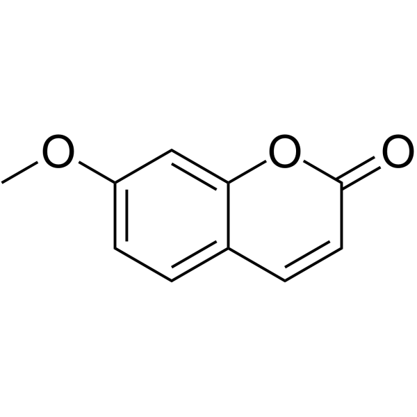
Herniarin is a natural coumarin occurs in some flowering plants with anticancer effects. Herniarin results in a significant decrease in cell viability by inducing apoptosis in MCF-7 cells. Herniarin also has anti-dermatophytic activity. Herniarin can be used for the study of bladder cancer and breast cancer.
For research use only. We do not sell to patients.
- Purity : 99.55%
- CAS No.: 531-59-9
- Formula: C10H8O3
- Molecular Weight:176.17
-
Storage:
4°C, protect from light
* In solvent : -80°C, 6 months; -20°C, 1 month (protect from light)

To place orders, for customer services and technical support, please contact: MedChemExpress USA
Tel: 609-228-6898 E-mail: [email protected] [email protected]


-
 Biological Activity
-
Chemical Information
-
Solvent & Solubility
- Protocols
- Purity & Documentation
- References
-
 Help & FAQs


-
HY-N0284
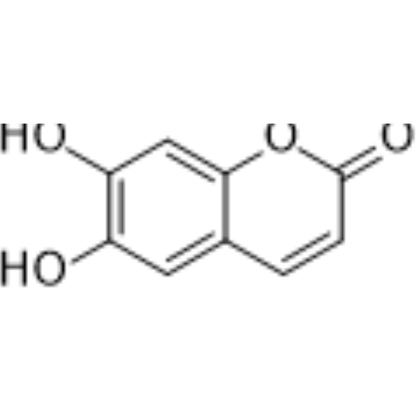
EsculetinPI3K Inhibitor -
HY-N0573
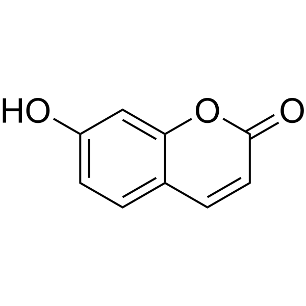
UmbelliferoneFluorescence Probe -
HY-N1365
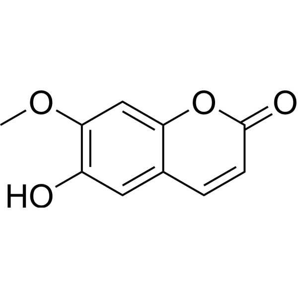
IsoscopoletinHBV Inhibitor -
HY-Y0320C
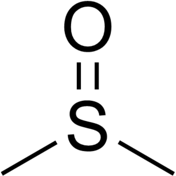
Dimethyl sulfoxideAprotic Solvent -
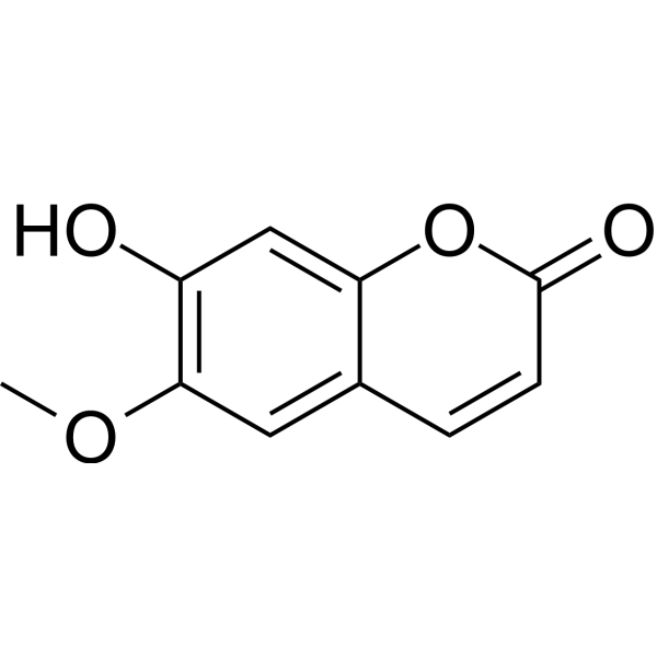
HY-N0342
ScopoletinCholinesterase (ChE) Inhibitor -
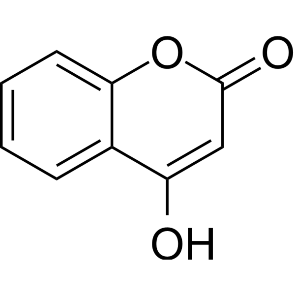
HY-N6856
4-HydroxycoumarinBacterial Inhibitor -
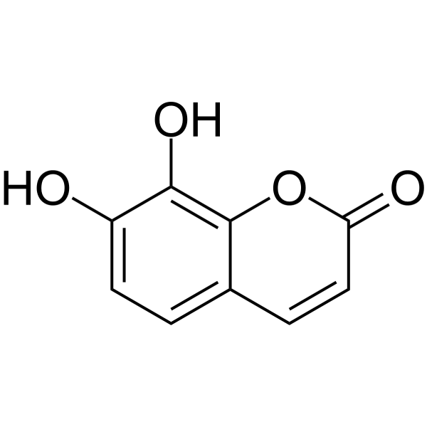
HY-N0281
DaphnetinProtein Kinase Inhibitor -
HY-N0709
CoumarinAnti-inflammatory Agent -
HY-133091
7-EthoxycoumarinCytochrome P451 Substrate -
HY-D0027
7-Amino-4-methylcoumarinFluorescent Probe -
HY-N0377
LiquiritigeninEstrogen Receptor/ERR Agonist -
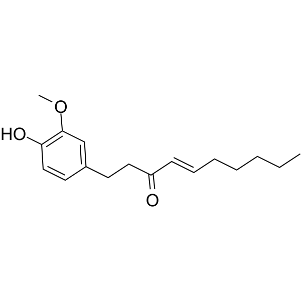
HY-14616
ShogaolAutophagy Inducer


Biological Activity
Description
Cellular Effect
|
Cell Line

|
Type | Value | Description | References |
|---|---|---|---|---|
| HeLa | IC50 |
>25 μM
Compound: 11
|
Inhibition of CRM1-mediated hemagglutininin-tagged HIV1 Rev protein nuclear export in human HeLa cells assessed as nucleus localization after 12 hrs by indirect fluorescent antibody technique
Inhibition of CRM1-mediated hemagglutininin-tagged HIV1 Rev protein nuclear export in human HeLa cells assessed as nucleus localization after 12 hrs by indirect fluorescent antibody technique
|
[PMID: 20493693] |
| Macrophage | IC50 |
84 μM
Compound: 1, herniarin
|
Antiinflammatory activity in NMRI mouse macrophages assessed as inhibition of A23187-induced PGE2 release by Ellman's method
Antiinflammatory activity in NMRI mouse macrophages assessed as inhibition of A23187-induced PGE2 release by Ellman's method
|
[PMID: 8988605] |
| Macrophage | IC50 |
84 μmol
Compound: 1, herniarin
|
Antiinflammatory activity in NMRI mouse macrophages assessed as inhibition of A23187-induced PGE2 release by Ellman's method
Antiinflammatory activity in NMRI mouse macrophages assessed as inhibition of A23187-induced PGE2 release by Ellman's method
|
[PMID: 8988605] |
| RAW264.7 | IC50 |
12.4 μM
Compound: 18
|
Antiinflammatory activity against mouse RAW264.7 cells assessed as reduction of LPS-induced NO production after 24 hrs
Antiinflammatory activity against mouse RAW264.7 cells assessed as reduction of LPS-induced NO production after 24 hrs
|
[PMID: 25621853] |
| V79 | IC50 |
>100 μM
Compound: 7-Methoxy-coumarin
|
Cytotoxicity against V-79 cells
Cytotoxicity against V-79 cells
|
10.1016/0960-894X(96)00315-0 |
In Vitro
Herniarin is cytotoxic to breast carcinoma cell line MCF-7 with an IC50 of 207.6 μM. Herniarin (100 μM) also induces apoptosis in MCF-7 cells[1]. Herniarin alone has no obvious cytotoxicity on transitional cell carcinoma (TCC) cells, but when in combination with 5 μg/mL cisplatin, Herniarin (80 μg/mL) potently enhances the antitumor effect of cisplatin, and increases chromatin condensation[2].
MedChemExpress (MCE) has not independently confirmed the accuracy of these methods. They are for reference only.
- Detection of Apoptosis
- TUNEL staining for apoptotic DNA fragmentation
- Cell Cytotoxicity Assay
- Mammalian live/dead viability and cytotoxicity staining
- Annexin V plus membrane-impermeant dye apoptosis staining
- Apoptosis Solutions
- CCK-8/WST-8 Cell Proliferation Assay
- MTT Cell Proliferation Assay
- Breast Cancer Modeling
- Directly Induced Neuron Culture
- Cell Viability Determination by MTT Colorimetric Assay


Chemical Information
-
CAS No. 531-59-9
-
Appearance Solid
-
Molecular Weight 176.17
-
Formula C10H8O3
-
SMILES
O=C1C=CC2=CC=C(OC)C=C2O1
-
Synonyms
Methylumbelliferone
-
Structure Classification
-
Initial Source
-
Shipping
Room temperature in continental US; may vary elsewhere.
-
Storage
4°C, protect from light
* In solvent : -80°C, 6 months; -20°C, 1 month (protect from light)


Solvent & Solubility
In Vitro:
DMSO : 100 mg/mL (567.63 mM; Need ultrasonic; Hygroscopic DMSO has a significant impact on the solubility of product, please use newly opened DMSO)
Please refer to the solubility information to select the appropriate solvent. Once prepared, please aliquot and store the solution to prevent product inactivation from repeated freeze-thaw cycles.
Storage method and period of stock solution: -80°C, 6 months; -20°C, 1 month (protect from light). When stored at -80°C, please use it within 6 months. When stored at -20°C, please use it within 1 month.
Please refer to the solubility information to select the appropriate solvent. Once prepared, please aliquot and store the solution to prevent product inactivation from repeated freeze-thaw cycles.
Storage method and period of stock solution: -80°C, 6 months; -20°C, 1 month (protect from light). When stored at -80°C, please use it within 6 months. When stored at -20°C, please use it within 1 month.
Concentration (start) × Volume (start) = Concentration (final) × Volume (final)
In Vivo:
Select the appropriate dissolution method based on your experimental animal and administration route.
- For the following dissolution methods, please ensure to first prepare a clear stock solution using an In Vitro approach and then sequentially add co-solvents:
- To ensure reliable experimental results, the clarified stock solution can be appropriately stored based on storage conditions. As for the working solution for In Vivo experiments, it is recommended to prepare freshly and use it on the same day.
- The percentages shown for the solvents indicate their volumetric ratio in the final prepared solution. If precipitation or phase separation occurs during preparation, heat and/or sonication can be used to aid dissolution.
Add each solvent one by one: 10% DMSO 40% PEG300 5% Tween-80 45% Saline
Solubility: ≥ 2.5 mg/mL (14.19 mM); Clear solution
This protocol yields a clear solution of ≥ 2.5 mg/mL (saturation unknown).
Taking 1 mL working solution as an example, add 100 μL DMSO stock solution (25.0 mg/mL) to 400 μL PEG300, and mix evenly; then add 50 μL Tween-80 and mix evenly; then add 450 μL Saline to adjust the volume to 1 mL.
Preparation of Saline: Dissolve 0.9 g sodium chloride in ddH₂O and dilute to 100 mL to obtain a clear Saline solution.
Add each solvent one by one: 10% DMSO 90% (20% SBE-β-CD in Saline)
Solubility: ≥ 2.5 mg/mL (14.19 mM); Clear solution
This protocol yields a clear solution of ≥ 2.5 mg/mL (saturation unknown).
Taking 1 mL working solution as an example, add 100 μL DMSO stock solution (25.0 mg/mL) to 900 μL 20% SBE-β-CD in Saline, and mix evenly.
Preparation of 20% SBE-β-CD in Saline (4°C, storage for one week): 2 g SBE-β-CD powder is dissolved in 10 mL Saline, completely dissolve until clear.
In Vivo Dissolution Calculator
Please enter the basic information of animal experiments:
-
-
-
-
Recommended: Prepare an additional quantity of animals to account for potential losses during experiments.
Please enter your animal formula composition:
-
%DMSO +
Recommended: Keep the proportion of DMSO in working solution below 2% if your animal is weak.
-
%+
-
+%Tween-80 + +
-
%Saline +
The co-solvents required include: DMSO, . All of co-solvents are available by MedChemExpress (MCE). , Tween 80. All of co-solvents are available by MedChemExpress (MCE).
Working solution concentration: 0.22 mg/mL
Method for preparing stock solution: mg drug dissolved in μL DMSO. Stock solution concentration: mg/mL. * In solvent : -80°C, 6 months; -20°C, 1 month (protect from light)
1. Take μL DMSO stock solution;
2. Add μL .
μL , mix evenly;
3. Then add μL Tween 80, mix evenly;
4. Then add μL
Please ensure that the stock solution in the first step is dissolved to a clear state, and add co-solvents in sequence. You can use ultrasonic heating (ultrasonic cleaner, recommended frequency 20-40 kHz), vortexing, etc. to assist dissolution.


Purity & Documentation
-
Data Sheet (273 KB)
-
SDS (393 KB)
- English - EN (393 KB)
- Français - FR (393 KB)
- Deutsch - DE (393 KB)
- Norwegian - NO (393 KB)
- Español - ES (393 KB)
- Swedish - SV (393 KB)
- Italian - IT (393 KB)
- Korean - KR (393 KB)
- Portuguese - PT (393 KB)
-
Handling Instructions (2659 KB)
References
[1]. Mousavi SH, et al. Comparative analysis of the cytotoxic effect of 7-prenyloxycoumarin compounds and herniarin on MCF-7 cell line. Avicenna J Phytomed. 2015 Nov-Dec;5(6):520-30. [Content Brief]
[2]. Haghighitalab A, et al. Enhancement of cisplatin cytotoxicity in combination with herniarin in vitro. Drug Chem Toxicol. 2014 Apr;37(2):156-62. [Content Brief]


Complete Stock Solution Preparation Table
Please refer to the solubility information to select the appropriate solvent. Once prepared, please aliquot and store the solution to prevent product inactivation from repeated freeze-thaw cycles.
Storage method and period of stock solution: -80°C, 6 months; -20°C, 1 month (protect from light). When stored at -80°C, please use it within 6 months. When stored at -20°C, please use it within 1 month.
| Optional Solvent | Concentration Solvent Mass | 1 mg | 5 mg | 10 mg | 25 mg |
|---|---|---|---|---|---|
| DMSO | 1 mM | 5.6763 mL | 28.3817 mL | 56.7634 mL | 141.9084 mL |
| 5 mM | 1.1353 mL | 5.6763 mL | 11.3527 mL | 28.3817 mL | |
| 10 mM | 0.5676 mL | 2.8382 mL | 5.6763 mL | 14.1908 mL | |
| 15 mM | 0.3784 mL | 1.8921 mL | 3.7842 mL | 9.4606 mL | |
| 20 mM | 0.2838 mL | 1.4191 mL | 2.8382 mL | 7.0954 mL | |
| 25 mM | 0.2271 mL | 1.1353 mL | 2.2705 mL | 5.6763 mL | |
| 30 mM | 0.1892 mL | 0.9461 mL | 1.8921 mL | 4.7303 mL | |
| 40 mM | 0.1419 mL | 0.7095 mL | 1.4191 mL | 3.5477 mL | |
| 50 mM | 0.1135 mL | 0.5676 mL | 1.1353 mL | 2.8382 mL | |
| 60 mM | 0.0946 mL | 0.4730 mL | 0.9461 mL | 2.3651 mL | |
| 80 mM | 0.0710 mL | 0.3548 mL | 0.7095 mL | 1.7739 mL | |
| 100 mM | 0.0568 mL | 0.2838 mL | 0.5676 mL | 1.4191 mL |


Herniarin Related Classifications
HY-N1366 Related Classifications
 Powered by Bioz