



Mouse CD8+ T Cells Negative Selection Kit

Based on 1 Customer Validation
MCE Mouse CD8+ T Cells Negative Selection Kit is designed for the isolation of CD8+ T cells from single cell suspensions of mouse spleen cells and lymph nodes.
-
Storage :
4°C,2 years. Do not freeze the magnetic beads.


To place orders, for customer services and technical support, please contact: MedChemExpress USA
Tel: 609-228-6898 E-mail: [email protected] [email protected]





Description & Advantages
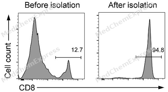
MCE Mouse CD8+ T Cells Negative Selection Kit is designed for the isolation of CD8+ T cells from single cell suspensions of mouse spleen cells and lymph nodes. Its principle hinges on utilizing a biotin-labeled monoclonal antibody to mark non-target cells (non-CD8+ T-cells), which are subsequently removed using streptavidin-labeled magnetic beads. This process effectively facilitates the sorting of human CD8+ T cells.
The kit is user-friendly and portable, with components that are non-toxic to cells. It enables the isolation of high-purity cells, and the resulting cells can be utilized in a variety of downstream applications. These include flow cytometry, cell culture, Western blot analysis, immunoprecipitation (IP), reporter gene detection, and DNA/RNA extraction.
Features of MCE Mouse CD8+ T Cells Negative Selection Kit
• Efficiency: Enables sorting of target cells in as little as 15 minutes;
• Separation Columns-free: Eliminates the need for separation columns by using a magnetic separator for swift cell separation;
• High Purity: Can yield sorted cells with a purity exceeding 97%;
• High Activity: The sorted cells are free of antibody and magnetic bead labeling, and retain high activity and functionality for downstream experiments.


Protocol
Recommended Buffer (Not Provided)
| Buffer | Composition |
|---|---|
| Isolation Buffer | PBS, 0.5% BSA, 2 mM EDTA, pH 7.0-7.4 |
Note: a. BSA may be replaced with human serum albumin (HSA) or 2% fetal bovine serum (FBS);
b. It is recommended to prepare the Isolation Buffer with water for injection, sterilize it through a 0.22 μm filter, and store it at 4°C.
Example: Isolation of mouse CD8+ T cells from mouse spleen or lymph nodes
1. Prepare a single-cell suspension from mouse spleen or lymph nodes: dissociate the tissue through a 70 μm cell strainer and rinse the strainer with pre-cooled PBS. Transfer the suspension to a 50 mL centrifuge tube, centrifuge at 500 × g for 5 min, and discard the supernatant.
2. Add 5 mL ACK Red Blood Cell Lysis Buffer and lyse at room temperature for 5 min. Add 20 mL PBS to resuspend the cells, centrifuge at 500 × g for 5 min, and discard the supernatant.
Note: a. Red blood cell lysis conditions may be adjusted according to the lysis buffer used;
b. A small number of residual red blood cells will not affect subsequent separation or purity.
3. Resuspend the cells in PBS, filter the suspension through a 70 μm cell strainer, and count the cells. Centrifuge at 500 × g for 5 min and discard the supernatant.
Note: This step removes tissue debris or cell clumps that may affect separation purity.
4. Resuspend the cells in Isolation Buffer and adjust the cell density to 1 × 10⁸ cells/mL.
5. Transfer 100 μL of cell suspension (1 × 10⁷ cells) to the bottom of a sterile centrifuge tube. Add 2 μL Biotin-Antibody Mix, mix thoroughly, and incubate at 4°C for 10 min.
Note: a. Add the cell suspension directly to the bottom of the tube rather than along the tube wall;
b. Scale the amount of Biotin-Antibody Mix proportionally when separating a larger number of cells.
6. Preparation of Streptavidin Magnetic Beads: thoroughly resuspend the beads and transfer 20 μL to a 1.5 mL microcentrifuge tube. Add 1 mL Isolation Buffer, mix, and centrifuge at 10,000 × g for 1 min. Discard the supernatant, repeat the wash 1-2 times, and finally resuspend the beads in 20 μL Isolation Buffer.
Note: The final volume of Isolation Buffer used to resuspend the beads should be equal to the initial volume of beads taken.
7. After antibody incubation, add 20 μL of pre-washed Streptavidin Magnetic Beads, mix thoroughly, and incubate at 4°C for 10 min.
Note: Scale all reagents proportionally for larger cell numbers. If fewer than 1 × 10⁷ cells are used, adjust the suspension volume to 100 μL and add 2 μL Biotin-Antibody Mix and 20 μL beads.
8. After incubation, add 2.5 mL Isolation Buffer and mix gently. Avoid vigorous shaking or repeated pipetting.
9. Place the tube on a magnetic separation rack and allow it to stand for 5 min.
10. While keeping the tube on the magnetic rack, carefully transfer the cell suspension to a new sterile centrifuge tube. The suspension contains enriched mouse CD8+ T cells. Centrifuge at 300 × g for 5 min, discard the supernatant, and collect the cells.
Note: Avoid touching the magnetic beads with the pipette tip. The suspension may also be collected by pouring.
11. Wash the cells as required and resuspend them in an appropriate buffer or culture medium. The isolated cells may be used for downstream molecular or cell biology experiments.
보관
4°C,2 years.
Do not freeze the magnetic beads.


Components
| Components | HY-K0309-0.2 mL (For 108 cells) |
HY-K0309-1 mL (For 5 × 10⁸ cells) |
HY-K0309-2 mL (For 109 cells) |
|---|---|---|---|
| Biotin-Antibody Mix | 20 μL | 100 μL | 200 μL |
| Streptavidin Magnetic Beads | 0.2 mL | 1 mL | 1 mL × 2 |


 Please enter the correct Lot Number


