



Faricimab

Based on 1 Customer Validation
Faricimab, an overall good safety and tolerability profile, is a bispecific antibody targeting Angiopoietin-2 (Ang-2) and vascular endothelial growth factor-A (VEGF-A). Faricimab prevents retinal vascular leakage, cell death and inflammation in retinal ischemia/reperfusion (I/R) injury and sCNV mouse models. Faricimab demonstrates statistically superior visual acuity gains versus Ranibizumab (HY-P9951). Faricimab can be used for retinal diseases, such as age-related macular degeneration (w-AMD), diabetic macular edema (DME) and macular edema following retinal vein occlusion (RVO).
For research use only. We do not sell to patients.
- Purity : 97.97%
- CAS No.: 1607793-29-2
- Molecular Weight:146.16 kDa
-
Storage:
Please store the product under the recommended conditions in the Certificate of Analysis.

To place orders, for customer services and technical support, please contact: MedChemExpress USA
Tel: 609-228-6898 E-mail: [email protected] [email protected]


-
 Biological Activity
-
Chemical Information
- Protocols
- Purity & Documentation
- References
-
 Help & FAQs


-
HY-P9906
BevacizumabVEGF-A Inhibitor -

HY-P9951A
Ranibizumab (anti-VEGF)Anti-VEGF Antibody Fragment -
HY-P9908
AdalimumabTNF-α Inhibitor -

HY-P9977
AmivantamabEGFR-MET Dual Antibody -

HY-P99024
GlofitamabT-Cell-Engaging Bispecific Antibody -

HY-P9909
Ustekinumab -

HY-P99392
TeclistamabBCMA/CD3 Dual Antibodies -

HY-P99033
MosunetuzumabCD20/CD3 Inhibitor -
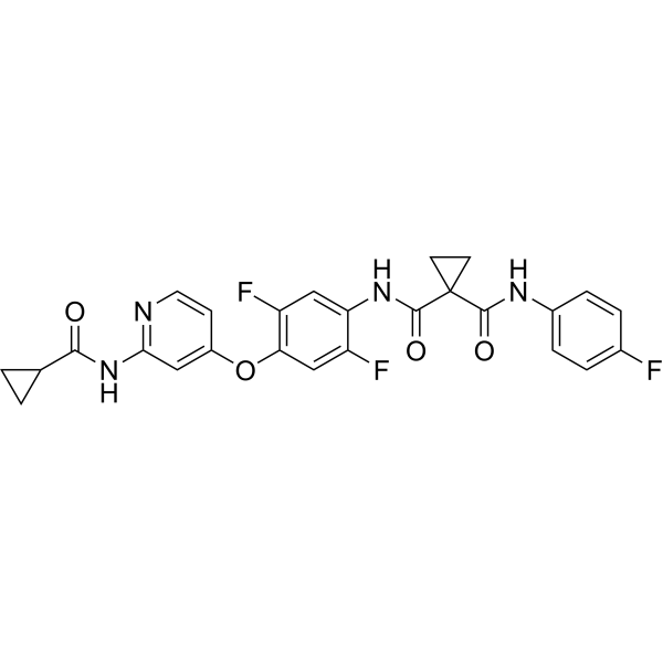
HY-B0791
AltiratinibMET/TIE2/VEGFR2/FLT3/Trk Inhibitor -

HY-P99001
Human IgG1 kappa, Isotype ControlIgG1κ Isotype Control -
HY-P99036
NesvacumabAng2 Inhibitor -

HY-P990876
Afimkibart (WT IgG1)Trimeric TL1A Antibody
All VEGFR Isoforms
More

Biological Activity
Description
Isotype
IgG1-kappa-lambda with half-IG VL-CH1/VH-CK crossover
Recommend Isotype Controls
Species Reactivity
Human
IC50 & Target
ANG2 & VEGFA
In Vitro
Faricimab (2000 ng/mL, 37 days) to pharmaceutical compounding and storage for up to 37 days has no impairment on its bispecific antigen-binding properties and has a structural integrity of the IgG1 Fc fragment[3].
MedChemExpress (MCE) has not independently confirmed the accuracy of these methods. They are for reference only.
In Vivo
Faricimab (3 μg/μL, intravitreal injections, a single dose for 48 h) prevents ischemia/reperfusion (I/R) injury-induced retinal permeability and cell death in retinal I/R injury mouse model[4].
MedChemExpress (MCE) has not independently confirmed the accuracy of these methods. They are for reference only.
-
Animal Model:Retinal I/R injury mouse model[4]
-
Dosage:3 μg/μL
-
Administration:intravitreal injections, a single dose for 48 h
-
Result:Significantly reduced retinal vascular permeability (by 64%) and cell death compared with IgG control-treated mice.
-
Animal Model:JR5558 mice[4]
-
Dosage:10 mg/kg
-
Administration:i.p., twice interval 1 week for 1 or 5 weeks
-
Result:Might promote vascular stability by inhibiting neovascularization and subsequent neovascular leakage and decreased immune cell accumulation associated with sCNV lesions in JR5558 mice.
Clinical Trial
| NCT Number | Sponsor | Condition | Start Date |
Phase

|
|---|---|---|---|---|
| NCT01329991 | Plexxikon| | 2011-05 | PHASE1 |
Gene ID
Accession
Conjugated
Unconjugated
Reconsititution
The product can be reconstituted/diluted with sterile PBS or saline.
Format
-
IgG1-kappa-lambda with half-IG VL-CH1/VH-CK crossover

Application
ELISA, FACS, Functional assay
Verified Bioactivity
-
 Immobilized Faricimab can bind VEGF165 Protein, Human.The EC50 for this effect is 3.418 ng/mL.
 -
 Immobilized Faricimab can bind Human Angiopoietin-2/ANGPT2 Protein (His Tag).The EC50 for this effect is 4.10 ng/mL.
 -
 Flow cytometric analysis of 1X10⁶ HeLa cells with Faricimab (HY-P99116, red). Cells were fixed with 4% paraformaldehyde. Then stained with the primary antibody at 1/200 dilution for an hour at 4℃. AF488-conjugated AffiniPure Goat Anti-Human IgG H&L (HY-P83776) was used as the secondary antibody at 1/1,000 dilution for 30 minutes at 4℃. Human IgG1 kappa (HY-P99001, blue) was used as the isotype control.


Chemical Information
-
CAS No. 1607793-29-2
-
Appearance Liquid
-
Molecular Weight 146.16 kDa
-
Color Colorless to light yellow
-
SMILES
[Faricimab]
-
Synonyms
RG7716; RO-6867461
-
Shipping
Shipping with dry ice.
-
Formulation
Please refer to the lot-specific COA for specific buffer information.
-
Storage
Please store the product under the recommended conditions in the Certificate of Analysis.


Purity & Documentation
-
Data Sheet (262 KB)
-
SDS (251 KB)
- English - EN (251 KB)
- Français - FR (251 KB)
- Deutsch - DE (251 KB)
- Norwegian - NO (251 KB)
- Español - ES (251 KB)
- Swedish - SV (251 KB)
- Italian - IT (251 KB)
- Korean - KR (251 KB)
- Portuguese - PT (251 KB)
-
Inhibitory Antibodies User Guide (603 KB)
References
[1]. Sahni J, et al. Simultaneous Inhibition of Angiopoietin-2 and Vascular Endothelial Growth Factor-A with Faricimab in Diabetic Macular Edema: BOULEVARD Phase 2 Randomized Trial. Ophthalmology. 2019 Aug;126(8):1155-1170. [Content Brief]
[2]. Nicolò M, et al. Faricimab: an investigational agent targeting the Tie-2/angiopoietin pathway and VEGF-A for the treatment of retinal diseases[J]. Expert Opin Investig Drugs. 2021 Mar;30(3):193-200. [Content Brief]
[3]. Penha FM, et al. Review of real-world evidence of dual inhibition of VEGF-A and ANG-2 with faricimab in NAMD and DME[J]. Int J Retina Vitreous. 2024 Jan 17;10(1):5. [Content Brief]
[4]. Canonica J, et al. Delineating effects of angiopoietin-2 inhibition on vascular permeability and inflammation in models of retinal neovascularization and ischemia/reperfusion[J]. Front Cell Neurosci. 2023 Jun 12;17:1192464. [Content Brief]
[5]. Jørstad ØK, et al. Pharmaceutical compounding and storage of faricimab in a syringe for intravitreal injection do not impair stability and bi-specific binding properties[J]. Int J Retina Vitreous. 2023 Nov 7;9(1):65. [Content Brief]

Calculators
Concentration (start) × Volume (start) = Concentration (final) × Volume (final)


Faricimab Related Classifications
HY-P99116 Related Classifications
 Powered by Bioz