



Peluntamig

Based on 1 Customer Validation
Peluntamig (PT-217) is a bispecific antibody targeting DLL3 and CD47. The antibody arms of Peluntamig specifically bind to DLL3 on small cell lung cancer tumor cells while blocking the CD47-SIRPa interaction, thereby effectively stimulating macrophage phagocytosis and inducing NK cell-mediated cytotoxicity. Peluntamig exhibits significant anti-tumor activity against small cell lung cancer and also has favorable safety profiles. Peluntamig can be used in a variety of tumor-related studies including those on small cell lung cancer and L1210 leukemia.
For research use only. We do not sell to patients.
- Purity : 99.9%
- CAS No.: 2928515-21-1
- Molecular Weight:145.027 kDa
-
Storage:
Please store the product under the recommended conditions in the Certificate of Analysis.


To place orders, for customer services and technical support, please contact: MedChemExpress USA
Tel: 609-228-6898 E-mail: [email protected] [email protected]


-
 Biological Activity
-
Chemical Information
- Protocols
- Purity & Documentation
- References
-
 Help & FAQs




Biological Activity
Description
Isotype
Human IgG1 kappa
Recommend Isotype Controls
Species Reactivity
Human
IC50 & Target
CD47 & DLL3
In Vivo
MedChemExpress (MCE) has not independently confirmed the accuracy of these methods. They are for reference only.
-
Animal Model:C57BL/6 × DBA/2 F1 (BDF1) (male, injected intraperitoneally with 106 L1210 leukemia cells on day 0)[2]
-
Dosage:15 mg/kg
-
Administration:i.p.; single injection; day 3 post-tumor cell inoculation
-
Result:Produced a percent increase in life span (% ILS) that contributed to an additive predicted % ILS for the triple combination (Peluntamig+cyclophosphamide + 5-fluorouracil).
Yielded an observed % ILS significantly exceeding the additive predicted value (p < 0.001) when used in the triple combination.
Resulted in 7 cures (≥60-day survival) out of 10 treated mice in the triple combination.
Caused a +2% mean weight change 5 days post-treatment when administered alone.
Caused a -9% mean weight change 5 days post-treatment in the triple combination.
Led to no toxic deaths in any treatment group.
Clinical Trial
| NCT Number | Sponsor | Condition | Start Date |
Phase

|
|---|---|---|---|---|
| NCT01329991 | Plexxikon| | 2011-05 | PHASE1 |
Gene ID
Accession
Q61735-1 & Q9NYJ7
Conjugated
Unconjugated
Reconsititution
The product can be reconstituted/diluted with sterile PBS or saline.
Format
-
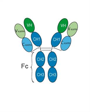
IgG1-kappa
Application
ELISA, FACS, Functional assay
Verified Bioactivity
-
 Immobilized Delta-like protein 3/DLL3 Protein, Human (HEK293, His, HY-P700456) can bind Peluntamig. The EC50 for this effect is 19.78 ng/mL.
 -
 Immobilized CD47 Protein, Human (HEK293, His, HY-P7336) can bind Peluntamig. The EC50 for this effect is 187.6 ng/mL.


Chemical Information
-
CAS No. 2928515-21-1
-
Appearance Liquid
-
Molecular Weight 145.027 kDa
-
Color Colorless to light yellow
-
SMILES
[Peluntamig]
-
Synonyms
PT-217
-
Shipping
Shipping with dry ice.
-
Formulation
Please refer to the lot-specific COA for specific buffer information.
-
Storage
Please store the product under the recommended conditions in the Certificate of Analysis.


Purity & Documentation
-
Data Sheet (260 KB)
-
SDS (251 KB)
- English - EN (251 KB)
- Français - FR (251 KB)
- Deutsch - DE (251 KB)
- Norwegian - NO (251 KB)
- Español - ES (251 KB)
- Swedish - SV (251 KB)
- Italian - IT (251 KB)
- Korean - KR (251 KB)
- Portuguese - PT (251 KB)
-
Inhibitory Antibodies User Guide (603 KB)
References

Calculators
Concentration (start) × Volume (start) = Concentration (final) × Volume (final)


Peluntamig Related Classifications
HY-P991016 Related Classifications
 Powered by Bioz