



Trabikibart

Based on 1 Customer Validation
Trabikibart (CSL311) is a specific inhibitor targeting the βc receptor (CSF2RB) that inhibits signal transduction mediated by GM-CSF, IL-5, and IL-3. Trabikibart exhibits significant anti-inflammatory and anti-edema effects, reduces myeloid cell infiltration, and inhibits inflammatory cell survival. Trabikibart also possesses antiviral immune functions, which alleviate pulmonary inflammation, reverse airway dysfunction and fibrosis, and thereby restore impaired pulmonary function. Trabikibart can be used in research on related diseases such as acute respiratory distress syndrome, viral pneumonia, asthma, and chronic rhinosinusitis with nasal polyps.
For research use only. We do not sell to patients.
- Purity : 99.17%
- CAS No.: 2643974-98-3
- Molecular Weight:144.66 kDa
-
Storage:
Please store the product under the recommended conditions in the Certificate of Analysis.




To place orders, for customer services and technical support, please contact: MedChemExpress USA
Tel: 609-228-6898 E-mail: [email protected] [email protected]


-
 Biological Activity
-
Chemical Information
- Protocols
- Purity & Documentation
- References
-
 Help & FAQs







Biological Activity
Description
Isotype
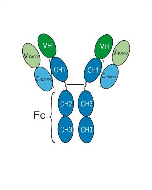
Human IgG4 kappa
Recommend Isotype Controls
Species Reactivity
Human
IC50 & Target
CSF2Rb/CD131
In Vitro
Trabikibart potently blocks human βc receptor signalling with high potency[1].
Trabikibart binds to the Site 2 region of the soluble βc-receptor subunit with a KD of 100 pM, blocking IL-3, IL-5, and GM-CSF binding[2].
MedChemExpress (MCE) has not independently confirmed the accuracy of these methods. They are for reference only.
In Vivo
Trabikibart (50 mg/kg; i.v.; single dose; administered day 4 post-infection) reduces systemic and lung myeloid inflammation, haemorrhage, and pro-inflammatory cytokine expression in IAV-infected hβcTg mice without compromising viral clearance or anti-viral immune cell function[1].
Trabikibart (50 mg/kg; i.v.; twice) potently inhibits mixed granulocytic inflammation, airway hyperresponsiveness, and airway fibrosis in an acute steroid-resistant asthma model, reducing inflammation scores and collagen deposition while normalizing lung function and gene expression[4].
Trabikibart (50 mg/kg; i.v.; once weekly; 4 weeks) effectively inhibits chronic allergen-induced airway inflammation, impaired lung function, and remodeling, normalizing fibrosis-associated extracellular matrix gene expression and outperforming single cytokine-targeting therapies[4].
MedChemExpress (MCE) has not independently confirmed the accuracy of these methods. They are for reference only.
-
Animal Model:hβcTg mice (8-to-12-week-old male and female; endogenous mouse βc and βIL-3 receptors knocked out, replaced with human βc receptor)[1]
-
Dosage:3 mg/kg; 10 mg/kg; 30 mg/kg; 50 mg/kg
-
Administration:i.v.; single dose
-
Result:Dose-dependently reduced peak blood neutrophil numbers, with levels declining to control values at the 10 mg/kg dose.
Maximally reduced peak blood monocyte numbers at the 50 mg/kg dose.
Dose-dependently reduced lung MPO activity, with maximal decrease observed at the 50 mg/kg dose.
Dose-dependently decreased peak BAL neutrophil numbers, with maximal reduction at the 10 mg/kg dose.
Significantly improved peripheral blood oxygen saturation (SpO2) levels at 24 and 72 hours post-LPS challenge with a single 50 mg/kg dose.
Significantly reduced lung injury scores (assessed by vascular, bronchiole, and alveolar inflammation).
Reduced BAL fluid total protein levels (a marker of oedema).
Significantly reduced peak BAL MPO activity and dsDNA levels (markers of netosis).
-
Animal Model:hβcTg mice (8-to-12-week-old male and female; endogenous mouse βc and βIL-3 receptors knocked out, replaced with human βc receptor)[1]
-
Dosage:50 mg/kg
-
Administration:i.v.; single dose; administered day 4 post-infection
-
Result:Did not alter IAV-induced body weight loss or lung viral load.
Significantly reduced elevated blood monocyte, blood neutrophil, and blood haemoglobin levels induced by IAV infection.
Significantly reduced BAL neutrophil and macrophage numbers.
Reduced lung haemorrhagic regions.
Significantly reduced lung neutrophils, alveolar macrophages, exudative macrophages, monocytes, and eosinophils; did not alter lung NK cells, NKT cells, regulatory T cells, CD4 T cells, and CD8 T cell numbers.
Significantly reduced lung Ccl2, Ccl24, and Il1α levels; did not alter Cxcl1, Cxcl10, Il6, Ifnb, Ifng, or Ifnl2/3 levels.
-
Animal Model:human common b transgenic (hbcTg) mice (8- to 12-week-old female, devoid of murine bc/bIL-3 receptors, expressing human bc receptor, acute steroid-resistant asthma model)[4]
-
Dosage:50 mg/kg
-
Administration:i.v.; twice (day 8 and day 10)
-
Result:Significantly reduced blood monocyte and neutrophil counts.
Reduced bronchoalveolar lavage (BAL) cell infiltration of macrophages, neutrophils, and eosinophils.
Reduced lung tissue infiltration of neutrophils, eosinophils, alveolar macrophages, and interstitial macrophages.
Decreased BAL markers of neutrophil extracellular traps (dsDNA, myeloperoxidase activity) and lung injury (total protein, lactate dehydrogenase).
Suppressed TH2/TH17 cytokines (Il4, Il13, Il17) and T-cell chemokine Ccl17 while preserving TH1 cytokines (Ifng, Il12b).
Abolished methacholine-induced airway hyperresponsiveness.
Rescued restrictive lung function abnormalities (partial restoration of pressure-volume curve, quasi-static compliance, and respiratory system elastance).
Reduced peribronchiolar/alveolar inflammation score and airway collagen deposition (Masson trichrome-positive area fraction).
Normalized Col1a1 gene expression to control levels.
-
Animal Model:human common b transgenic (hbcTg) mice (8- to 12-week-old female, devoid of murine bc/bIL-3 receptors, expressing human bc receptor, chronic asthma model)[4]
-
Dosage:50 mg/kg
-
Administration:i.v.; once weekly; 4 weeks
-
Result:Rescued ASP-induced restrictive lung function abnormalities (normalized respiratory system elastance, static compliance, and pressure-volume curves).
Reduced lung inflammation score.
Decreased airway collagen deposition (Masson trichrome-positive area fraction).
Normalized ASP-induced global gene expression changes, including downregulating 268 ASP-upregulated genes.
Suppressed hallmark inflammatory response gene signatures.
Inhibited TH2 and IL-17 signaling pathways.
Normalized extracellular matrix (ECM) gene expression (including reducing ASP-upregulated ECM regulators [Mmp12, Mmp13, Tgfb1, Thbs4] and matrisome-associated genes).
Shifted collagen gene expression from fibril-forming to non-fibril-forming species.
Reduced both BAL eosinophils and neutrophils (unlike single cytokine blockers targeting only eosinophils or neutrophils).
- Fibrosis/Collagen Morphometry
- Research Protocol for Infectious Diseases
- Collagen: Sirius Red Staining
- Cell Cytotoxicity Assay
- Mammalian live/dead viability and cytotoxicity staining
- Connective Tissue: Masson's Trichrome/Collagen Trichrome Staining
- Research Protocol for Inflammation-related Diseases
- Cell Viability Determination by MTT Colorimetric Assay
Gene ID
Accession
Conjugated
Unconjugated
Reconsititution
The product can be reconstituted/diluted with sterile PBS or saline.
Format
-
Human IgG4 kappa
Application
ELISA, FACS, Functional assay
Verified Bioactivity


Chemical Information
-
CAS No. 2643974-98-3
-
Appearance Liquid
-
Molecular Weight 144.66 kDa
-
Color Colorless to light yellow
-
SMILES
[Trabikibart]
-
Synonyms
CSL311
-
Shipping
Shipping with dry ice.
-
Formulation
Please refer to the lot-specific COA for specific buffer information.
-
Storage
Please store the product under the recommended conditions in the Certificate of Analysis.


Purity & Documentation
-
Data Sheet (268 KB)
-
SDS (251 KB)
- English - EN (251 KB)
- Français - FR (251 KB)
- Deutsch - DE (251 KB)
- Norwegian - NO (251 KB)
- Español - ES (251 KB)
- Swedish - SV (251 KB)
- Italian - IT (251 KB)
- Korean - KR (251 KB)
- Portuguese - PT (251 KB)
-
Inhibitory Antibodies User Guide (603 KB)
References
[1]. Wang H, et al. Blocking the human common beta subunit of the GM-CSF, IL-5 and IL-3 receptors markedly reduces hyperinflammation in ARDS models. Cell Death Dis. 2022;13(2):137. Published 2022 Feb 10. [Content Brief]
[2]. Kan WL, et al. The β Common Cytokine Receptor Family Reveals New Functional Paradigms From Structural Complexities. Immunol Rev. 2025;329(1):e13430. [Content Brief]
[4]. Wang H, et al. Dual inhibition of airway inflammation and fibrosis by common β cytokine receptor blockade. J Allergy Clin Immunol. 2024;153(3):672-683.e6. [Content Brief]

Calculators
Concentration (start) × Volume (start) = Concentration (final) × Volume (final)


Trabikibart Related Classifications
HY-P990094 Related Classifications
 Powered by Bioz