



AGX51
Based on 11 publication(s) in Google Scholar
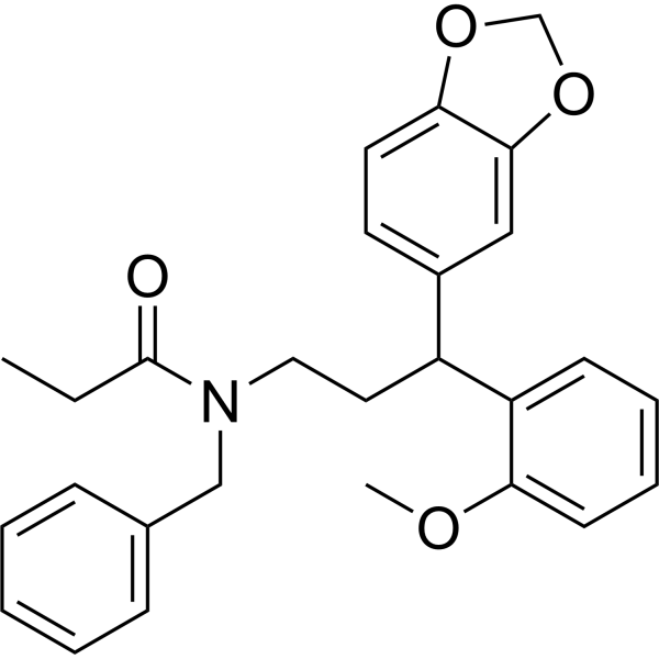
AGX51 is a pan-Id (inhibitors of DNA-binding/differentiation proteins) antagonist that disrupts ID-E protein interactions, thereby triggering ubiquitin-mediated proteasomal degradation of ID1, ID2, ID3 and ID4. AGX51 induces ROS production, G0/G1 cell cycle arrest, non-apoptotic cell death, and regulates EMT-related proteins. AGX51 reduces the proliferation, migration and myofibroblast differentiation of lung fibroblasts, alleviates pulmonary fibrosis, inhibits cancer cell viability and tumor growth, impairs neovascularization, and shows no obvious toxicity in mice. AGX51 can be used in research related to idiopathic pulmonary fibrosis, triple-negative breast cancer, breast cancer, pancreatic ductal adenocarcinoma, colorectal tumors, wet age-related macular degeneration and retinopathy of prematurity.
商品は「研究用試薬」です。人や動物の医療用・臨床診断用・食品用の製品ではありません。
研究用途以外に使用した場合、当社は一切の責任を負いかねます。
- 純度 : 98.71%
- CAS 番号: 330834-54-3
- 分子式: C27H29NO4
- 分子量:431.52
-
保管条件:Pure form -20°C, 3 years , 4°C, 2 years ; In solvent -80°C, 2 years , -20°C, 1 year

To place orders, for customer services and technical support, please contact: MedChemExpress USA
Tel: 609-228-6898 E-mail: [email protected] [email protected]


-
 生物活性
生物活性
-
化学情報
-
溶剤 & 溶解度
- プロトコル
- 純度とドキュメンテーション
- 参考文献
-
 Help & FAQs


-

HY-13259
MG-132Proteasome Inhibitor -
HY-13585
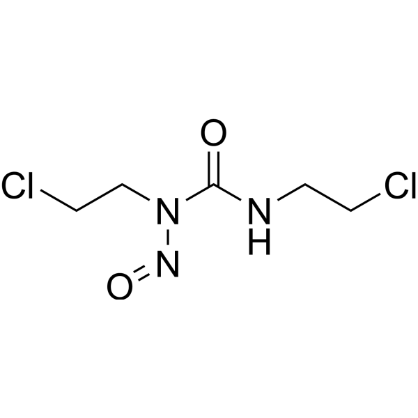
CarmustineDNA/RNA Alkylator -
HY-10201
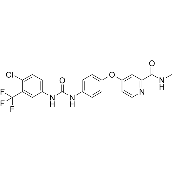
SorafenibMultikinase Inhibitor -
HY-100941
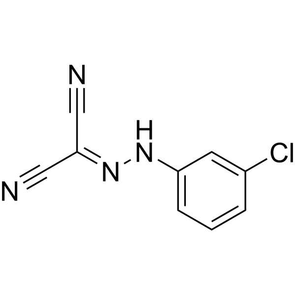
CCCPOXPHOS Uncoupler -

HY-10182
LaduviglusibGSK-3 Inhibitor -
HY-13027
DAPTγ-secretase Inhibitor -
HY-10261
AfatinibEGFR/HER2 Inhibitor -
HY-34758
N-Nitroso-N-methylureaCarcinogen Agent -
HY-13016
CabozantinibVEGFR2/MET Inhibitor -

HY-Y0320
Dimethyl sulfoxide, meets analytical specification of Ch.P.Aprotic Solvent -

HY-18739
Phorbol 12-myristate 13-acetatePKC Activator -
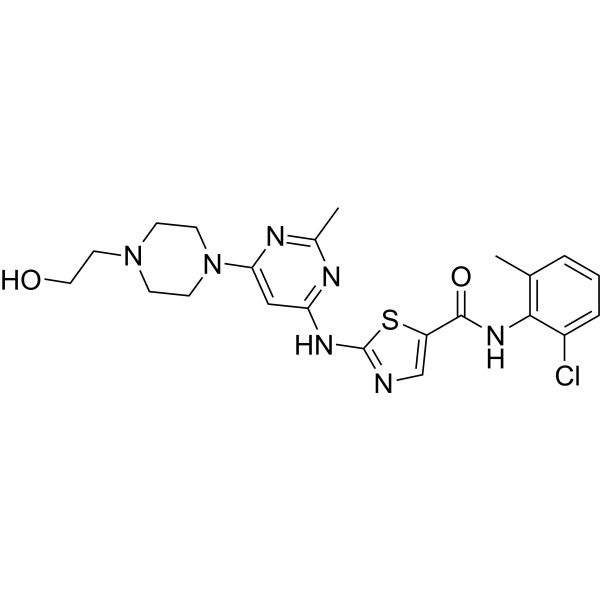
HY-10181
DasatinibSrc/Bcr-Abl Inhibitor
MedChemExpress(MCE)の使用を引用している文献 AGX51
More- Cell Mol Immunol. 2025 Mar;22(3):300-315. [Abstract]
- J Exp Clin Cancer Res. 2024 Aug 10;43(1):222. [Abstract]
- Glia. 2024 Jul;72(7):1236-1258. [Abstract]
- Front Cell Dev Biol. 2021 Feb 12:9:616610. [Abstract]
- Cancers (Basel). 2026 Apr 8;18(8):1186. [Abstract]
- Mol Oncol. 2025 Mar 21. [Abstract]
- bioRxiv. 2026 Aug 5.
- San Diego State University
- Norwegian University of Science and Technology. 2024 May.
- bioRxiv. 2023 Oct 4.
- Leiden University. 9 (2021): 182.
DNA/RNA Synthesis アイソフォーム固有の製品をすべて表示
More

生物活性
製品説明
Cellular Effect
|
Cell Line

|
Type | Value | Description | References |
|---|---|---|---|---|
| PANC-1 | IC50 |
5.5 μM
|
Cell viability inhibition against pancreatic ductal adenocarcinoma cell line Panc1 assessed via Cell Titer-Glo assay or trypan blue exclusion after 120 h treatment.
Cell viability inhibition against pancreatic ductal adenocarcinoma cell line Panc1 assessed via Cell Titer-Glo assay or trypan blue exclusion after 120 h treatment.
|
34031428 |
体外実験
AGX51 (0-60 μM; 24-72 h) inhibits cell viability and growth in breast cancer cell lines, including 4T1 murine mammary cancer cells and human TNBC cell lines, with an IC50 of ~25 μM in TNBC cells, by inducing G0/G1 arrest and non-apoptotic cell death[3].
AGX51 (40 μM; 4 h) treatment of 4T1 murine mammary cancer cells for 4 h reduces ID1 protein levels and alters the expression of 83 other proteins, including multiple regulators of cell cycle progression[3].
AGX51 (40 μM; 2, 4, 48 h) increases Id1 mRNA expression in 4T1 murine mammary cancer cells, with a 20-fold increase observed after 48 h of 40 μM treatment[3].
AGX51 (40 μM; 1, 24 h) increases E protein-DNA binding in 4T1 murine mammary cancer cells within 1 h of treatment, prior to detectable ID1 protein loss[3].
AGX51 (4-60 μM; 24, 48, 120 h) reduces ID1 and ID3 protein levels and inhibits cell viability in pancreatic ductal adenocarcinoma cells and organoids, with IC50 values ranging from 5.5-19.5 μM[3].
AGX51 (10, 50 μM; 24 h) shows that quiescent 4T1 murine mammary cancer cells, which lack ID protein expression, are resistant to AGX51-mediated cell killing, supporting ID proteins as the primary targets of AGX51 in cycling cells[3].
AGX51 (20-40 μM; up to 7 weeks) shows that 4T1 murine mammary cancer cells do not acquire stable resistance to AGX51 after prolonged in vitro exposure, as surviving cells show reduced growth and fail to maintain sustained proliferation in the presence of the compound[3].
AGX51 (20 μM) reduces ID1 and ID3 protein levels and inhibits serum-induced proliferation of IPF-derived human lung fibroblasts at a concentration of 20 μM[1].
AGX51 (20 μM) reduces migration of healthy human lung fibroblasts and IPF-derived human lung fibroblasts[1].
AGX51 (20 μM; 48 h) reduces TGF-β-induced differentiation of healthy human lung fibroblasts and IPF-derived human lung fibroblasts into myofibroblasts by suppressing fibrotic marker expression[1].
AGX51 (20 μM) inhibits MEK1 phosphorylation in human lung fibroblasts, and MEK1 activation partially counteracts AGX51's suppression of fibrotic marker expression[1].
AGX51 (10-50 μM) directly interacts with purified Id1 and Id3 proteins (with a dissociation constant around 20 μM) to alter their secondary structure, but does not interact with purified E47 protein[4].
AGX51 directly engages ID1 in digitonin-permeabilized 293T cells expressing a NanoLuc luciferase-ID1 fusion protein, as shown by dose-dependent BRET signal changes and competition with an AGX51 tracer[4].
AGX51 (5-40 μM; 24 h) reduces ID1 and ID3 protein levels in HUVECs after 24 hours of treatment, with significant ID1 loss observed at 10 μM[4].
AGX51 (5-40 μM; 24 h) reduces Cyclin D1 protein levels in HUVECs in vitro in a dose-dependent manner after 24 hours of treatment[4].
AGX51 (60 μM; 2-24 h) reduces FLAG-tagged ID1 protein levels in HCT116 cells in a time-dependent manner over 24 hours of treatment with 60 μM AGX51[4].
AGX51 (60 μM; 2 h) induces polyubiquitylation of ID1 in HCT116 and U87 glioma cells, indicating ID1 degradation occurs via the ubiquitin-proteasome system[4].
AGX51 (60 μM; 1 h) disrupts the endogenous ID1-E47 protein-protein interaction in HCT116 cells after 1 hour of treatment with 60 μM AGX51[4].
AGX51 (20 μM; 1-5 days) completely inhibits the growth of HUVECs over 5 days of treatment[4].
AGX51 (5-40 μM; 18-20 h) dose-dependently impairs vascular branching of HUVECs on Matrigel after 18-20 hours of treatment[4].
AGX51 (5-20 μM; 24 h) impairs the migration of HUVECs in a scratch assay after 24 hours of treatment[4].
AGX51 (24 h) reduces the levels of all four ID family proteins (ID1, ID2, ID3, ID4) in HCT116 cells after 24 hours of treatment, while increasing ID1 mRNA levels[4].
MedChemExpress (MCE) has not independently confirmed the accuracy of these methods. They are for reference only.
-
Cell Line:healthy human lung fibroblasts (HLFs)
-
Concentration:20 μM
-
Incubation Time:48 h
-
Result:Attenuated TGF-β-induced upregulation of fibrotic markers Acta2, Col1a1, Ctgf, Vimentin, Fibronectin, Col3A1, and Snail1 in healthy HLFs, as measured by qPCR and immunoblot.
-
Cell Line:IPF-derived human lung fibroblasts (HLFs)
-
Concentration:20 μM
-
Incubation Time:48 h
-
Result:Attenuated TGF-β-induced upregulation of fibrotic markers Acta2, Col1a1, Ctgf, Vimentin, Fibronectin, Col3A1, and Snail1 in IPF-derived HLFs, as measured by qPCR and immunoblot.
-
Cell Line:4T1 murine mammary cancer cells
-
Concentration:5 μM, 10 μM, 20 μM, 40 μM, 80 μM
-
Incubation Time:2 h, 4 h, 8 h,12 h, 24 h, 48 h, 72 h
-
Result:Reduced ID1 protein levels with near-complete loss observed by 24 h at 40 μM.
Decreased ID3 and ID4 levels with delayed kinetics.
Recovered ID1 protein levels to untreated levels within 24 h of AGX51 removal.
Showed slower ID3 recovery after AGX51 removal.
-
Cell Line:4T1 murine mammary cancer cells
-
Concentration:40 μM
-
Incubation Time:2, 4, 48 h
-
Result:Increased Id1 mRNA levels by a modest 5-fold at 4 h.
Increased Id1 mRNA levels by a dramatic 20-fold at 48 h.
-
Cell Line:4T1 murine mammary cancer cells, multiple human breast cancer cell lines, breast cancer patient-derived xenograft (PDX) cell lines
-
Concentration:40 μM; 0-60 μM
-
Incubation Time:24 h, 24-48 h; 24-72 h
-
Result:Reduced 4T1 cell viability by ~3-fold at 40 μM for 24 h.
Achieved an IC50 of ~25 μM in TNBC cell lines.
Induced G0/G1 cell cycle arrest.
Reduced BrdU incorporation and phospho-histone H3 levels.
Increased the Annexin V+/PI+ fraction from 12.7% to 40.7% after 24 h of treatment.
-
Cell Line:Pancreatic ductal adenocarcinoma (PDA) cells (806, NB44, 4279, Panc1, A21), PDA organoid lines (T7, T8)
-
Concentration:4-40 μM (24 h in 806 cells); 0-60 μM (120 h in organoids and cell lines); 4, 40 μM (48 h in organoids)
-
Incubation Time:24 h (806 cells); 120 h (organoids and cell lines); 48 h (organoids)
-
Result:Reduced ID1 and ID3 protein levels in 806 PDA cells starting at 4 μM.
Decreased cell viability in PDA organoid lines with an IC50 of 19.5 μM for T7 and 12.1 μM for T8.
Decreased cell viability in PDA cell lines with an IC50 of 5.5 μM for Panc1 and 9.2 μM for A21.
Caused collapse of well-formed organoid structures at 40 μM for 48 h.
-
Cell Line:Quiescent and cycling 4T1 murine mammary cancer cells
-
Concentration:10, 50 μM
-
Incubation Time:24 h
-
Result:Showed dramatically reduced sensitivity to AGX51-mediated killing in quiescent 4T1 cells (with near-complete loss of ID expression) compared to cycling cells.
Caused minimal viability loss in quiescent 4T1 cells at 50 μM.
-
Cell Line:primary human umbilical vein endothelial cells (HUVECs)
-
Concentration:5, 10, 20, 30, 40 μM
-
Incubation Time:24 h
-
Result:Caused a significant decrease in ID1 protein levels at 10 μM, with further reduction at higher concentrations.
Reduced ID3 protein levels in a dose-dependent manner, with diminished effect at 20-40 μM compared to ID1.
Showed ID2 and ID4 were undetectable in HUVECs.\nReduced Cyclin D1 protein levels in a dose-dependent manner with increasing concentrations.
-
Cell Line:HCT116 colorectal cancer cells expressing FLAG-tagged ID1
-
Concentration:60 μM
-
Incubation Time:2-24 h
-
Result:Induced a time-dependent decrease in FLAG-ID1 protein levels over 24 hours.
-
Cell Line:primary human umbilical vein endothelial cells (HUVECs)
-
Concentration:20 μM
-
Incubation Time:1, 3, 5 days
-
Result:Completely inhibited HUVEC growth over 5 days, while vehicle-treated cells showed robust proliferation.
-
Cell Line:primary human umbilical vein endothelial cells (HUVECs)
-
Concentration:20 μM
-
Incubation Time:24 h
-
Result:Induced G0/G1 cell cycle arrest, with 84.73% of cells in G0/G1 phase compared to 44.48% in vehicle-treated cells.
Reduced the percentage of cells in S phase from 34.31% to 4.07%, and G2 phase from 21.21% to 11.20%.
-
Cell Line:HUVECs
-
Concentration:5, 10, 20 μM
-
Incubation Time:24 h
-
Result:Significantly impaired HUVEC migration into the scratched area after 24 hours.
Parmacokinetics
| Species | Dose | Route | T1/2 |
|---|---|---|---|
| Mice[4] | 30 mg/kg | i.p. | 3.7 h |
体内実験
AGX51 (6.7-60 mg/kg; i.p.; twice daily; 7-19 days) alone does not suppress orthotopic MDA-MB-231 breast tumor growth, but when combined with Paclitaxel (HY-B0015), doses of 6.7, 20, and 60 mg/kg (twice daily) almost completely inhibit or regress tumor growth, with even a 7-day course of 60 mg/kg AGX51 plus Paclitaxel inducing regression[3].
AGX51 (50 mg/kg; i.p.; twice daily) combined with Paclitaxel significantly reduces the growth of established 4T1 breast cancer lung metastases, while single-agent AGX51 or paclitaxel has no effect[3].
AGX51 (15 mg/kg; i.p.; twice daily; 3 weeks) significantly reduces colon tumor number and size in an Azoxymethane (HY-111375)-induced mouse model of sporadic colorectal neoplasia[3].
AGX51 (10 μg; intravitreal; single dose at post-natal day 12) inhibits retinal neovascularization in a mouse model of ROP[4].
MedChemExpress (MCE) has not independently confirmed the accuracy of these methods. They are for reference only.
-
Animal Model:Wild-type mice[1]
-
Dosage:50 mg/kg
-
Administration:i.p.; twice daily; three days per week; two weeks
-
Result:Reduced bleomycin-induced upregulation of ID1 and ID3 protein levels in lung homogenates.
Increased inspiratory capacity and static compliance, and reduced respiratory elastance.
Significantly decreased pulmonary mRNA levels of Col1a1, Col3a1, and Fn1, and reduced collagen I and collagen III protein levels in lung homogenates.
Markedly attenuated fibrotic remodeling, with significantly lower Ashcroft scores and reduced hydroxyproline content compared with untreated bleomycin-challenged mice.
Exhibited no effect in non-fibrotic mice.
-
Animal Model:Athymic nu/nu (female, 8-12 weeks old, orthotopic xenograft via injection of 5 × 10⁶ MDA-MB-231 cells into right caudal mammary fat pad)[3]
-
Dosage:6.7 mg/kg; 20 mg/kg; 60 mg/kg
-
Administration:i.p.; twice daily; 19 days (monotherapy, 6.7/20/60 mg/kg + Paclitaxel); i.p.; twice daily; 7 days (60 mg/kg + Paclitaxel); i.p.; once daily; days 1-5 (Paclitaxel component)
-
Result:Did not suppress tumor growth.
Almost completely inhibited tumor growth when combined with paclitaxel at 6.7 mg/kg.
Almost completely inhibited tumor growth when combined with paclitaxel at 20 mg/kg.
Resulted in tumor regression when combined with paclitaxel at 60 mg/kg for 19 days.
Resulted in tumor regression when combined with paclitaxel at 60 mg/kg for 7 days.
-
Animal Model:Balb/c (female, 6-8 weeks old, experimental metastasis via tail vein injection of 5 × 10⁴ luciferase-labeled 4T1 cells)[3]
-
Dosage:50 mg/kg
-
Administration:i.p.; twice daily; 4 weeks; i.p.; once daily; 4 weeks
-
Result:Significantly reduced the number of lung metastases at twice daily dosing.
Significantly reduced the number of lung metastases at once daily dosing, with a less dramatic effect than the twice-daily regimen.
Decreased ID1 staining in small lung metastases relative to control.
Significantly increased Vimentin staining in small metastatic tumors.
Significantly reduced Cyclin D1 staining in lung metastases, with about half of metastases showing negative staining.
-
Animal Model:Balb/c (female, 6-8 weeks old, experimental metastasis via tail vein injection of 4T1 cells, treatment initiated when lung metastases were detectable)[3]
-
Dosage:50 mg/kg (in combination with 15 mg/kg Paclitaxel)
-
Administration:i.p.; twice daily
-
Result:Did not affect lung tumor growth as a single agent.
Significantly reduced the continued expansion of established lung tumors when combined with paclitaxel.
Resulted in smaller tumor size when combined with Paclitaxel.
-
Animal Model:A/J (male, 4 weeks old, chemically induced via weekly i.p. injection of 10 mg/kg azoxymethane for 6 weeks followed by 3-week break)[3]
-
Dosage:15 mg/kg
-
Administration:i.p.; twice daily; 3 weeks
-
Result:Significantly reduced the total number of colon tumors.
Significantly reduced the number of tumors measuring >3 mm.
-
Animal Model:C57BL/6 (female, 4-6 weeks old, laser-induced rupture of Bruch’s membrane to induce choroidal neovascularization)[4]
-
Dosage:10 μg (intravitreal); 5 μg (intravitreal); 500 μg (i.p.)
-
Administration:intravitreal; 2 doses (immediately and 7 days post-laser); i.p.; twice daily; 14 days
-
Result:Significantly reduced CNV area relative to vehicle control (10 μg intravitreal).
Significantly reduced CNV area relative to vehicle control (5 μg intravitreal).
Significantly reduced CNV area relative to vehicle control (500 μg i.p.).
Eliminated Id1 staining in CNV regions, with no Id1-positive cells observed in regions without CNV, and rare CNV regions showing no Id1 staining.
- Collagen: Sirius Red Staining
- Mammalian live/dead viability and cytotoxicity staining
- ROS/oxidative-stress fluorescent staining
- BrdU Incorporation Assay
- Cell migration
- Connective Tissue: Masson's Trichrome/Collagen Trichrome Staining
- Patient-Derived Orthotopic Xenograft (PDOX)
- Protocol for Cell Cycle
- CCK-8/WST-8 Cell Proliferation Assay
- Breast Cancer Modeling
- Cell Viability Determination by MTT Colorimetric Assay
- Fibrosis/Collagen Morphometry
- Detection of Apoptosis
- Flow cytometric DNA-content cell-cycle staining
- Cell Cytotoxicity Assay
- Patient-Derived Xenograft (PDX)
- Apoptosis Solutions
- MTT Cell Proliferation Assay
- Subcutaneous Cell-Line-Derived Xenograft
- Orthotopic Cell-Line Xenograft


化学情報
-
CAS 番号 330834-54-3
-
性状 Liquid to low melt solid
-
分子量 431.52
-
分子式 C27H29NO4
-
Color Light yellow to yellow
-
SMILES
CCC(N(CCC(C1=CC=C(OCO2)C2=C1)C3=CC=CC=C3OC)CC4=CC=CC=C4)=O
-
輸送条件
Room temperature in continental US; may vary elsewhere.
-
保管条件
Pure form -20°C 3 years 4°C 2 years In solvent -80°C 2 years -20°C 1 year


Publications (11)
-
Journal Impact Factor
-
Most Recent
-
Cell Mol Immunol
The AhR-Ovol1-Id1 regulatory axis in keratinocytes promotes epidermal and immune homeostasis in atopic dermatitis-like skin inflammation. [Abstract]2025 Mar;22(3):300-315. PMID: 39939818 -
J Exp Clin Cancer Res
Autophagy-mediated ID1 turnover dictates chemo-resistant fate in ovarian cancer stem cells. [Abstract]2024 Aug 10;43(1):222. PMID: 39123206 -
Glia
ID factors regulate the ability of Müller glia to become proliferating neurogenic progenitor-like cells. [Abstract]2024 Jul;72(7):1236-1258. PMID: 38515287 -
Front Cell Dev Biol
TGF-β-Induced Endothelial to Mesenchymal Transition Is Determined by a Balance Between SNAIL and ID Factors. [Abstract]2021 Feb 12:9:616610. PMID: 33644053 -
Cancers (Basel)
Chemotherapy Enrichment of ID Family Expression Is Associated with IL-6 Signaling in Ovarian Cancer. [Abstract]2026 Apr 8;18(8):1186. PMID: 42073515 -
Mol Oncol
2025 Mar 21. PMID: 40116596 -
-
San Diego State University
-
-
-


溶剤 & 溶解度
体外:
DMSO : 100 mg/mL (231.74 mM; Need ultrasonic; Hygroscopic DMSO has a significant impact on the solubility of product, please use newly opened DMSO)
Ethanol : 100 mg/mL (231.74 mM; Need ultrasonic)
Please refer to the solubility information to select the appropriate solvent. Once prepared, please aliquot and store the solution to prevent product inactivation from repeated freeze-thaw cycles.
Storage method and period of stock solution: -80°C, 2 years; -20°C, 1 year. When stored at -80°C, please use it within 2 years. When stored at -20°C, please use it within 1 year.
Please refer to the solubility information to select the appropriate solvent. Once prepared, please aliquot and store the solution to prevent product inactivation from repeated freeze-thaw cycles.
Storage method and period of stock solution: -80°C, 2 years; -20°C, 1 year. When stored at -80°C, please use it within 2 years. When stored at -20°C, please use it within 1 year.
濃度 (開始) × 体積 (開始) = 濃度 (終了) × 体積 (終了)
体内:
Select the appropriate dissolution method based on your experimental animal and administration route.
- For the following dissolution methods, please ensure to first prepare a clear stock solution using an In Vitro approach and then sequentially add co-solvents:
- To ensure reliable experimental results, the clarified stock solution can be appropriately stored based on storage conditions. As for the working solution for In Vivo experiments, it is recommended to prepare freshly and use it on the same day.
- The percentages shown for the solvents indicate their volumetric ratio in the final prepared solution. If precipitation or phase separation occurs during preparation, heat and/or sonication can be used to aid dissolution.
Add each solvent one by one: 10% DMSO 40% PEG300 5% Tween-80 45% Saline
Solubility: ≥ 2.08 mg/mL (4.82 mM); Clear solution
This protocol yields a clear solution of ≥ 2.08 mg/mL (saturation unknown).
Taking 1 mL working solution as an example, add 100 μL DMSO stock solution (20.8 mg/mL) to 400 μL PEG300, and mix evenly; then add 50 μL Tween-80 and mix evenly; then add 450 μL Saline to adjust the volume to 1 mL.
Preparation of Saline: Dissolve 0.9 g sodium chloride in ddH₂O and dilute to 100 mL to obtain a clear Saline solution.
Add each solvent one by one: 10% DMSO 90% (20% SBE-β-CD in Saline)
Solubility: 2.08 mg/mL (4.82 mM); Suspended solution; Need ultrasonic
This protocol yields a suspended solution of 2.08 mg/mL. Suspended solution can be used for oral and intraperitoneal injection.
Taking 1 mL working solution as an example, add 100 μL DMSO stock solution (20.8 mg/mL) to 900 μL 20% SBE-β-CD in Saline, and mix evenly.
Preparation of 20% SBE-β-CD in Saline (4°C, storage for one week): 2 g SBE-β-CD powder is dissolved in 10 mL Saline, completely dissolve until clear.
In Vivo Dissolution Calculator
Please enter the basic information of animal experiments:
-
-
-
-
Recommended: Prepare an additional quantity of animals to account for potential losses during experiments.
Please enter your animal formula composition:
-
%DMSO +
Recommended: Keep the proportion of DMSO in working solution below 2% if your animal is weak.
-
%+
-
+%Tween-80 + +
-
%Saline +
The co-solvents required include: DMSO, . All of co-solvents are available by MedChemExpress (MCE). , Tween 80. All of co-solvents are available by MedChemExpress (MCE).
Working solution concentration: 0.22 mg/mL
Method for preparing stock solution: mg drug dissolved in μL DMSO. Stock solution concentration: mg/mL.
1. Take μL DMSO stock solution;
2. Add μL .
μL , mix evenly;
3. Then add μL Tween 80, mix evenly;
4. Then add μL
Please ensure that the stock solution in the first step is dissolved to a clear state, and add co-solvents in sequence. You can use ultrasonic heating (ultrasonic cleaner, recommended frequency 20-40 kHz), vortexing, etc. to assist dissolution.


純度とドキュメンテーション
-
データシート (326 KB)
-
SDS (252 KB)
- English - EN (252 KB)
- Français - FR (252 KB)
- Deutsch - DE (252 KB)
- Norwegian - NO (252 KB)
- Español - ES (252 KB)
- Swedish - SV (252 KB)
- Italian - IT (252 KB)
- Korean - KR (252 KB)
- Portuguese - PT (252 KB)
-
取扱説明書 (2659 KB)
参考文献


Complete Stock Solution Preparation Table
Please refer to the solubility information to select the appropriate solvent. Once prepared, please aliquot and store the solution to prevent product inactivation from repeated freeze-thaw cycles.
Storage method and period of stock solution: -80°C, 2 years; -20°C, 1 year. When stored at -80°C, please use it within 2 years. When stored at -20°C, please use it within 1 year.
| Optional Solvent | Concentration Solvent Mass | 1 mg | 5 mg | 10 mg | 25 mg |
|---|---|---|---|---|---|
| DMSO / Ethanol | 1 mM | 2.3174 mL | 11.5869 mL | 23.1739 mL | 57.9347 mL |
| 5 mM | 0.4635 mL | 2.3174 mL | 4.6348 mL | 11.5869 mL | |
| 10 mM | 0.2317 mL | 1.1587 mL | 2.3174 mL | 5.7935 mL | |
| 15 mM | 0.1545 mL | 0.7725 mL | 1.5449 mL | 3.8623 mL | |
| 20 mM | 0.1159 mL | 0.5793 mL | 1.1587 mL | 2.8967 mL | |
| 25 mM | 0.0927 mL | 0.4635 mL | 0.9270 mL | 2.3174 mL | |
| 30 mM | 0.0772 mL | 0.3862 mL | 0.7725 mL | 1.9312 mL | |
| 40 mM | 0.0579 mL | 0.2897 mL | 0.5793 mL | 1.4484 mL | |
| 50 mM | 0.0463 mL | 0.2317 mL | 0.4635 mL | 1.1587 mL | |
| 60 mM | 0.0386 mL | 0.1931 mL | 0.3862 mL | 0.9656 mL | |
| 80 mM | 0.0290 mL | 0.1448 mL | 0.2897 mL | 0.7242 mL | |
| 100 mM | 0.0232 mL | 0.1159 mL | 0.2317 mL | 0.5793 mL |


AGX51 関連分類
HY-129241 関連分類
 Powered by Bioz