



PKH 67
Based on 21 publication(s) in Google Scholar
PKH67 is a fluorescent cell binding dye with green fluorescence. PKH67 can stain the cell membrane and the Ex/Em is 490/502 nm. PKH67 is often used in combination with the non-specific red fluorescent dye PKH26 (Ex/Em=551/567 nm) to label cells, detect cell proliferation in vitro, and trace cells in vitro and in vivo.
商品は「研究用試薬」です。人や動物の医療用・臨床診断用・食品用の製品ではありません。
研究用途以外に使用した場合、当社は一切の責任を負いかねます。
- CAS 番号: 257277-27-3
-
保管条件:
-80°C, protect from light


To place orders, for customer services and technical support, please contact: MedChemExpress USA
Tel: 609-228-6898 E-mail: [email protected] [email protected]


-
 生物活性
生物活性
-
化学情報
- プロトコル
- 純度とドキュメンテーション
- 参考文献
-
 Help & FAQs


-
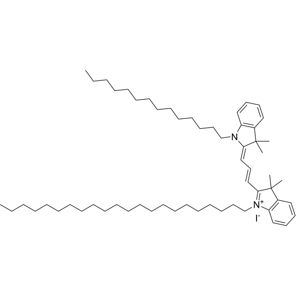
HY-D1451
PKH 26Fluorescent -
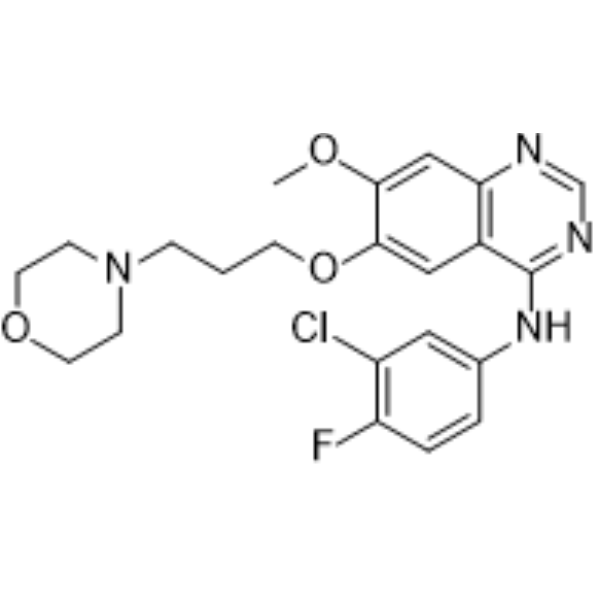
HY-50895
GefitinibEGFR Inhibitor -
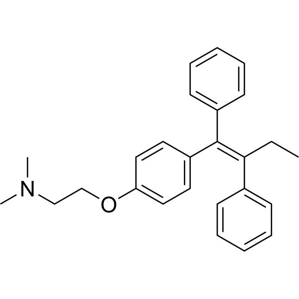
HY-13757A
TamoxifenEstrogen Receptor Modulator -
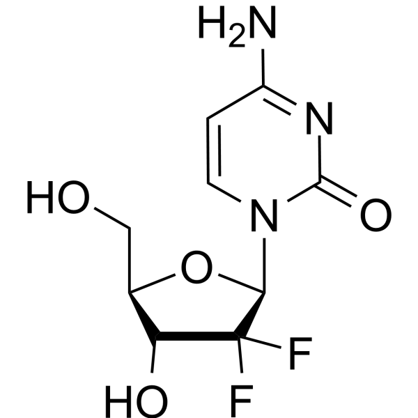
HY-17026
GemcitabineDNA Synthesis Inhibitor -
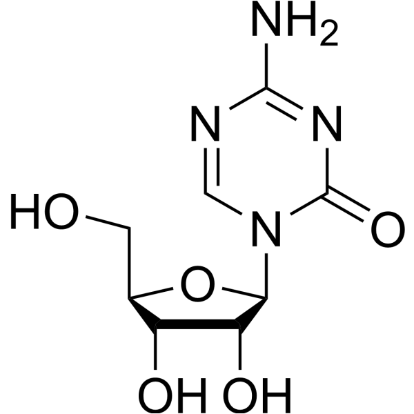
HY-10586
5-AzacytidineDNMT Inhibitor -
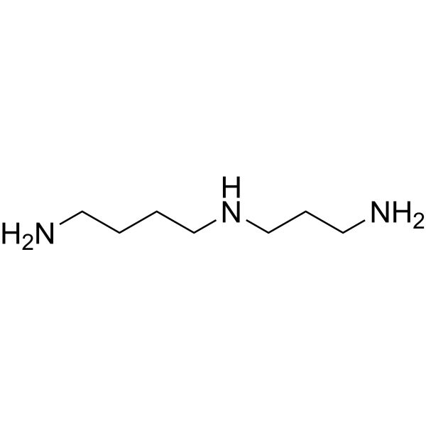
HY-B1776
SpermidinePolyamine Compound -
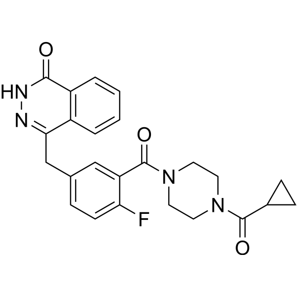
HY-10162
OlaparibPARP Inhibitor -
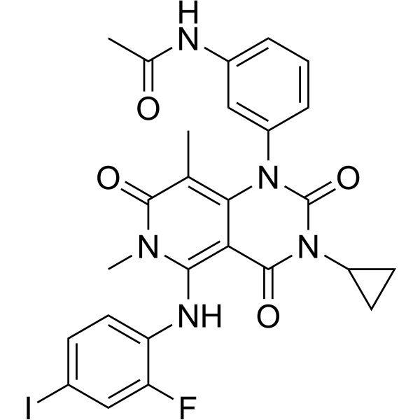
HY-10999
TrametinibMEK Inhibitor -

HY-18739
Phorbol 12-myristate 13-acetatePKC Activator -

HY-14648
DexamethasoneGR Agonist -
HY-17031
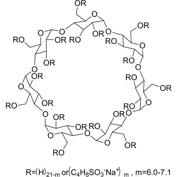
SBE-β-CDExcipient -
HY-D1783
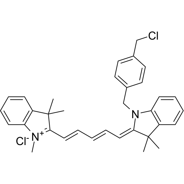
MitoTracker Deep Red FMRed Fluorescent Dye
MedChemExpress(MCE)の使用を引用している文献 PKH 67
More- Cancer Cell. 2026 Jan 8:S1535-6108(25)00545-8. [Abstract]
- Gut Microbes. 2025 Dec 31;17(1):2599543. [Abstract]
- J Nanobiotechnology. 2026 Jun 30.
- J Nanobiotechnology. 2025 Jun 13;23(1):443. [Abstract]
- Theranostics. 2025 Jan 1;15(1):86-102. [Abstract]
- J Cachexia Sarcopenia Muscle. 2025 Dec;16(6):e70159. [Abstract]
- Nano Res. 2024 Aug 13;17:9898–9907.
- Oncogene. 2026 Aug;45(32):3327-3341.
- Int J Surg. 2025 Dec 17. [Abstract]
- Life Sci. 2024 Aug 15:351:122787. [Abstract]
- Drug Des Devel Ther. 2025 Mar 24:19:2189-2203. [Abstract]
- Cancer Immunol Immunother. 2025 Dec 22;75(1):23. [Abstract]
- Tissue Eng Regen Med. 2026 Jul;23(5):753-770. [Abstract]
- Front Biosci-Landmrk. 2026 Aug 13;31(8).
- J Immunol. 2026 Apr 15;215(4):vkaf376. [Abstract]
- J Neurotrauma. 2025 Oct 6. [Abstract]
- Ann Surg Oncol. 2025 Oct 7. [Abstract]
- J Bioenerg Biomembr. 2024 Feb;56(1):31-44. [Abstract]
- Immun Inflamm Dis. 2024 Mar;12(3):e1155. [Abstract]
- Andrology. 2026 Apr 16. [Abstract]
- SSRN. 2025 Nov 25.
 Customer Validation & Images

-
Cell Imaging/Staining
-
IF
-
Cell Imaging/Staining
-
In Vivo Imaging
-
Cell Imaging/Staining


生物活性
製品説明
体外実験
Guidelines (Following is our recommended protocol. This protocol only provides a guideline, and should be modified according to your specific needs).
1. Preparation of dyeing solution
1.1 Take out the PKH 67 reagent from the refrigerator, let it stand for a few minutes to reach room temperature, or after a while in a 37°C water bath, centrifuge the tube containing PKH 67. Please be sure to centrifuge the tube for a few minutes to allow the reagent to fully dissolve before opening the cap. The lid can only be opened after it falls into the bottom of the tube.
1.2 According to the number of cell samples to be detected, dilute the probe 10 times with diluent (The product comes with a buffer solution (100 μL PKH 67 comes with 6 mL Dilutiuon Buffer), which can be used together), and then dilute the PKH 67 solution 25 times with a suitable solution (such as serum-free medium, HBSS or PBS) to prepare Dyeing working solution. Please adjust the optimal working solution concentration according to different cells and your own experimental system. Generally, cells can be diluted 250 times according to the final concentration of the liquid in the kit. Some cells may need to increase the concentration appropriately.
2. Cell staining
2.1 Resuspend the prepared cells to be tested in 100 μL of staining solution to a cell concentration of approximately 107/mL. In situ staining can also be performed, as long as the staining solution is enough to cover the cells.
2.2 Culture cells at 2-8°C for 15-30 minutes. The optimal culture time is different for different cells.
(Note: It is recommended that the cells to be labeled be incubated in the dyeing working solution at 37°C for 5 minutes, and then incubated at 4°C for 15 minutes. Incubation at low temperature can reduce the endocytosis of the dye by the cells and help the dye to bind to the plasma membrane. labeling and reduce the likelihood of the dye localizing to cytoplasmic vesicles).
2.3 After centrifugation, remove the supernatant, collect the cells, wash the cells 1-2 times with PBS or serum-free medium, and finally add PBS or serum-free medium to resuspend the cells.
2.4 Take 500 μL cell suspension and detect it with flow cytometer. Ex/Em=490/502 nm.
2.5 Subsequently, the cells can be cultured according to the normal culture method.
2.6 The labeling effect can be directly observed under a fluorescence microscope, or the cell proliferation can be detected by flow cytometry after culturing for an appropriate period, or used for cell fluorescence tracing for other specific experimental purposes.
Precautions
1. The stain concentration varies depending on the cell type and the number of cells in each well.
2. The prepared PKH 67 liquid is very easy to hydrolyze. It is recommended to store it in separate packages and freeze and dry it at -20°C.
3. PKH 67 working solution should be prepared for immediate use and cannot be prepared in advance, because PKH 67 will decompose when absorbing water and affect the dyeing effect.
4. PKH 67 is easily hydrolyzed and will deteriorate quickly in aqueous solution. Please avoid contact with water during use. The working fluid is in contact with water within the permitted time range during the process of labeling cells.
5. PKH 67 fluorescent dye is an alcoholic solution. It will solidify and stick to the bottom, wall or lid of the centrifugation tube at lower temperatures such as 4°C or ice bath. It will recover after being taken out of the refrigerator. After it reaches room temperature and becomes liquid, centrifuge it to the bottom of the tube before opening the lid. It can be used after immersing it in a 37°C water bath until it is completely dissolved.
6. The passages or times that can be traced after labeling for different cell types vary greatly. Please conduct testing based on the actual situation or reference literature.
7. The product comes with a buffer solution (100 μL PKH 67 comes with 6 mL Dilutiuon Buffer), which can be used together.
MedChemExpress (MCE) has not independently confirmed the accuracy of these methods. They are for reference only.
- BrdU Incorporation Assay
- Somatic Cell Culture
- CFSE Dye Dilution Proliferation Assay
- Dye-dilution cell tracking and proliferation staining
- CCK-8/WST-8 Cell Proliferation Assay
- Protocol for Cell Cycle
- Cell Counting-Based Growth Curve Assay
- Colony Formation (Clonogenic) Assay
- Cell Viability Determination by MTT Colorimetric Assay
- EdU Incorporation Assay (Click Chemistry-Based DNA Synthesis Measurement)
- Ki-67 Immunostaining Proliferation Assay
- PCNA Immunodetection Proliferation Assay
- Protocol for Cell Counting and Cell Density Analysis
- MTT Cell Proliferation Assay


化学情報
-
CAS 番号 257277-27-3
-
性状 Liquid
-
Color Light yellow to yellow
-
SMILES
[P].[KH6].[KH6].[KH6].[KH6].[KH6].[KH6].[KH6]
-
輸送条件
Shipping with dry ice.
-
保管条件
-80°C, protect from light


Publications (21)
-
Journal Impact Factor
-
Most Recent
-
Cancer Cell
2026 Jan 8:S1535-6108(25)00545-8. PMID: 41512868 -
Gut Microbes
Helicobacter pylori infection aggravates hepatic steatosis by lactylation-driven WTAP-mediated m6A modification. [Abstract]2025 Dec 31;17(1):2599543. PMID: 41384837
PKH 67 purchased from MedChemExpress. Usage Cited in: Gut Microbes. 2025 Dec 31;17(1):2599543. [Abstract]
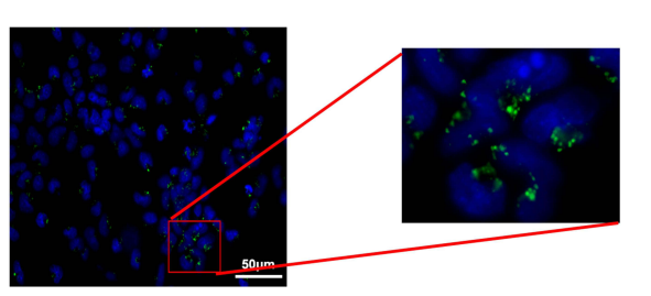
Immunofluorescence images (200μm) of H. pylori OMVs internalized into HepG2 cells: The green signals represent PKH67 labeled OMVs, while the blue represents DAPI; OMVs were incubated with 2 μL PKH 67 dye (MCE, 257277-27-3) in cold PBS for 15 min.
PKH 67 purchased from MedChemExpress. Usage Cited in: Gut Microbes. 2025 Dec 31;17(1):2599543. [Abstract]
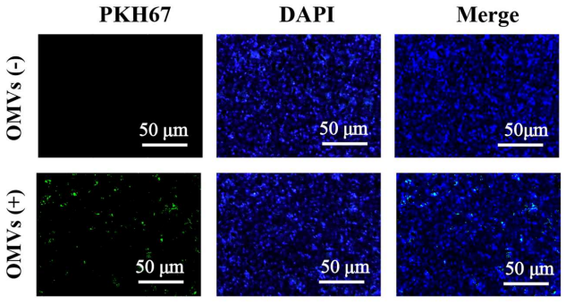
Representative images of immunofluorescence with mean fluorescence intensity of liver tissues (50μm) in C57/BL6 mice with or without H. pylori OMVs gavage. Left: Signal for PKH67 labeled OMVs (green). Middle: DAPI counterstain (blue) identifying cell nuclei. Right: Merged image; OMVs were incubated with 2 μL PKH 67 dye (MCE, 257277-27-3) in cold PBS for 15 min.
-
-
J Nanobiotechnology
Overcoming acquired immunotherapy resistance in non-small cell lung cancer using ginsenoside Rb1-loaded, peptide-enhanced exosome delivery systems. [Abstract]2025 Jun 13;23(1):443. PMID: 40514658
PKH 67 purchased from MedChemExpress. Usage Cited in: J Nanobiotechnology. 2025 Jun 13;23(1):443. [Abstract]
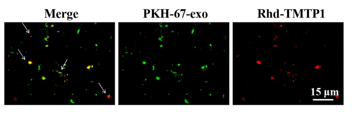
Colocalization of PKH-67-exo and Rhd-TMTP1 observed under a confocal laser scanning microscope (white arrows indicate colocalized PKH-67-exo and Rhd-TMTP1, scale bar = 15 μm). Rhd (Rhodamine), HY-Y0016; PKH-67, HY-D1421.
-
Theranostics
iRGD-TRP-PK1-modified red blood cell membrane vesicles as a new chemotherapeutic drug delivery and targeting system in head and neck cancer. [Abstract]2025 Jan 1;15(1):86-102. PMID: 39744235 -
J Cachexia Sarcopenia Muscle
C2C12-Derived ApoVs Promote Skeletal Muscle Development and Ameliorate Age-Related Muscle Loss Through Igf1r/PI3K/AKT/mTOR Pathway. [Abstract]2025 Dec;16(6):e70159. PMID: 41344911
PKH 67 purchased from MedChemExpress. Usage Cited in: J Cachexia Sarcopenia Muscle. 2025 Dec;16(6):e70159. [Abstract]
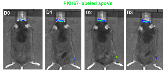
Representative whole-body bioluminescence images of mouse after injecting PKH67-labelled apoVs in tibialis anterior (TA) muscle. 100μL of PKH26 or PKH 67-labelled apoVs at a concentration of 2 μg/g were injected into the tibialis anterior (TA) muscle in C57BL/6J male mice at 8weeks of age.
-
-
-
Int J Surg
Apoptotic extracellular vesicles from peripancreatic adipose-derived mesenchymal stem cells ameliorate severe acute pancreatitis through the transcription factor EB-mediated autophagy-lysosomal pathway: an experimental study. [Abstract]2025 Dec 17. PMID: 41403290 -
Life Sci
Exosomal miR-1228-5p down-regulates DUSP22 to promotes cell proliferation and migration in small cell lung cancer. [Abstract]2024 Aug 15:351:122787. PMID: 38851418 -
Drug Des Devel Ther
Quercetin Improves Hippocampal Neurogenesis in Depression by Regulating the Level of Let-7e-5p in Microglia Exosomes. [Abstract]2025 Mar 24:19:2189-2203. PMID: 40160967
PKH 67 purchased from MedChemExpress. Usage Cited in: Drug Des Devel Ther. 2025 Mar 24:19:2189-2203. [Abstract]
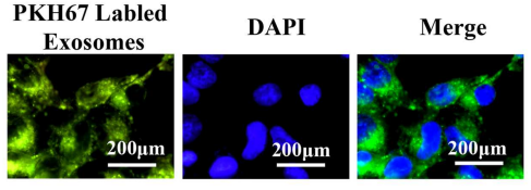
The uptake of PKH-67-labeled exosomes by NSCs was observed. The green fluorescence represents PKH-67-labeled exosomes, while the blue fluorescence corresponds to nucleus.
-
Cancer Immunol Immunother
FUBP3 mediates MXI1 stability to silence RRAS and hinder MAPK signaling in acute megakaryoblastic leukemia progression. [Abstract]2025 Dec 22;75(1):23. PMID: 41428087 -
Tissue Eng Regen Med
2026 Jul;23(5):753-770. PMID: 42068522 -
-
J Immunol
miR-449b-5p from adipose-derived mesenchymal stem cell-derived exosomes targets IGF1R to alleviate airway inflammation and improve airway smooth muscle cell dysfunction in children with asthma. [Abstract]2026 Apr 15;215(4):vkaf376. PMID: 41986878 -
J Neurotrauma
Neuroprotective Effects of Salvia Miltiorrhiza-Derived Extracellular Nanovesicles in Traumatic Brain Injury. [Abstract]2025 Oct 6. PMID: 41052963 -
Ann Surg Oncol
Experimental Study on the Inhibitory Effect of Eupatilin on Osteosarcoma by the NBR2/miR-129-5p/FKBP11 Regulatory Axis. [Abstract]2025 Oct 7. PMID: 41057744 -
J Bioenerg Biomembr
Chondroprotective effects of bone marrow mesenchymal stem cell-derived exosomes in osteoarthritis. [Abstract]2024 Feb;56(1):31-44. PMID: 38012335 -
Immun Inflamm Dis
Exosomes secreted from induced pluripotent stem cell ameliorate the lipopolysaccharide induced neuroinflammatory response via lncRNA-0949. [Abstract]2024 Mar;12(3):e1155. PMID: 38533916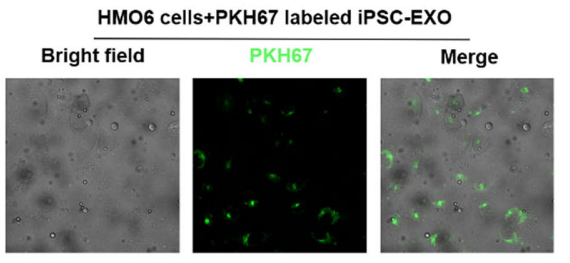
PKH 67 purchased from MedChemExpress. Usage Cited in: Immun Inflamm Dis. 2024 Mar;12(3):e1155. [Abstract]
After PKH 67 labeling of iPSC‐Exos, the uptake of iPSC‐Exos by HMO6 cells was detected by fluorescence microscope. HMO6 cells cocultured with iPSC‐Exos labeled with PKH 67 for 12 h.
-
Andrology
Smooth Muscle Cell-Derived Exosomal miR-30a-5p as a Novel Therapeutic Strategy for Erectile Dysfunction in Diabetes Mellitus. [Abstract]2026 Apr 16. PMID: 41987693 -


純度とドキュメンテーション
-
データシート (275 KB)
-
SDS (251 KB)
- English - EN (251 KB)
- Français - FR (251 KB)
- Deutsch - DE (251 KB)
- Norwegian - NO (251 KB)
- Español - ES (251 KB)
- Swedish - SV (251 KB)
- Italian - IT (251 KB)
- Korean - KR (251 KB)
- Portuguese - PT (251 KB)
-
取扱説明書 (2659 KB)
参考文献

Calculators
濃度 (開始) × 体積 (開始) = 濃度 (終了) × 体積 (終了)


PKH 67 関連分類
HY-D1421 関連分類
 Powered by Bioz