



AP-6
Based on 1 publication(s) in Google Scholar
AP-6 is a selective inhibitor of TMEM175 with activity in modulating lysosomal function. Acute inhibition of TMEM175 by AP-6 increases lysosomal macromolecular catabolism, thereby accelerating macrophage and other digestive processes. AP-6 may be used in Parkinson's disease research.
연구목적의 판매만을 진행합니다. 환자를 대상으로 한 판매는 하지 않습니다.
- Purity : 99.81%
- CAS No.: 1369963-51-8
- 화학식: C16H14N4
- 분자량:262.31
-
보관:Powder -20°C, 3 years , 4°C, 2 years ; In solvent -80°C, 6 months , -20°C, 1 month


To place orders, for customer services and technical support, please contact: MedChemExpress USA
Tel: 609-228-6898 E-mail: [email protected] [email protected]


-
 Biological Activity
-
Chemical Information
-
용액&용해도
- Protocol
- 순도&문서
- References
-
 Help & FAQs



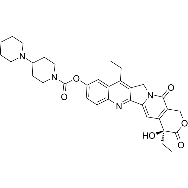
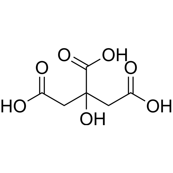
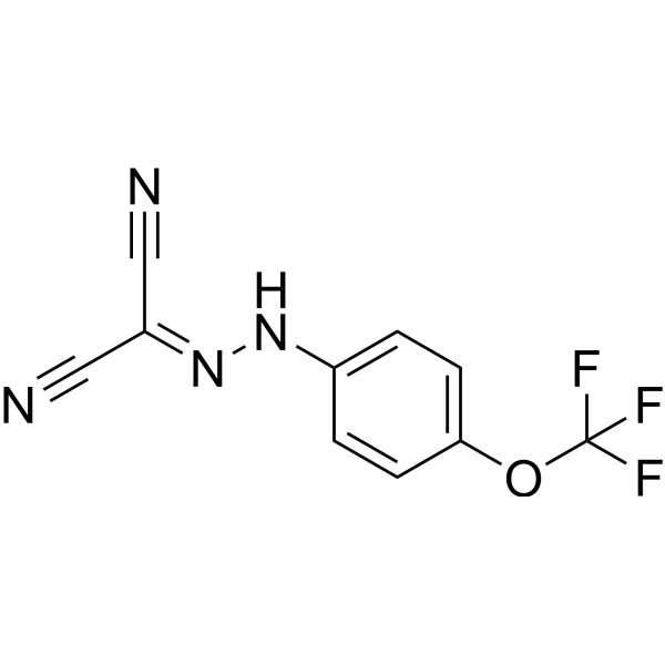
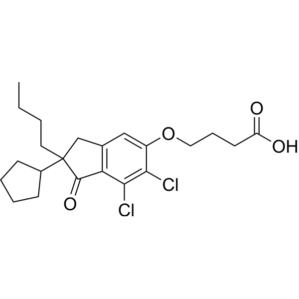

Publications Citing Use of MedChemExpress (MCE) AP-6
More

Biological Activity
제품 설명


Chemical Information
-
CAS No. 1369963-51-8
-
Appearance Solid
-
분자량 262.31
-
화학식 C16H14N4
-
Color White to off-white
-
SMILES
NC1=CC(C2=CC=CC(C3=CC(N)=CC=N3)=C2)=NC=C1
-
선적
Room temperature in continental US; may vary elsewhere.
-
보관
Powder -20°C 3 years 4°C 2 years In solvent -80°C 6 months -20°C 1 month


Publications (1)
-
Journal Impact Factor
-
Most Recent


용액&용해도
In Vitro:
DMSO : 0.93 mg/mL (3.55 mM; ultrasonic and warming and adjust pH to 3 with 1 M HCl and heat to 60°C; Hygroscopic DMSO has a significant impact on the solubility of product, please use newly opened DMSO)
H2O : < 0.1 mg/mL (insoluble)
Please refer to the solubility information to select the appropriate solvent. Once prepared, please aliquot and store the solution to prevent product inactivation from repeated freeze-thaw cycles.
Storage method and period of stock solution: -80°C, 6 months; -20°C, 1 month. When stored at -80°C, please use it within 6 months. When stored at -20°C, please use it within 1 month.
Please refer to the solubility information to select the appropriate solvent. Once prepared, please aliquot and store the solution to prevent product inactivation from repeated freeze-thaw cycles.
Storage method and period of stock solution: -80°C, 6 months; -20°C, 1 month. When stored at -80°C, please use it within 6 months. When stored at -20°C, please use it within 1 month.
Concentration (start) × Volume (start) = Concentration (final) × Volume (final)


순도&문서
-
Data Sheet (272 KB)
-
SDS (251 KB)
- English - EN (251 KB)
- Français - FR (251 KB)
- Deutsch - DE (251 KB)
- Norwegian - NO (251 KB)
- Español - ES (251 KB)
- Swedish - SV (251 KB)
- Italian - IT (251 KB)
- Korean - KR (251 KB)
- Portuguese - PT (251 KB)
-
Handling Instructions (2659 KB)
References


Complete Stock Solution Preparation Table
Please refer to the solubility information to select the appropriate solvent. Once prepared, please aliquot and store the solution to prevent product inactivation from repeated freeze-thaw cycles.
Storage method and period of stock solution: -80°C, 6 months; -20°C, 1 month. When stored at -80°C, please use it within 6 months. When stored at -20°C, please use it within 1 month.
| Optional Solvent | Concentration Solvent Mass | 1 mg | 5 mg | 10 mg | 25 mg |
|---|---|---|---|---|---|
| DMSO | 1 mM | 3.8123 mL | 19.0614 mL | 38.1228 mL | 95.3071 mL |


AP-6 Related Classifications
HY-161949 Related Classifications
 Powered by Bioz