



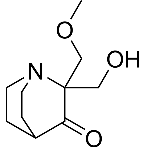
Eprenetapopt (GMP) (APR-246(GMP)) is Eprenetapopt (HY-19980) in GMP grade. GMP small molecules works appropriately as an auxiliary reagent for cell therapy manufacture. Eprenetapopt (APR-246) is a first-in-class, small molecule that restores wild-type p53 functions in TP53-mutant cells. Eprenetapopt triggers apoptosis in tumor cells. Eprenetapopt also targets the selenoprotein thioredoxin reductase 1 (TrxR1), a key regulator of cellular redox balance.
연구목적의 판매만을 진행합니다. 환자를 대상으로 한 판매는 하지 않습니다.
- CAS No.: 5291-32-7
- 화학식: C10H17NO3
- 분자량:199.25
-
보관:
Please store the product under the recommended conditions in the Certificate of Analysis.


To place orders, for customer services and technical support, please contact: MedChemExpress USA
Tel: 609-228-6898 E-mail: [email protected] [email protected]


-
 Biological Activity
-
Chemical Information
- Protocol
- 순도&문서
- References
-
 Help & FAQs


-
HY-19980
Eprenetapoptp53 Activator -
HY-10029
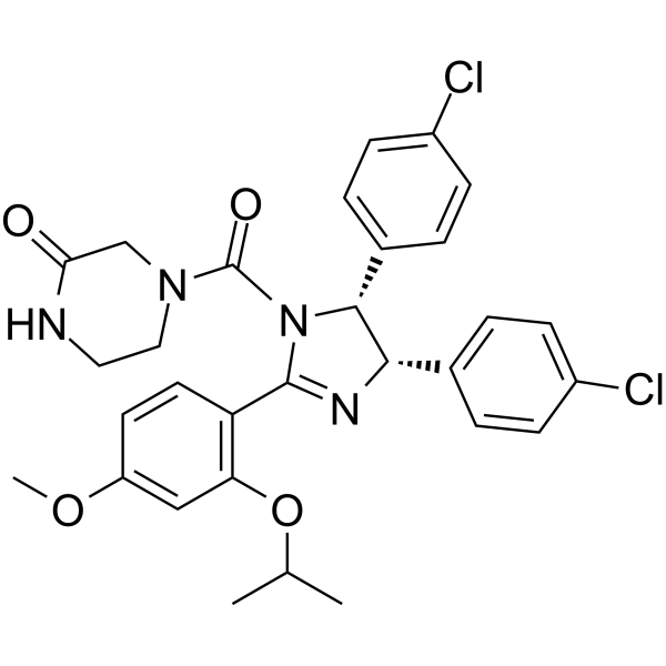
Nutlin-3aMDM2 Antagonist -
HY-19980A
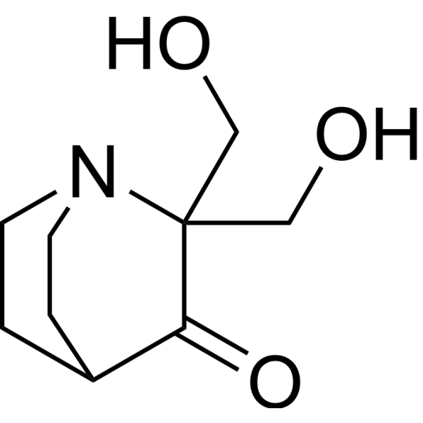
PRIMA-1Mutant p53 Reactivator -
HY-15484
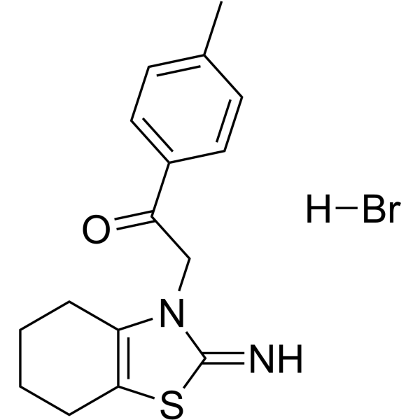
Pifithrin-α hydrobromidep53 Inhibitor -
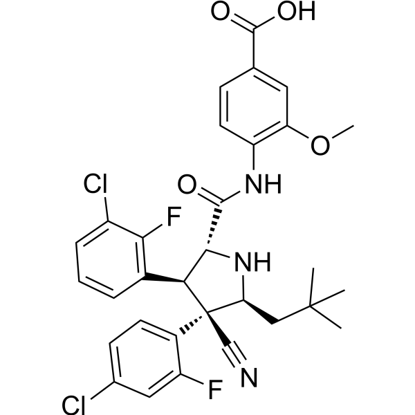
HY-15676
IdasanutlinMDM2 Inhibitor -
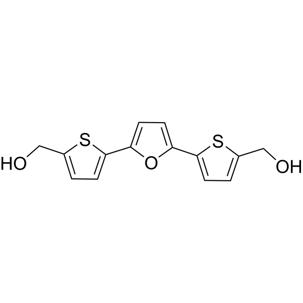
HY-13424
RITAp53-HDM-2 Interaction Inhibitor -
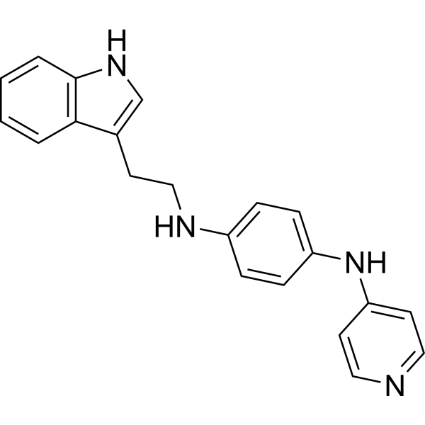
HY-12025
SerdemetanAnticancer Aagent -
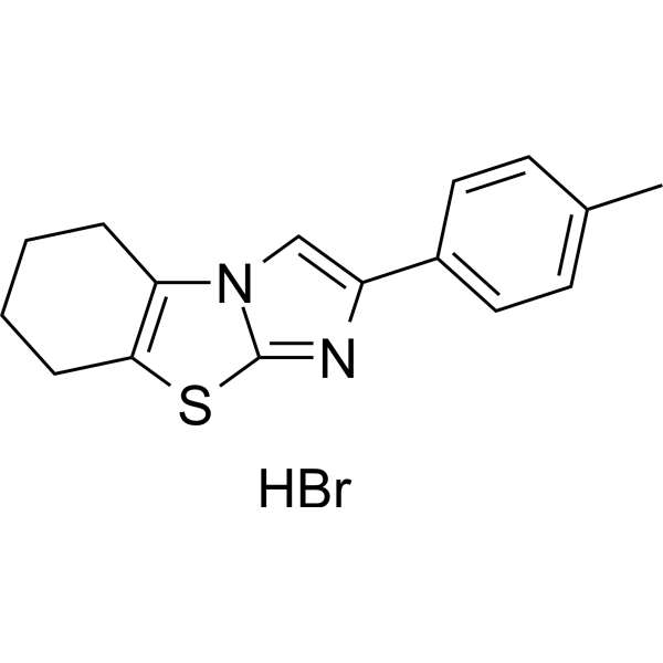
HY-16702A
Pifithrin-β hydrobromideMDM-2/p53 p53 Inhibitor -
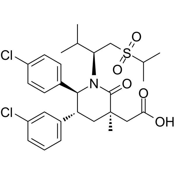
HY-12296
NavtemadlinMDM2-p53 Interaction Inhibitor -
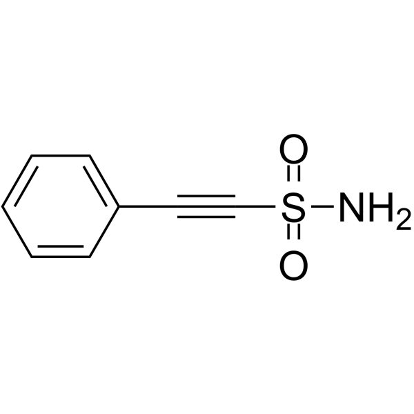
HY-10940
Pifithrin-μp53 and HSP70 Inhibitor -
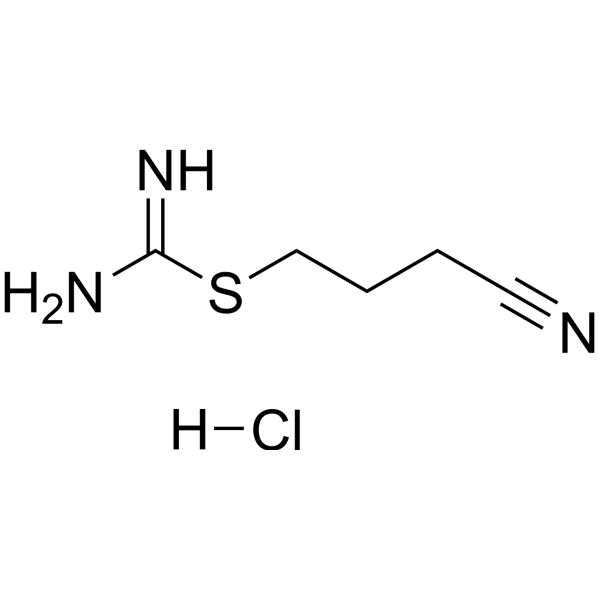
HY-16271
Kevetrin hydrochloridep53 Activator -
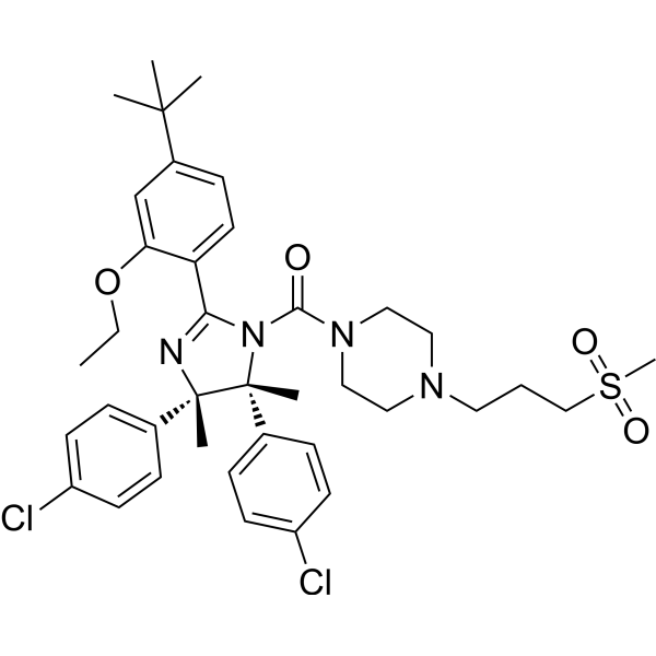
HY-10959
RG7112MDM2 Antagonist


Biological Activity
제품 설명
In Vitro
Eprenetapopt inhibits both recombinant TrxR1 in vitro and TrxR1 in cells. Cellular TrxR1 activity is inhibited by Eprenetapopt irrespective of p53 status. Eprenetapopt can directly affect cellular redox status via targeting of TrxR1. Several small molecules have been shown to restore wild-type activity to mutant p53, including CP-31398, PRIMA-1 and Eprenetapopt, MIRA, STIMA, PhiKan-083 and NSC319726. PRIMA-1 and its methylated analog Eprenetapopt promote correct folding of mutant p53, induce cell death by apoptosis, and inhibit tumor growth in mice. Eprenetapopt has also been shown to reactivate mutant forms of the p63 and p73 proteins that share high structural homology with p53[1].
Eprenetapopt is a powerful apoptosis-inducing agent. Eprenetapopt can enhance apoptosis in mutant p53 carrying cells, compared to the p53 null parental cells. Most p53 mutants are in complex with Hsp70 proteins. Eprenetapopt treatment increases Hsp70 expression and nucleolar translocation, in parallel with the induction of nucleolar accumulation of mutant p53. Several lines of evidence suggest that Eprenetapopt can also act independently of the p53 status of the cell. It can radiosensitize prostate carcinoma cell lines with mutant or wild type p53 and p53-/- cells as well. Introduction of mutant p53 (p53ser249 or p53gln248) into p53-/- hepatocarcinoma cells increases sensitivity to Eprenetapopt without the induction of p53 target genes.Eprenetapopt regularly induces apoptosis in mutant p53 expressing cells[2].
MedChemExpress (MCE) has not independently confirmed the accuracy of these methods. They are for reference only.
Clinical Trial
| NCT Number | Sponsor | Condition | Start Date |
Phase

|
|---|---|---|---|---|
| NCT01329991 | Plexxikon| | 2011-05 | PHASE1 |


Chemical Information
-
CAS No. 5291-32-7
-
분자량 199.25
-
화학식 C10H17NO3
-
SMILES
O=C1C(COC)(CO)N2CCC1CC2
-
Synonyms
APR-246 (GMP); PRIMA-1Met (GMP)
-
선적
Room temperature in continental US; may vary elsewhere.
-
보관
Please store the product under the recommended conditions in the Certificate of Analysis.


순도&문서
References
[1]. Peng X, et al. APR-246/PRIMA-1MET inhibits thioredoxin reductase 1 and converts the enzyme to a dedicated NADPH oxidase. Cell Death Dis. 2013 Oct 24;4:e881 [Content Brief]
[2]. Stuber G, et al. PRIMA-1MET induces nucleolar translocation of Epstein-Barr virus-encoded EBNA-5 protein. Mol Cancer. 2009 Mar 26;8:23. [Content Brief]
[3]. Sallman DA, et al. Eprenetapopt (APR-246) and Azacitidine in TP53-Mutant Myelodysplastic Syndromes. J Clin Oncol. 2021;39(14):1584-1594. [Content Brief]

Calculators
Concentration (start) × Volume (start) = Concentration (final) × Volume (final)


Eprenetapopt (GMP) Related Classifications
HY-19980G Related Classifications
 Powered by Bioz