



Resigratinib

Based on 1 Customer Validation
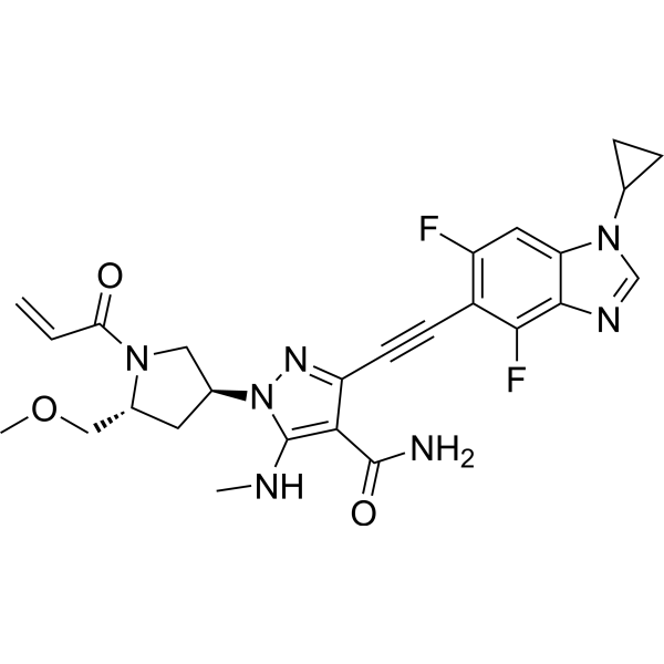
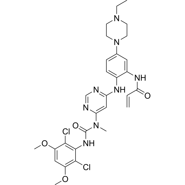
Resigratinib (KIN-3248) is an irreversible and orally active covalent inhibitor of FGFR1-4 that effectively inhibits wild-type and drug-resistant mutations (such as FGFR2 V565F, FGFR3 V555M). Resigratinib covalently binds to the Cys492 site of FGFR, blocks the FGFR signaling pathway, inhibits tumor cell proliferation and induces apoptosis. Resigratinib can be used for the study of FGFR2/3-driven solid tumors (such as cholangiocarcinoma and bladder cancer).
연구목적의 판매만을 진행합니다. 환자를 대상으로 한 판매는 하지 않습니다.
- Purity : 99.43%
- CAS No.: 2750709-91-0
- 화학식: C26H27F2N7O3
- 분자량:523.53
-
보관:Powder -20°C, 3 years , 4°C, 2 years ; In solvent -80°C, 6 months , -20°C, 1 month

To place orders, for customer services and technical support, please contact: MedChemExpress USA
Tel: 609-228-6898 E-mail: [email protected] [email protected]


-
 Biological Activity
-
Chemical Information
-
용액&용해도
- Protocol
- 순도&문서
- References
-
 Help & FAQs


-
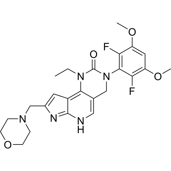
HY-109099
PemigatinibFGFR Inhibitor -
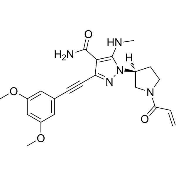
HY-132817
GunagratinibFGFR Inhibitor -
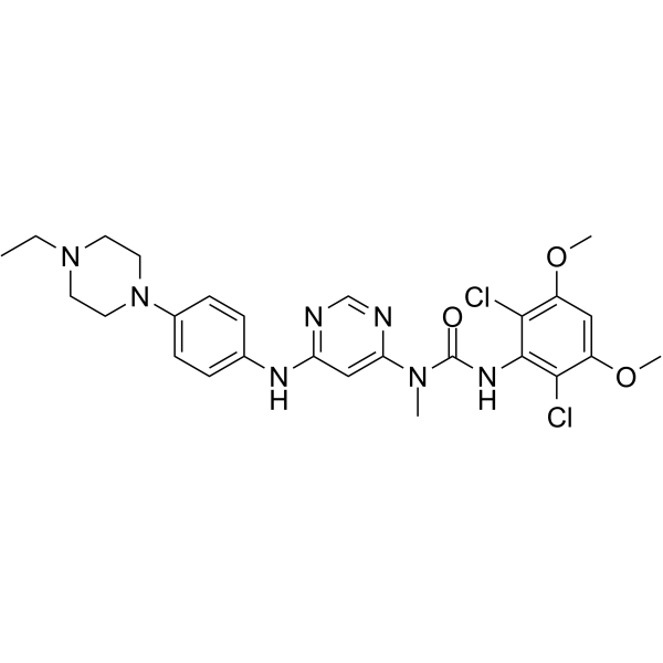
HY-13311
InfigratinibFGFR Inhibitor -
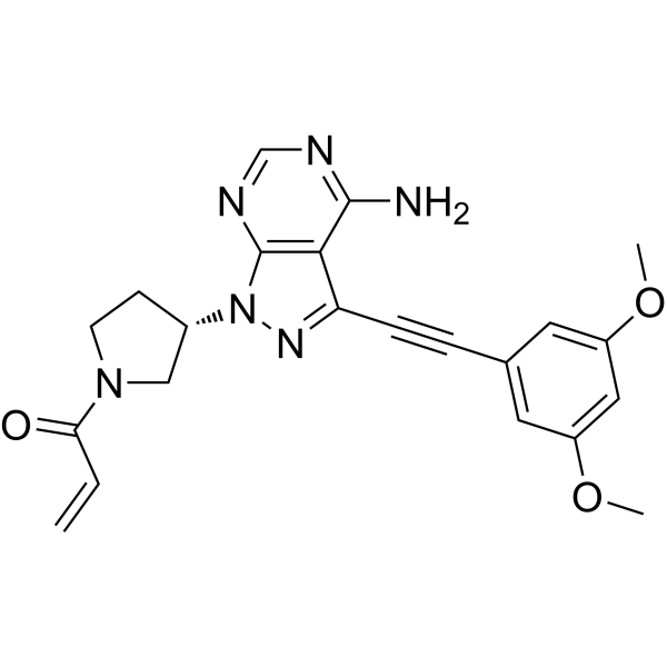
HY-100818
FutibatinibFGFR Inhibitor -
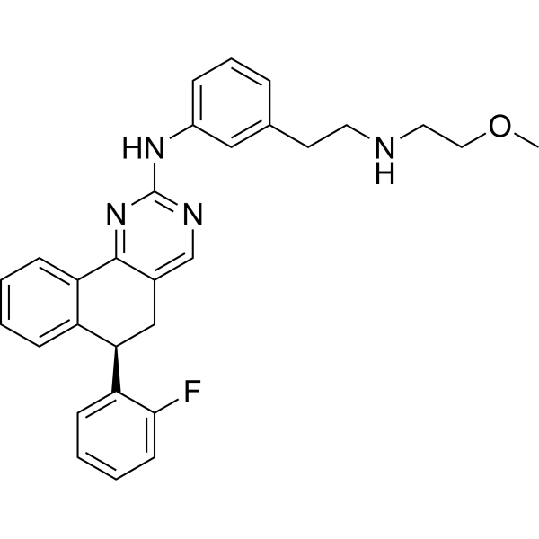
HY-19981
Derazantinib -
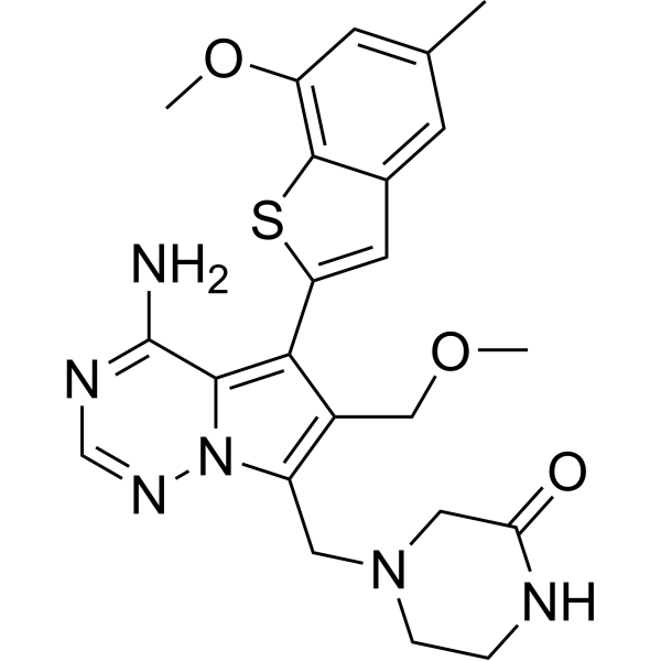
HY-100019
RogaratinibFGFR1-4 Inhibitor -
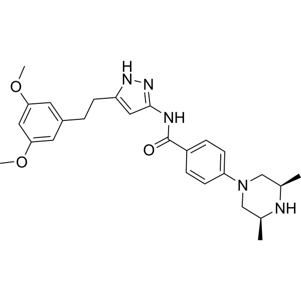
HY-13330
FexagratinibFGFR Inhibitor -
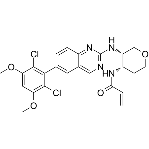
HY-100492
FisogatinibFGFR4 Inhibitor -
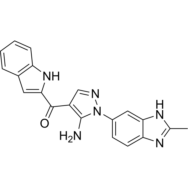
HY-19957
Zoligratinib -
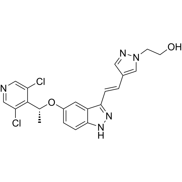
HY-13304
LY2874455FGFR Inhibitor -
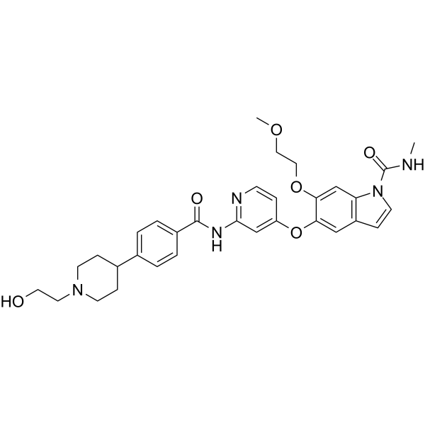
HY-101466
E7090FGFR1/2/3 Inhibitor -
HY-100491
H3B-6527FGFR4 Inhibitor


Biological Activity
제품 설명
Cellular Effect
|
Cell Line

|
Type | Value | Description | References |
|---|---|---|---|---|
| RT-112 | IC50 |
16.2 nM
Compound: 11; KIN-3248
|
Antiproliferative activity against human RT-112 cells expressing FGFR3 K650M mutant assessed as inhibition of cell growth incubated for 5 days by Cell-titer glo assay
Antiproliferative activity against human RT-112 cells expressing FGFR3 K650M mutant assessed as inhibition of cell growth incubated for 5 days by Cell-titer glo assay
|
[PMID: 38267212] |
| RT-112 | IC50 |
22.4 nM
Compound: 11; KIN-3248
|
Antiproliferative activity against human RT-112 cells expressing FGFR3 N540K mutant assessed as inhibition of cell growth incubated for 5 days by Cell-titer glo assay
Antiproliferative activity against human RT-112 cells expressing FGFR3 N540K mutant assessed as inhibition of cell growth incubated for 5 days by Cell-titer glo assay
|
[PMID: 38267212] |
| RT-112 | IC50 |
4 nM
Compound: 11; KIN-3248
|
Antiproliferative activity against human RT-112 cells assessed as inhibition of cell growth incubated for 5 days by Cell-titer glo assay
Antiproliferative activity against human RT-112 cells assessed as inhibition of cell growth incubated for 5 days by Cell-titer glo assay
|
[PMID: 38267212] |
| RT-112 | IC50 |
45.2 nM
Compound: 11; KIN-3248
|
Antiproliferative activity against human RT-112 cells expressing FGFR3 V555M mutant assessed as inhibition of cell growth incubated for 5 days by Cell-titer glo assay
Antiproliferative activity against human RT-112 cells expressing FGFR3 V555M mutant assessed as inhibition of cell growth incubated for 5 days by Cell-titer glo assay
|
[PMID: 38267212] |
| RT-112 | IC50 |
8.2 nM
Compound: 11; KIN-3248
|
Antiproliferative activity against human RT-112 cells expressing FGFR3 assessed as inhibition of cell growth incubated for 5 days by Cell-titer glo assay
Antiproliferative activity against human RT-112 cells expressing FGFR3 assessed as inhibition of cell growth incubated for 5 days by Cell-titer glo assay
|
[PMID: 38267212] |
| SNU-16 | IC50 |
3.6 nM
Compound: 11; KIN-3248
|
Antiproliferative activity against human SNU-16 cells assessed as inhibition of cell growth incubated for 5 days by Cell-titer glo assay
Antiproliferative activity against human SNU-16 cells assessed as inhibition of cell growth incubated for 5 days by Cell-titer glo assay
|
[PMID: 38267212] |
In Vitro
Resigratinib has an IC50 of 0.6-2.3 nM for wild-type FGFR1-4 and an IC50 of <25 nM for drug-resistant mutations such as FGFR2 V565F, FGFR3 V555M, and FGFR3 N550K. Resigratinib has a selectivity of >90% for FGFR family kinase activity and weak inhibitory effects on other kinases[1].
Resigratinib (compound 11) (0.1 nM-10 μM; 72 h) inhibits cell viability of cancer cell lines expressing FGFR2 or FGFR3 (e.g., SNU-16, RT112)[1].
MedChemExpress (MCE) has not independently confirmed the accuracy of these methods. They are for reference only.
-
Cell Line:FGFR2 or FGFR3 expressing Cellular Viability in:
(FGFR2) SNU16, CCLP-1, CCLP-1V565F, CCLP-1N550H, CCLP-1N550K, ICC13-7, ICC13-7V565F, ICC13-7N550K
(FGFR3) RT112 (WT), RT112:FGFR3-TACC3, RT112:FGFR3-TACC3, V555M RT112:FGFR3-TACC3N540K, RT112:FGFR3-TACC3K650M -
Concentration:0.1 nM-10 μM
-
Incubation Time:72 h
-
Result:Showed IC50s of 3.6 nM (SNU16), 3.5 nM (CCLP-1), 5.2 nM (CCLP-1V565F), 2.9 nM (CCLP-1N550H), 10.2 nM (CCLP-1N550K), 2.0 nM (ICC13-7), 4.3 nM (ICC13-7V565F), 8.0 nM (ICC13-7N550K); 4.0 nM (RT112-WT), 8.2 nM (RT112:FGFR3-TACC3), 45.2 nM (RT112:FGFR3-TACC3), 22.4 nM (V555M RT112:FGFR3-TACC3N540K), 16.2 nM (RT112:FGFR3-TACC3K650M), respectively.
In Vivo
MedChemExpress (MCE) has not independently confirmed the accuracy of these methods. They are for reference only.
-
Animal Model:Female NSG mice (6-10 weeks old) with SNU-16 (FGFR2-amplified gastric cancer) xenograft model[1]
-
Dosage:2, 5, 15 mg/kg
-
Administration:Oral gavage, once daily, 21 days
-
Result:Dose-dependent tumor growth inhibition, with TGI of 69%, 81%, and 93% at 2, 5, and 15 mg/kg, respectively.
No significant body weight loss was noted, and phospho-ERK signaling was suppressed in tumor tissues.
- Annexin V plus membrane-impermeant dye apoptosis staining
- BrdU Incorporation Assay
- Somatic Cell Culture
- CFSE Dye Dilution Proliferation Assay
- Dye-dilution cell tracking and proliferation staining
- CCK-8/WST-8 Cell Proliferation Assay
- Protocol for Cell Cycle
- Cell Counting-Based Growth Curve Assay
- Colony Formation (Clonogenic) Assay
- Cell Viability Determination by MTT Colorimetric Assay
- EdU Incorporation Assay (Click Chemistry-Based DNA Synthesis Measurement)
- Ki-67 Immunostaining Proliferation Assay
- Detection of Apoptosis
- TUNEL staining for apoptotic DNA fragmentation
- PCNA Immunodetection Proliferation Assay
- Protocol for Cell Counting and Cell Density Analysis
- Apoptosis Solutions
- MTT Cell Proliferation Assay
Clinical Trial
| NCT Number | Sponsor | Condition | Start Date |
Phase
|
|---|---|---|---|---|
| NCT01329991 | Plexxikon| | 2011-05 | PHASE1 |


Chemical Information
-
CAS No. 2750709-91-0
-
Appearance Solid
-
분자량 523.53
-
화학식 C26H27F2N7O3
-
Color White to off-white
-
SMILES
CNC1=C(C(N)=O)C(C#CC2=C(F)C=C(N(C3CC3)C=N4)C4=C2F)=NN1[C@H]5C[C@@H](N(C5)C(C=C)=O)COC
-
Synonyms
KIN-3248
-
선적
Room temperature in continental US; may vary elsewhere.
-
보관
Powder -20°C 3 years 4°C 2 years In solvent -80°C 6 months -20°C 1 month


용액&용해도
In Vitro:
DMSO : 100 mg/mL (191.01 mM; Need ultrasonic; Hygroscopic DMSO has a significant impact on the solubility of product, please use newly opened DMSO)
Please refer to the solubility information to select the appropriate solvent. Once prepared, please aliquot and store the solution to prevent product inactivation from repeated freeze-thaw cycles.
Storage method and period of stock solution: -80°C, 6 months; -20°C, 1 month. When stored at -80°C, please use it within 6 months. When stored at -20°C, please use it within 1 month.
Please refer to the solubility information to select the appropriate solvent. Once prepared, please aliquot and store the solution to prevent product inactivation from repeated freeze-thaw cycles.
Storage method and period of stock solution: -80°C, 6 months; -20°C, 1 month. When stored at -80°C, please use it within 6 months. When stored at -20°C, please use it within 1 month.
Concentration (start) × Volume (start) = Concentration (final) × Volume (final)
In Vivo:
Select the appropriate dissolution method based on your experimental animal and administration route.
- For the following dissolution methods, please ensure to first prepare a clear stock solution using an In Vitro approach and then sequentially add co-solvents:
- To ensure reliable experimental results, the clarified stock solution can be appropriately stored based on storage conditions. As for the working solution for In Vivo experiments, it is recommended to prepare freshly and use it on the same day.
- The percentages shown for the solvents indicate their volumetric ratio in the final prepared solution. If precipitation or phase separation occurs during preparation, heat and/or sonication can be used to aid dissolution.
Add each solvent one by one: 10% DMSO 40% PEG300 5% Tween-80 45% Saline
Solubility: ≥ 5 mg/mL (9.55 mM); Clear solution
This protocol yields a clear solution of ≥ 5 mg/mL (saturation unknown).
Taking 1 mL working solution as an example, add 100 μL DMSO stock solution (50.0 mg/mL) to 400 μL PEG300, and mix evenly; then add 50 μL Tween-80 and mix evenly; then add 450 μL Saline to adjust the volume to 1 mL.
Preparation of Saline: Dissolve 0.9 g sodium chloride in ddH₂O and dilute to 100 mL to obtain a clear Saline solution.
Add each solvent one by one: 10% DMSO 90% (20% SBE-β-CD in Saline)
Solubility: ≥ 5 mg/mL (9.55 mM); Clear solution
This protocol yields a clear solution of ≥ 5 mg/mL (saturation unknown).
Taking 1 mL working solution as an example, add 100 μL DMSO stock solution (50.0 mg/mL) to 900 μL 20% SBE-β-CD in Saline, and mix evenly.
Preparation of 20% SBE-β-CD in Saline (4°C, storage for one week): 2 g SBE-β-CD powder is dissolved in 10 mL Saline, completely dissolve until clear.
In Vivo Dissolution Calculator
Please enter the basic information of animal experiments:
-
-
-
-
Recommended: Prepare an additional quantity of animals to account for potential losses during experiments.
Please enter your animal formula composition:
-
%DMSO +
Recommended: Keep the proportion of DMSO in working solution below 2% if your animal is weak.
-
%+
-
+%Tween-80 + +
-
%Saline +
The co-solvents required include: DMSO, . All of co-solvents are available by MedChemExpress (MCE). , Tween 80. All of co-solvents are available by MedChemExpress (MCE).
Working solution concentration: 0.22 mg/mL
Method for preparing stock solution: mg drug dissolved in μL DMSO. Stock solution concentration: mg/mL.
1. Take μL DMSO stock solution;
2. Add μL .
μL , mix evenly;
3. Then add μL Tween 80, mix evenly;
4. Then add μL
Please ensure that the stock solution in the first step is dissolved to a clear state, and add co-solvents in sequence. You can use ultrasonic heating (ultrasonic cleaner, recommended frequency 20-40 kHz), vortexing, etc. to assist dissolution.


순도&문서
-
Data Sheet (277 KB)
-
SDS (252 KB)
- English - EN (252 KB)
- Français - FR (252 KB)
- Deutsch - DE (252 KB)
- Norwegian - NO (252 KB)
- Español - ES (252 KB)
- Swedish - SV (252 KB)
- Italian - IT (252 KB)
- Korean - KR (252 KB)
- Portuguese - PT (252 KB)
-
Handling Instructions (2659 KB)
References
[1]. Tyhonas JS, et al. Discovery of KIN-3248, An Irreversible, Next Generation FGFR Inhibitor for the Treatment of Advanced Tumors Harboring FGFR2 and/or FGFR3 Gene Alterations. J Med Chem. 2024 Feb 8;67(3):1734-1746. [Content Brief]
[2]. Balasooriya ER, et al. The Irreversible FGFR Inhibitor KIN-3248 Overcomes FGFR2 Kinase Domain Mutations. Clin Cancer Res. 2024 May 15;30(10):2181-2192. [Content Brief]


Complete Stock Solution Preparation Table
Please refer to the solubility information to select the appropriate solvent. Once prepared, please aliquot and store the solution to prevent product inactivation from repeated freeze-thaw cycles.
Storage method and period of stock solution: -80°C, 6 months; -20°C, 1 month. When stored at -80°C, please use it within 6 months. When stored at -20°C, please use it within 1 month.
| Optional Solvent | Concentration Solvent Mass | 1 mg | 5 mg | 10 mg | 25 mg |
|---|---|---|---|---|---|
| DMSO | 1 mM | 1.9101 mL | 9.5506 mL | 19.1011 mL | 47.7528 mL |
| 5 mM | 0.3820 mL | 1.9101 mL | 3.8202 mL | 9.5506 mL | |
| 10 mM | 0.1910 mL | 0.9551 mL | 1.9101 mL | 4.7753 mL | |
| 15 mM | 0.1273 mL | 0.6367 mL | 1.2734 mL | 3.1835 mL | |
| 20 mM | 0.0955 mL | 0.4775 mL | 0.9551 mL | 2.3876 mL | |
| 25 mM | 0.0764 mL | 0.3820 mL | 0.7640 mL | 1.9101 mL | |
| 30 mM | 0.0637 mL | 0.3184 mL | 0.6367 mL | 1.5918 mL | |
| 40 mM | 0.0478 mL | 0.2388 mL | 0.4775 mL | 1.1938 mL | |
| 50 mM | 0.0382 mL | 0.1910 mL | 0.3820 mL | 0.9551 mL | |
| 60 mM | 0.0318 mL | 0.1592 mL | 0.3184 mL | 0.7959 mL | |
| 80 mM | 0.0239 mL | 0.1194 mL | 0.2388 mL | 0.5969 mL | |
| 100 mM | 0.0191 mL | 0.0955 mL | 0.1910 mL | 0.4775 mL |


Resigratinib Related Classifications
HY-156632 Related Classifications
 Powered by Bioz