



Latrunculin A
Based on 14 publication(s) in Google Scholar
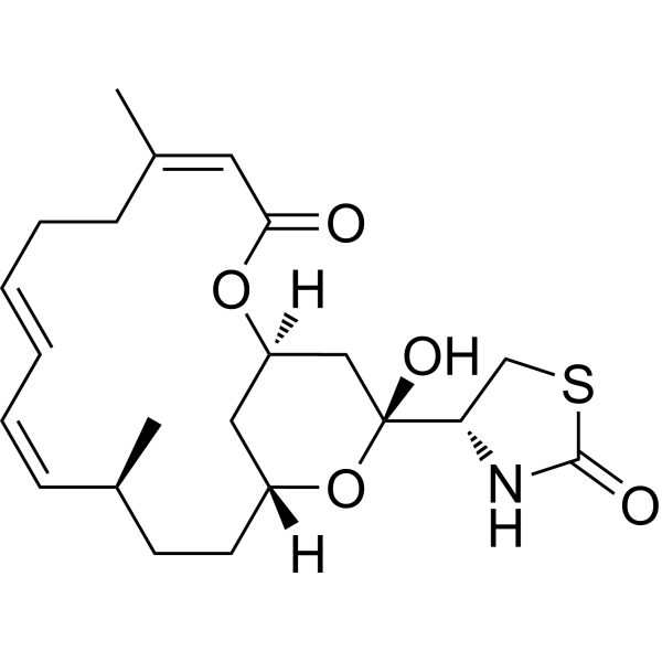
Latrunculin A (LAT-A), found in the red sea sponge Latrunculia magnifica, is a G-actin polymerization inhibitor. Latrunculin A binds to actin monomers and inhibits polymerization of actin with Kds of 0.1, 0.4, 4.7 μM and 0.19 μM for ATP-actin, ADP-Pi-actin, ADP-actin and G-actin, respectively. Latrunculin A has effective anti-metastatic properties for cancer research. Latrunculin A blocks cell migration.
For research use only. We do not sell to patients.
- Purity : 98.9%
- CAS No.: 76343-93-6
- Formula: C22H31NO5S
- Molecular Weight:421.55
-
Storage:
Solution, -20°C, 2 years

To place orders, for customer services and technical support, please contact: MedChemExpress USA
Tel: 609-228-6898 E-mail: [email protected] [email protected]


-
 Biological Activity
-
Chemical Information
- Protocols
- Purity & Documentation
- References
-
 Help & FAQs


-
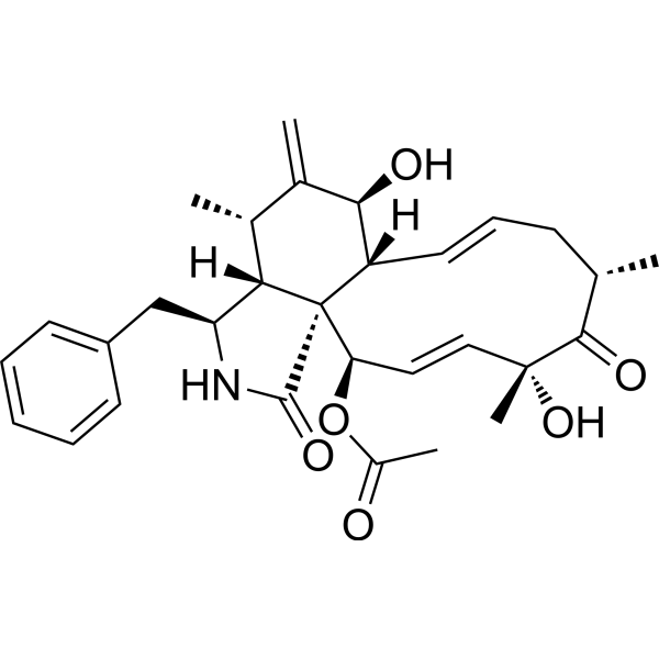
HY-N6682
Cytochalasin DActin Polymerization Inhibitor -
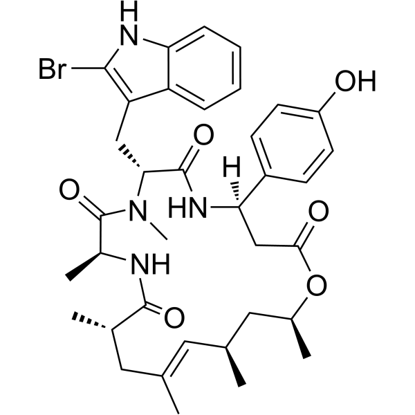
HY-P0027
JasplakinolideActin Polymerization Inducer -
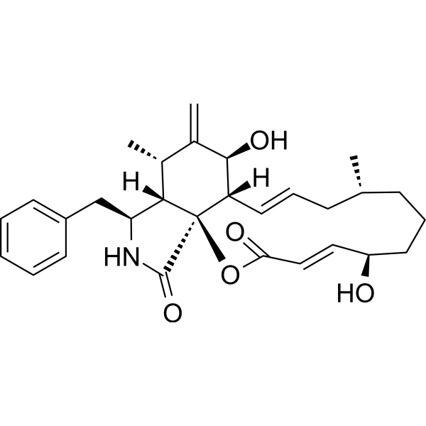
HY-16928
Cytochalasin BMycotoxin -
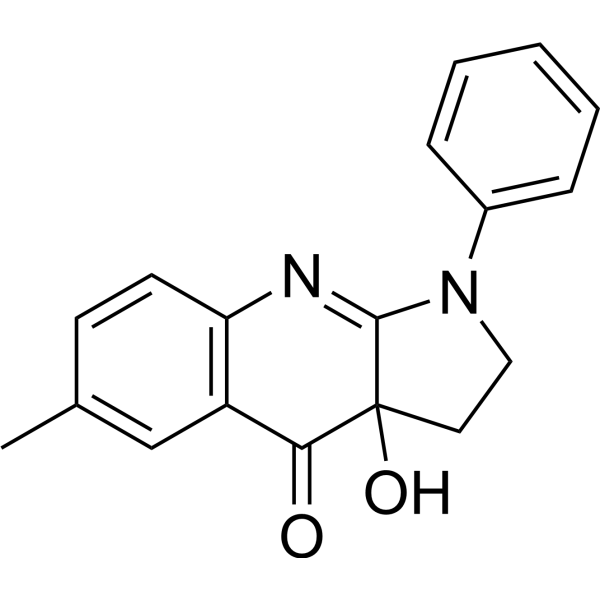
HY-13813
BlebbistatinMyosin II Inhibitor -
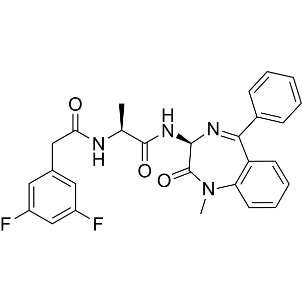
HY-14176
Compound Eγ-secretase Inhibitor -
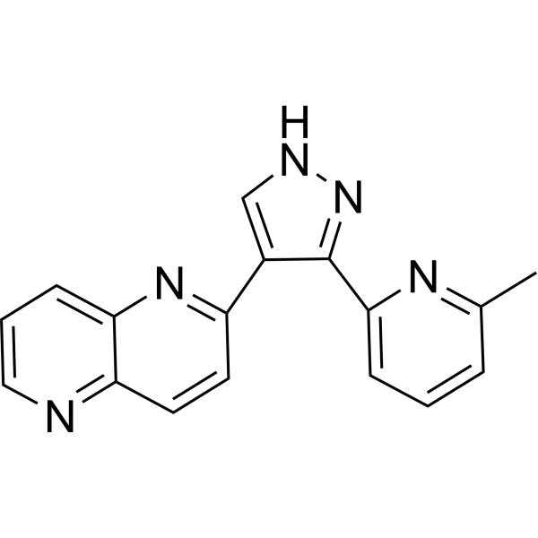
HY-13012
RepSoxTGFβR-1/ALK5 Inhibitor -
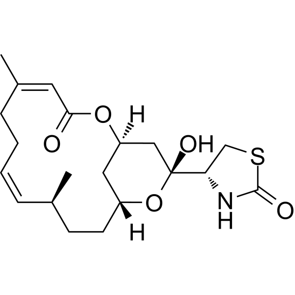
HY-101848
Latrunculin BActin Polymerization Inhibitor -
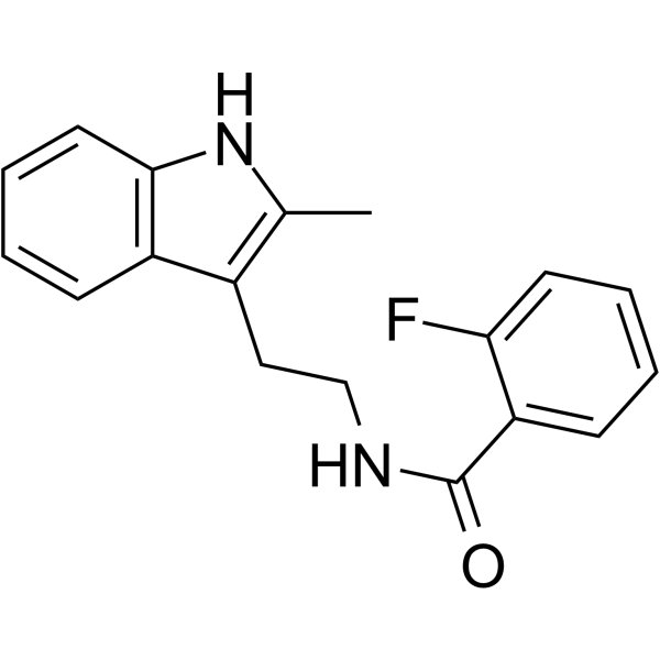
HY-16926
CK-666Arp2/3 Complex Inhibitor -
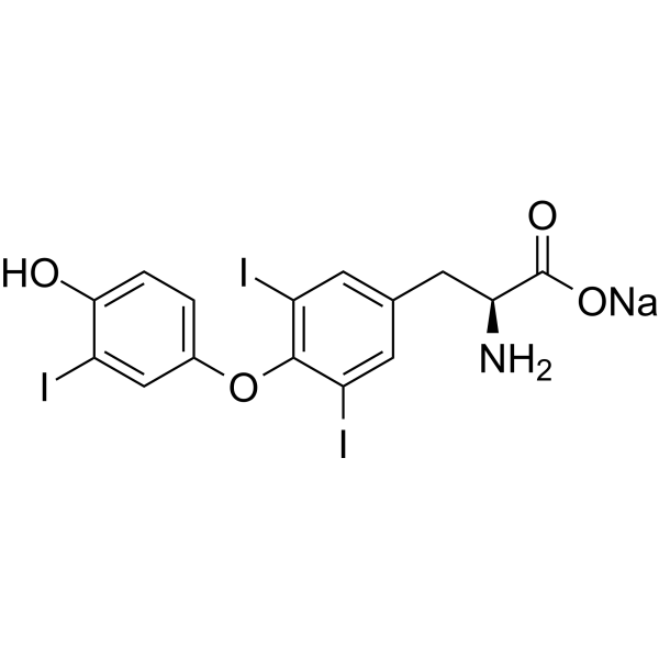
HY-A0070
Liothyronine sodiumTRα/TRβ Agonist -
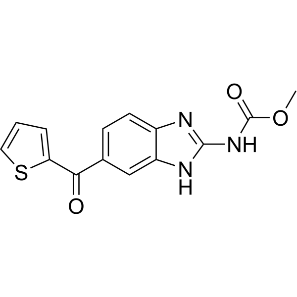
HY-13520
NocodazoleMicrotubule Inhibitor -
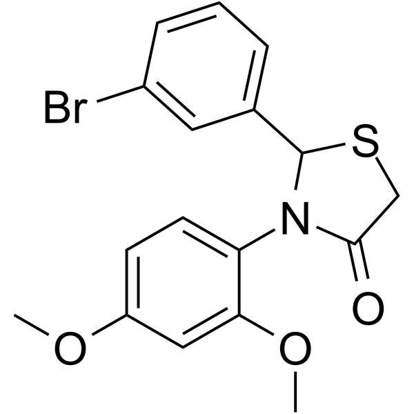
HY-16927
CK-869Arp2/3 Complex Inhibitor -

HY-15371
ForskolinAdenylate Cyclase Activator
Publications Citing Use of MedChemExpress (MCE) Latrunculin A
More- J Extracell Vesicles. 2022 May;11(5):e12218. [Abstract]
- Nat Commun. 2022 Sep 26;13(1):5657. [Abstract]
- Adv Sci (Weinh). 2026 Jun 15:e76077. [Abstract]
- Adv Sci (Weinh). 2022 Aug 28;e2203173. [Abstract]
- Carbon. 2025 Dec 22;248:121179.
- Oncogene. 2026 Aug;45(30):3083-3101.
- Apoptosis. 2023 Aug;28(7-8):1048-1059. [Abstract]
- Talanta. 2024 Feb 1;268(Pt 1):125286. [Abstract]
- Ecotoxicol Environ Saf. 2025 Jan 15:290:117598. [Abstract]
- mBio. 2025 Oct 8;16(10):e0194525. [Abstract]
- PLoS Pathog. 2026 Feb 27;22(2):e1014000. [Abstract]
- Exp Cell Res. 2025 Feb 21;446(1):114461. [Abstract]
- New J Chem. 2023 Jun 28.
- bioRxiv. 2023 Feb 5.
 Customer Validation & Images

-
Flow Cytometry
-
ELISA
-
Cell Imaging/Staining
-
In Vivo Efficacy Study

-
Flow Cytometry


Biological Activity
Description
IC50 & Target
Kd: 0.1 μM (ATP-actin), 0.4 μM (ADP-Pi-actin), 4.7 μM (ADP-actin), 0.19 μM (G-actin)[2]
Cellular Effect
|
Cell Line

|
Type | Value | Description | References |
|---|---|---|---|---|
| A549 | IC50 |
0.06 μg/mL
Compound: 1
|
Cytotoxicity against human A549 cells after 48 hrs by MTT assay
Cytotoxicity against human A549 cells after 48 hrs by MTT assay
|
[PMID: 8350092] |
| HCT-116 | IC50 |
1.1 μM
Compound: 1, NSC-613011
|
Cytotoxicity against human HCT116 cells after 3 days by trypan blue assay
Cytotoxicity against human HCT116 cells after 3 days by trypan blue assay
|
[PMID: 18942825] |
| HT-29 | IC50 |
0.06 μg/mL
Compound: 1
|
Cytotoxicity against human HT-29 cells after 48 hrs by MTT assay
Cytotoxicity against human HT-29 cells after 48 hrs by MTT assay
|
[PMID: 8350092] |
| HT-29 | IC50 |
0.08 μg/mL
Compound: 10
|
Antiproliferative activity against human HT-29 cells assessed as cell viability after 96 hrs by CellTiter-Glo luminescence assay
Antiproliferative activity against human HT-29 cells assessed as cell viability after 96 hrs by CellTiter-Glo luminescence assay
|
[PMID: 22129061] |
| MCF7 | IC50 |
0.48 μM
Compound: 99a
|
Anticancer activity against human MCF7 cells assessed as inhibition of cell growth incubated for 72 hrs by MTT assay
Anticancer activity against human MCF7 cells assessed as inhibition of cell growth incubated for 72 hrs by MTT assay
|
[PMID: 33940466] |
| MDA-MB-231 | IC50 |
4.19 μM
Compound: 99a
|
Anticancer activity against human MDA-MB-231 cells assessed as inhibition of cell growth incubated for 72 hrs by MTT assay
Anticancer activity against human MDA-MB-231 cells assessed as inhibition of cell growth incubated for 72 hrs by MTT assay
|
[PMID: 33940466] |
| MDA-MB-435 | IC50 |
0.04 μg/mL
Compound: 10
|
Antiproliferative activity against human MDA-MB-435 cells assessed as cell viability after 96 hrs by CellTiter-Glo luminescence assay
Antiproliferative activity against human MDA-MB-435 cells assessed as cell viability after 96 hrs by CellTiter-Glo luminescence assay
|
[PMID: 22129061] |
| MDA-MB-435 | IC50 |
2.8 μM
Compound: 1, NSC-613011
|
Cytotoxicity against human MDA-MB-435 cells 48 hrs by SRB assay
Cytotoxicity against human MDA-MB-435 cells 48 hrs by SRB assay
|
[PMID: 18942825] |
| P388 | IC50 |
4.1 μg/mL
Compound: 1
|
Cytotoxicity against mouse P388 cells after 48 hrs by MTT assay
Cytotoxicity against mouse P388 cells after 48 hrs by MTT assay
|
[PMID: 8350092] |
| P388 | IC50 |
4.1 μg/mL
Compound: Latrunculin A
|
Antiproliferative activity against mouse P388 cells
Antiproliferative activity against mouse P388 cells
|
10.1021/np50082a019 |
| T47D | IC50 |
25 μM
Compound: 1,Latrunculin A
|
Inhibition of 1,10-phenanthroline-induced HIF1 activation in human T47D cells by cell based reporter gene assay
Inhibition of 1,10-phenanthroline-induced HIF1 activation in human T47D cells by cell based reporter gene assay
|
[PMID: 18298079] |
| T47D | IC50 |
6.7 μM
Compound: 1,Latrunculin A
|
Inhibition of hypoxia-induced HIF1 activation in human T47D cells by cell based reporter gene assay
Inhibition of hypoxia-induced HIF1 activation in human T47D cells by cell based reporter gene assay
|
[PMID: 18298079] |
| U-937 | IC50 |
0.07 μg/mL
Compound: 10
|
Antiproliferative activity against human U937 cells assessed as cell viability after 96 hrs by CellTiter-Glo luminescence assay
Antiproliferative activity against human U937 cells assessed as cell viability after 96 hrs by CellTiter-Glo luminescence assay
|
[PMID: 22129061] |
In Vitro
Latrunculin A (50-1000 nM) exhibits potent anti-invasive activity against human prostate cancer PC-3M cells, inhibits PC-3M-CT+ spheroids disaggregation and cell migration[3].
?
Latrunculin A (3-30 μM) inhibits hypoxia-induced HIF-1 activation with an IC50 value of 6.7 μM in human breast carcinoma T47D cells[3].
?
Latrunculin A (0-0.2 μM, 4 hours) has a significant inhibitory effect on HuR levels at high concentrations such as 0.2 μM in human hepatoma HepG2 cells while inhibits HuR only at 0.02 μM but no inhibitory effect at high concentrations in human hepatoma Huh7 cells[4].
?
Latrunculin A (0.1 μM, 24 hours) can lead to a significant decrease in cell migration and has an inhibitory effect on cell proliferation in human hepatoma cell lines HepG2[4].
MedChemExpress (MCE) has not independently confirmed the accuracy of these methods. They are for reference only. Further protocols information, click here.
In Vivo
MedChemExpress (MCE) has not independently confirmed the accuracy of these methods. They are for reference only.
-
Animal Model:Male BALB/c nude mice models infected with adenocarcinoma (MKN45) or carcinoma (NUGC-4)[5]
-
Dosage:0.05 mg/kg
-
Administration:Intraperitoneal injection; three doses in the first 20 days; 120 days
-
Result:Extended the mean life expectancy to 23.5 days comparing to control of 16 days in adenocarcinoma (MKN45) mice and the mean survival time was 42 days comparing to untreated of 31 days in carcinoma (NUGC-4) mice.
Clinical Trial
| NCT Number | Sponsor | Condition | Start Date |
Phase
|
|---|---|---|---|---|
| NCT01329991 | Plexxikon| | 2011-05 | PHASE1 |


Chemical Information
-
CAS No. 76343-93-6
-
Appearance Liquid
-
Molecular Weight 421.55
-
Formula C22H31NO5S
-
Color Colorless to light yellow
-
SMILES
O=C1SC[C@@H]([C@]2(O)O[C@]3([H])CC[C@H](C)/C=C\C=C\CC/C(C)=C\C(O[C@@](C3)([H])C2)=O)N1
-
Synonyms
LAT-A
-
Structure Classification
-
Initial Source
-
Shipping
Room temperature in continental US; may vary elsewhere.
-
Storage
Solution, -20°C, 2 years


Publications (14)
-
Journal Impact Factor
-
Most Recent
-
J Extracell Vesicles
CD73 in small extracellular vesicles derived from HNSCC defines tumour-associated immunosuppression mediated by macrophages in the microenvironment. [Abstract]2022 May;11(5):e12218. PMID: 35524455 -
Nat Commun
Graphdiyne oxide nanosheets display selective anti-leukemia efficacy against DNMT3A-mutant AML cells. [Abstract]2022 Sep 26;13(1):5657. PMID: 36163326 -
Adv Sci (Weinh)
The E3 Ligase RNF115 Aggravates Pathological Cardiac Hypertrophy via Ubiquitin-Mediated Degradation of SPTBN1. [Abstract]2026 Jun 15:e76077. PMID: 42294585 -
Adv Sci (Weinh)
Synthetic Retinoid Kills Drug-Resistant Cancer Stem Cells via Inducing RARγ-Translocation-Mediated Tension Reduction and Chromatin Decondensation. [Abstract]2022 Aug 28;e2203173. PMID: 36031407
Latrunculin A purchased from MedChemExpress. Usage Cited in: Adv Sci (Weinh). 2022 Aug 28;e2203173. [Abstract]
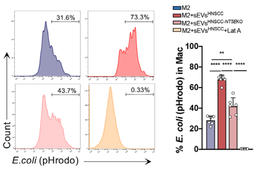
The sEVs (50 μg) were cocultured with macrophages (1 × 10⁶) for 24 h. The sEVsHNSCC derived from two HNSCC lines: SCC25 and HN6. The sEVshBMSC-NT5EOE derived from hBMSC cells with NT5E overexpression. Latrunculin A (Lat A, 30 μM; 24 h) was used as the inhibitor of sEVs uptaken showed downregulated phagocytosis. The percentage of pHrodo dyes of M2 macrophages was analyzed by flow cytometry.
Latrunculin A purchased from MedChemExpress. Usage Cited in: Adv Sci (Weinh). 2022 Aug 28;e2203173. [Abstract]
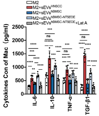
The expression of cytokines also increased when macrophages were cocultured with sEVs derived from hBMSCs overexpressing CD73, whereas this trend was reversed by Latrunculin A (Lat A, 30 μM; 24 h).
Latrunculin A purchased from MedChemExpress. Usage Cited in: Adv Sci (Weinh). 2022 Aug 28;e2203173. [Abstract]
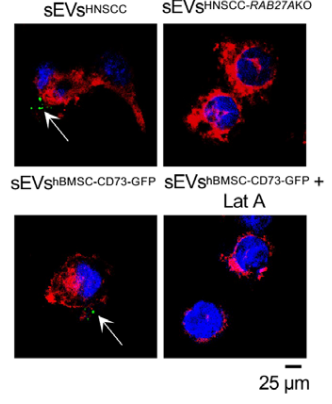
Macrophages were cocultured with anti-CD73-FITC labelled sEVs from HNSCC cell lines control or RABAKO, CD73-GFP labelled sEVs from hBMSC treated with or without Latrunculin A (Lat A, 30 μM; 24 h) were cocultured with macrophages for 1 h, and Laser Scanning Confocal Microscopy was used to analyze the internalization of HNSCC-derived sEVs into macrophages (Scale bar = 25 μm).
Latrunculin A purchased from MedChemExpress. Usage Cited in: Adv Sci (Weinh). 2022 Aug 28;e2203173. [Abstract]
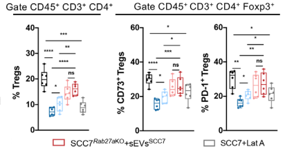
Absence of CD73 in sEVs rescues immune suppression and restrains tumour growth in C3H/He mice aged 6–8 weeks. (a) Schematic of subcutaneous tumorigenesis in vivo experiment, followed with intratumoral injection of sEVs which were collected from SCC7, SCC7-Nt5eKO or SCC7-Nt5eOE cells grown in vitro. (b) The exhibition of isolated tumours. (c and d) The tumour weight and the time course of tumour growth in grams for 15 days postinjection with SCC7 or SCC7Rab27aKO cells with or without CD73 in sEVs. Latrunculin A (Lat A, 0.15 mg/kg; ip; every 2 days for 15 days) was used as inhibitor of sEVs uptaken.
Latrunculin A purchased from MedChemExpress. Usage Cited in: Adv Sci (Weinh). 2022 Aug 28;e2203173. [Abstract]
Latrunculin A (Lat A, 0.15 mg/kg; ip; every 2 days for 15 days) was used as an inhibitor of sEVs uptake in C3H/He mice aged 6–8 weeks. Flow cytometry analysis for infiltration of Tregs and percentage of CD73+/PD-1+ Tregs in tumours.
-
-
-
Apoptosis
TFAM-Mediated mitochondrial transfer of MSCs improved the permeability barrier in sepsis-associated acute lung injury. [Abstract]2023 Aug;28(7-8):1048-1059. PMID: 37060506 -
Talanta
Super-resolution imaging of folate receptor alpha on cell membranes using peptide-based probes. [Abstract]2024 Feb 1;268(Pt 1):125286. PMID: 37832456 -
Ecotoxicol Environ Saf
Lead-induced actin polymerization aggravates neutrophil extracellular trap formation and contributes to vascular inflammation. [Abstract]2025 Jan 15:290:117598. PMID: 39721424 -
mBio
PRRSV promotes bacterial infection by remodeling actin cytoskeleton and cell membrane proteins. [Abstract]2025 Oct 8;16(10):e0194525. PMID: 40937850 -
PLoS Pathog
2026 Feb 27;22(2):e1014000. PMID: 41758870 -
Exp Cell Res
Mechanical stretch promotes the migration of mesenchymal stem cells via Piezo1/F-actin/YAP axis. [Abstract]2025 Feb 21;446(1):114461. PMID: 39988125 -
-

Protocols
-
Scratch/Wound-Healing Migration Assay
The scratch/wound-healing migration assay measures collective migration of adherent cells into an experimentally created cell-free gap in a confluent monolayer. The readout is generated by imaging the gap immediately after scratching and at later time points, then quantifying reduction in wound area, wound width, or percentage closure as cells move into the denuded region. Gap closure reflects cell migration but may also include cell proliferation, so interpretation should distinguish migration-focused conditions from proliferation-driven closure when possible, such as by using short assay windows, serum-controlled conditions, cell counting, or proliferation controls reported in published protocols.
-
Cell migration
Cell migration is a method that plays an important role in wound healing, cell differentiation, embryonic development, etc.
-
Chemotaxis Gradient Chamber Assay 1
The chemotaxis gradient chamber assay is based on the principle of observing directional cell migration in response to a stable, linear or quasi-linear concentration gradient of a chemoattractant formed between two fluid reservoirs separated by a narrow observation chamber. Cells placed within the chamber respond to the gradient by polarized movement toward higher chemoattractant concentrations, allowing quantification of chemotactic behavior in real time under microscopy. The classic Zigmond chamber design enables simultaneous visualization of gradient formation and individual cell trajectories, making it suitable for studying leukocyte chemotaxis and other motile cell types in vitro.
-
Under-Agarose Cell Migration Assay
The under-agarose cell migration assay is a classical in vitro chemotaxis method designed to evaluate directed cell movement through a semi-solid agarose matrix toward soluble chemoattractant gradients, enabling visualization and quantification of leukocyte or motile cell migration in a confined 2D-like environment. In this system, cells and chemoattractants are placed in separate wells cut into an agarose gel, allowing diffusion-driven gradient formation that guides directional migration, which is typically assessed by measuring migration distance, cell morphology changes, and accumulation toward the chemoattractant source. This assay has been widely used to study neutrophil and leukocyte chemotaxis as a simple alternative to filter-based migration systems and allows direct microscopic observation of migrating cells under near-physiological confinement conditions.
-
Neural Crest/Neuronal Cell Migration Explant Assay
Neural crest (NC) and neuronal cell migration explant assays are in vitro systems in which neural tube-derived tissues are cultured to allow premigratory or newly emigrated neural crest cells to undergo epithelial-to-mesenchymal transition (EMT), migrate away from the explant, and form a measurable radial outgrowth that reflects migratory capacity and environmental responsiveness. These assays typically quantify migration by measuring the expansion of cell outgrowth from neural tube or neural plate border explants over time, often comparing early and later timepoints to derive a migration index such as a radius ratio, which reflects net cell dispersal from the explant core. Neural tube explant cultures preserve key aspects of neural crest behavior, including EMT, migration, and early differentiation, making them suitable for assessing intrinsic migratory ability and extrinsic cue dependence. However, studies emphasize that migratory outgrowth from neural tube explants may include non-n
-
Transwell/Boyden Chamber Migration Assay
The Transwell/Boyden chamber migration assay measures cell movement through a porous membrane separating an upper and lower chamber, usually after a chemoattractant gradient is established by placing cells in the upper chamber and chemoattractant-containing medium in the lower chamber. The readout is generated by quantifying cells that traverse the membrane and appear on the lower membrane surface or in the lower chamber, depending on whether the cell type is adherent or non-adherent. This assay reflects chemotactic or haptotactic migration rather than matrix invasion unless an extracellular-matrix barrier is added to the membrane.
-
3D Collagen/Hydrogel Matrix Migration Assay
The 3D collagen/hydrogel matrix migration assay is based on embedding cells within or on top of a fibrillar collagen type I-rich three-dimensional matrix to model in vivo-like extracellular matrix (ECM) architecture, enabling analysis of cell migration through a physically and biochemically relevant scaffold. In contrast to 2D migration systems, cells in 3D matrices interact with fibrillar collagen networks, requiring coordinated adhesion remodeling and proteolytic or non-proteolytic deformation mechanisms to move through confined spaces, thereby providing a more physiologically relevant readout of invasive and migratory behavior in tissue-like environments. Cell movement in 3D collagen matrices is typically quantified by tracking single-cell trajectories, invasion depth, or matrix penetration over time, reflecting combined effects of cytoskeletal dynamics, cell-ECM adhesion turnover, and ECM remodeling. These systems are widely used to study tumor cell invasion and stromal cell motili
-
Cell-Exclusion Zone Migration Assay
The Cell-Exclusion Zone (CEZ) migration assay is an in vitro 2D cell migration method in which a defined cell-free area is created using removable physical barriers such as silicone stoppers, allowing cells to be seeded around the barrier and subsequently migrate into the cleared zone after barrier removal. This approach enables quantification of collective cell migration by monitoring repopulation of the initially cell-free region over time using microscopy-based imaging. Compared with scratch-based wound healing assays, barrier-based exclusion methods are designed to avoid mechanical damage to the extracellular matrix and reduce injury-induced effects on boundary cells, thereby improving interpretability of migration behavior in vitro. The assay readout is typically the progressive reduction in the cell-free area or the number of cells invading the exclusion zone, reflecting coordinated cell motility relevant to physiological processes such as wound healing, epithelial repair, and ca
-
Chemotaxis Gradient Chamber Assay 2
Chemotaxis gradient chamber assays measure directional cell migration in response to a soluble chemical gradient by imaging cells as they move across a defined observation region; the readout is generated from time-lapse cell trajectories, displacement toward the gradient, forward migration index, trajectory plots, rose/polar plots, and statistical tests of non-random directionality. The Dunn chamber is a direct-viewing glass chamber in which cells migrate across a bridge between control and chemoattractant wells, allowing observation of cells in a linear concentration gradient; related direct-viewing formats include the Insall chamber, which supports defined unidirectional gradients and high numerical-aperture microscopy, and the μ-Slide Chemotaxis chamber, which supports long-term live-cell imaging and gradient characterization with fluorescent dye.
-
Phalloidin F-actin cytoskeleton staining
Phalloidin F-actin staining detects polymerized filamentous actin in fixed and permeabilized specimens by using fluorescent phalloidin or phalloidin-derived phallotoxins that bind actin filaments and generate a fluorescence microscopy readout corresponding to F-actin organization, including stress fibers, cortical actin, filament bundles, and tissue-specific actin networks. Phalloidin stabilizes F-actin by reducing actin subunit dissociation from filament ends, and fluorescent phallotoxins were established as tools for visualizing actin-containing structures in eukaryotic cells.


Purity & Documentation
-
Data Sheet (279 KB)
-
SDS (394 KB)
- English - EN (394 KB)
- Français - FR (394 KB)
- Deutsch - DE (394 KB)
- Norwegian - NO (394 KB)
- Español - ES (394 KB)
- Swedish - SV (394 KB)
- Italian - IT (394 KB)
- Korean - KR (394 KB)
- Portuguese - PT (394 KB)
-
Handling Instructions (2659 KB)
References
[1]. Fujiwara I, et al. Latrunculin A Accelerates Actin Filament Depolymerization in Addition to Sequestering Actin Monomers. Curr Biol. 2018 Oct 8;28(19):3183-3192.e2. [Content Brief]
[2]. Coué M, et al. Inhibition of actin polymerization by latrunculin A. FEBS Lett. 1987 Mar 23;213(2):316-8. [Content Brief]
[3]. Khalid A El Sayed, et al. Latrunculin A and its C-17-O-carbamates inhibit prostate tumor cell invasion and HIF-1 activation in breast tumor cells. J Nat Prod. 2008 Mar;71(3):396-402. [Content Brief]
[4]. Anke Doller, et al. The cytoskeletal inhibitors latrunculin A and blebbistatin exert antitumorigenic properties in human hepatocellular carcinoma cells by interfering with intracellular HuR trafficking. Exp Cell Res. 2015 Jan 1;330(1):66-80. [Content Brief]
[5]. Hiroo Konishi, et al. Latrunculin a has a strong anticancer effect in a peritoneal dissemination model of human gastric cancer in mice. Anticancer Res. 2009 Jun;29(6):2091-7. [Content Brief]
[6]. Liang Ma, et al. Discovery of the migrasome, an organelle mediating release of cytoplasmic contents during cell migration. Cell Res. 2015 Jan;25(1):24-38. [Content Brief]

Calculators
Concentration (start) × Volume (start) = Concentration (final) × Volume (final)


Latrunculin A Related Classifications
HY-16929 Related Classifications
 Powered by Bioz