



Lirentelimab

Based on 1 Customer Validation
Lirentelimab (AK002) is a humanized IgG1 monoclonal antibody that targets sialic acid-binding Ig-like lectin 8 (SIGLEC8). Lirentelimab induces cell apoptosis of IL-5-activated eosinophils and inhibits IgE-mediated mast cell activation. Lirentelimab can be used for the research of eosinophilic gastritis and duodenitis.
Nur für Forschungszwecke. Wir verkaufen nicht an Patienten.
- Reinheit : 99.91%
- CAS. Nr.: 2283348-97-8
- Molecular Weight:144.31 kDa
-
Speicherung:
Please store the product under the recommended conditions in the Certificate of Analysis.


To place orders, for customer services and technical support, please contact: MedChemExpress USA
Tel: 609-228-6898 E-mail: [email protected] [email protected]


-
 Biologische Aktivität
-
Chemical Information
- Protokoll
- Reinheit & Dokumentation
- Verweise
-
 Help & FAQs





Biologische Aktivität
Beschreibung
Isotype
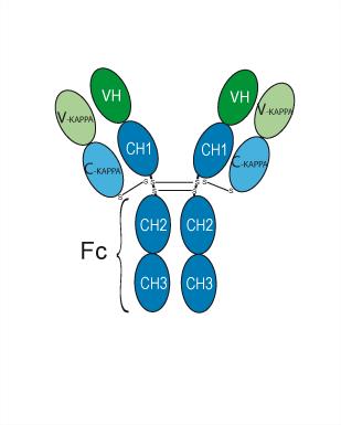
Human IgG1 kappa
Recommend Isotype Controls
Species Reactivity
Human
IC50 & Target
Siglec-8
In Vitro
Monovalent Lirentelimab fragment antigen-binding (fab) binds to recombinant SIGLEC8 extracellular domain (ECD) is determined to be 464 pM[1]. Lirentelimab shows high affinity to SIGLEC8 in vitro, to SIGLEC8 expressed on eosinophils, and to NK cells via its Fc region in human blood[1]. Lirentelimab (1 µg/mL) selectively binds to eosinophils in human peripheral blood, and eosinophils and mast cells from human lung tissue[1]. Lirentelimab (0.0001-100 μg/mL; 30 min) induces apoptosis of IL-5-activated eosinophils[1]. Lirentelimab (30 min) shows potent antibody-dependent cell-mediated cytotoxicity (ADCC) Activity on human eosinophils with an EC50 value of 1.9 ng/mL to eosinophils in peripheral blood leukocytes (PBL) preparations from healthy donors[1]. Lirentelimab reduces eosinophil numbers in ex vivo human tissue[1].
MedChemExpress (MCE) has not independently confirmed the accuracy of these methods. They are for reference only.
-
Cell Line:Eosinophils
-
Concentration:10 μg/mL-1 pg/mL
-
Incubation Time:30 min
-
Result:Dose-dependently induced apoptosis of IL-5-activated (50 ng/mL) eosinophils.
In Vivo
MedChemExpress (MCE) has not independently confirmed the accuracy of these methods. They are for reference only.
-
Animal Model:Humanized mice (NSG-SGM3) engrafts with human thymus, liver, and HSC[1]
-
Dosage:100 μg
-
Administration:Intravenous injection; 100 μg, once
-
Result:Completely prevented passive systemic anaphylaxis (PSA) as shown by a lack of change in rectal temperature and symptom scores in mice.
Clinical Trial
| NCT Number | Sponsor | Condition | Start Date |
Phase

|
|---|---|---|---|---|
| NCT01329991 | 2011-05 | PHASE1 |
Gene ID
Accession
Conjugated
Unconjugated
Reconsititution
The product can be reconstituted/diluted with sterile PBS or saline.
Format
-
Human IgG1 kappa
Anwendung
ELISA, FACS, Functional assay
Verified Bioactivity
-
 Immobilized Siglec-8 Protein, Human (HEK293, His, HY-P71313) can bind Lirentelimab. The EC50 for this effect is 2.468 ng/mL.


Chemical Information
-
CAS. Nr. 2283348-97-8
-
Appearance Liquid
-
Molecular Weight 144.31 kDa
-
Color Colorless to light yellow
-
SMILES
[Lirentelimab]
-
Synonyms
AK002; Antolimab
-
Versand
Shipping with dry ice.
-
Formulation
Please refer to the lot-specific COA for specific buffer information.
-
Speicherung
Please store the product under the recommended conditions in the Certificate of Analysis.


Reinheit & Dokumentation
-
Data Sheet (260 KB)
-
SDS (251 KB)
- English - EN (251 KB)
- Français - FR (251 KB)
- Deutsch - DE (251 KB)
- Norwegian - NO (251 KB)
- Español - ES (251 KB)
- Swedish - SV (251 KB)
- Italian - IT (251 KB)
- Korean - KR (251 KB)
- Portuguese - PT (251 KB)
-
Inhibitory Antibodies User Guide (603 KB)
Verweise
[1]. Youngblood BA, et al. AK002, a Humanized Sialic Acid-Binding Immunoglobulin-Like Lectin-8 Antibody that Induces Antibody-Dependent Cell-Mediated Cytotoxicity against Human Eosinophils and Inhibits Mast Cell-Mediated Anaphylaxis in Mice. Int Arch Allergy Immunol. 2019;180(2):91-102. [Content Brief]

Calculators
Konzentration (Stammlösung) × Volumen (Stammlösung) = Konzentration (Ziellösung) × Volumen (Ziellösung)


Lirentelimab Related Classifications
HY-P99371 Related Classifications
 Powered by Bioz