



PXS-5505 free base
Based on 3 publication(s) in Google Scholar
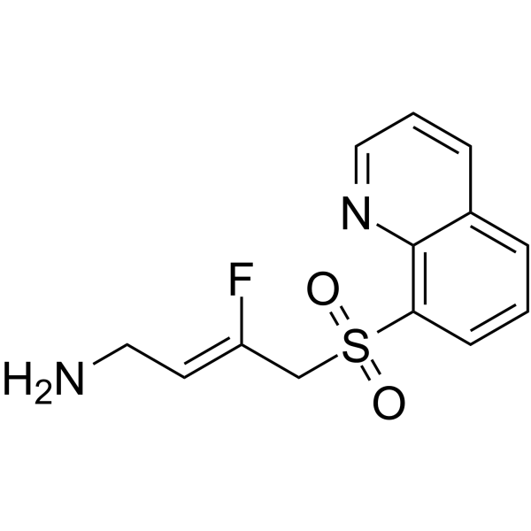
PXS-5505 free base (LOX-IN-3) is an orally active lysyl oxidase (LOX) inhibitor. PXS-5505 free base can be used for fibrosis, cancer and angiogenesis research.
For research use only. We do not sell to patients.
- Purity : 99.36%
- CAS No.: 2409963-83-1
- Formula: C13H13FN2O2S
- Molecular Weight:280.32
-
Storage:
4°C, protect from light
* In solvent : -80°C, 6 months; -20°C, 1 month (protect from light)

To place orders, for customer services and technical support, please contact: MedChemExpress USA
Tel: 609-228-6898 E-mail: [email protected] [email protected]


-
 Biological Activity
-
Chemical Information
-
Solvent & Solubility
- Protocols
- Purity & Documentation
- References
-
 Help & FAQs


-
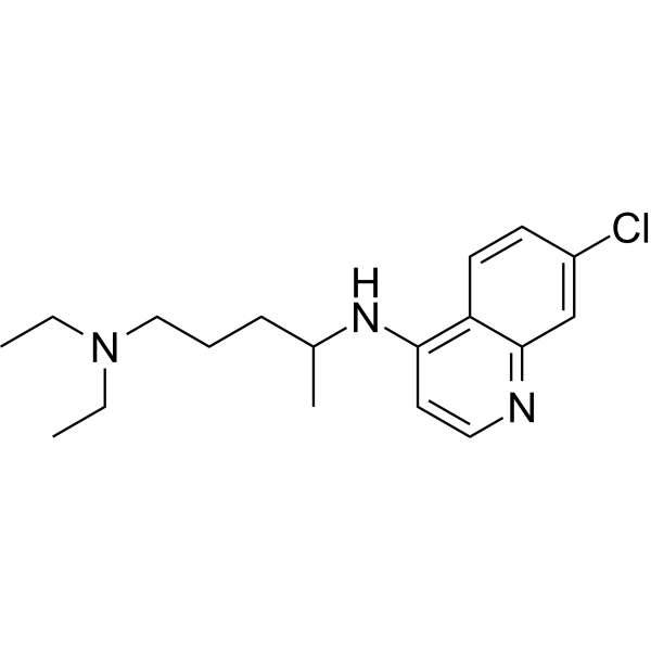
HY-17589A
ChloroquineAntimalarial Agent -
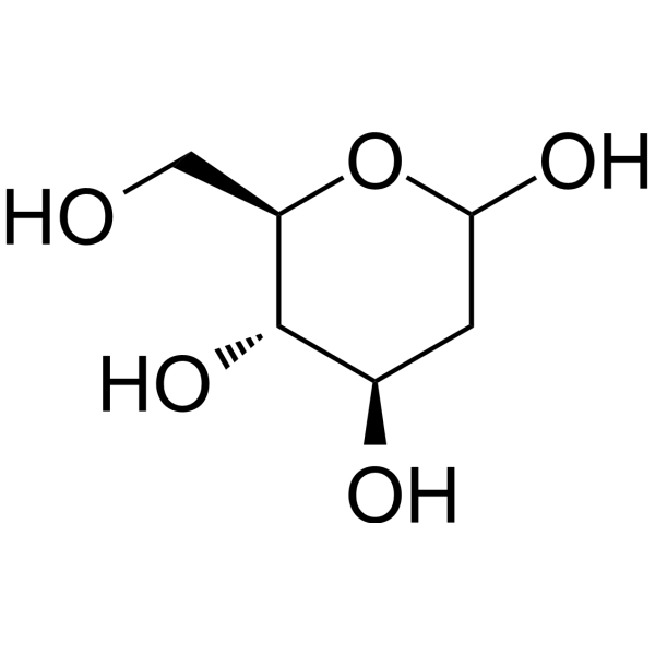
HY-13966
2-Deoxy-D-glucoseGlycolysis Inhibitor -
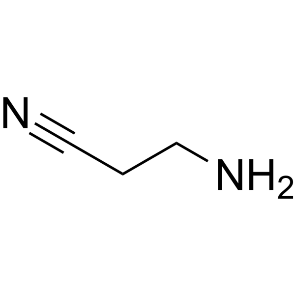
HY-Y1750
β-AminopropionitrileLysyl Oxidase Inhibitor -
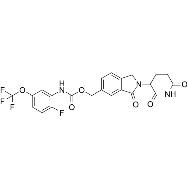
HY-153356
MRT-2359GSPT1 Molecular Glue Degrader -
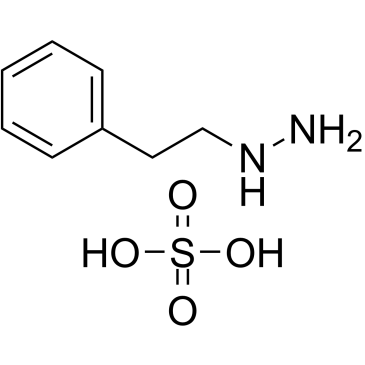
HY-B1018A
Phenelzine sulfateMAO Inhibitor -
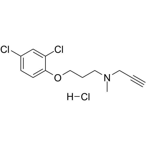
HY-14197A
Clorgyline hydrochlorideMAO-A Inhibitor -
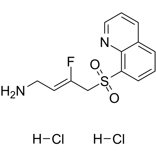
HY-138625A
PXS-5505 anhydrousLysyl Oxidase Inhibitor -
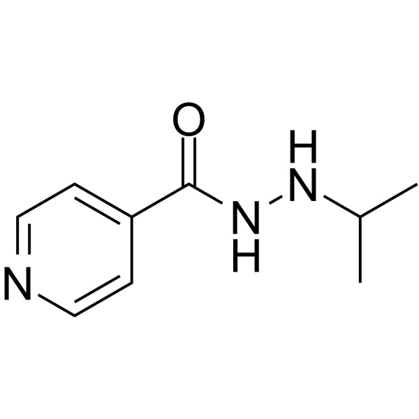
HY-B0886A
IproniazidMAO Inhibitor -
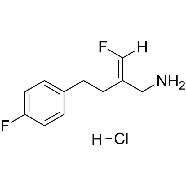
HY-16677A
Mofegiline hydrochlorideMAO-B Inhibitor -
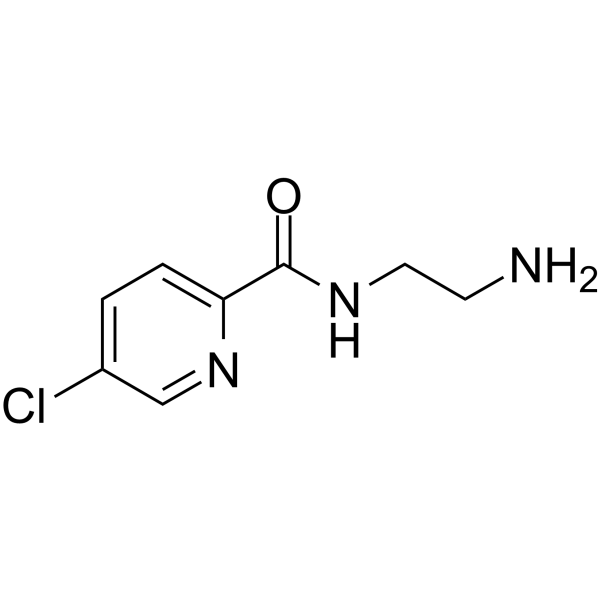
HY-14201
LazabemideMAO-B Inhibitor -

HY-Y0882
Hydroxylamine hydrochlorideMAO Inhibitor -
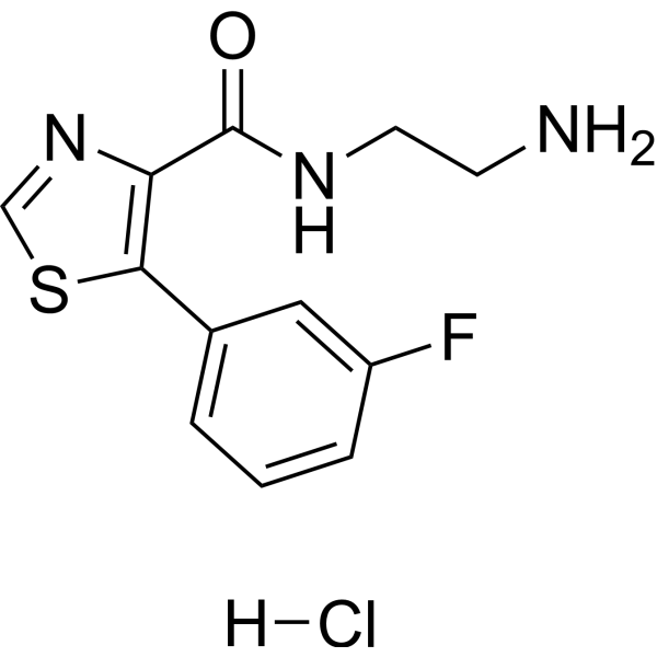
HY-100027A
Ro 41-1049 hydrochlorideMAO-A Inhibitor
Publications Citing Use of MedChemExpress (MCE) PXS-5505 free base
More

Biological Activity
Description
IC50 & Target
IC50: <1 μM (human LOXL2), <10 μM (bovine LOX)[1]
In Vitro
PXS-5505 free base (Compound 33) inhibits the bovine LOX and human LOXL2 activities with IC50 values of <10 μM and <1 μM, respectively[1].
PXS-5505 free base exhibits sustained inhibition of LOXL1 and LOXL2[1].
PXS-5505 free base is less active against SSAO/VAP-1 and MAO-B activities[1].
MedChemExpress (MCE) has not independently confirmed the accuracy of these methods. They are for reference only. Further protocols information, click here.
In Vivo
PXS-5505 free base (10 mg/kg; orally; daily for 14 days) reduces kidney fibrosis in unilateral ureteric obstruction (UUO) mice model[1].
PXS-5505 free base (15 mg/kg; orally; daily for 21 days) reduces lung fibrosis in mice[1].
MedChemExpress (MCE) has not independently confirmed the accuracy of these methods. They are for reference only.
-
Animal Model:Male Wistar rats[1]
-
Dosage:30 mg/kg
-
Administration:Oral administration, single dose
-
Result:Completely abolished lysyl oxidase activity. Plasma concentrations of tested compound are far below the IC50 after 8 hours, the half-life of recovery is between 2-3 days (ear) and 24 hours (aorta).
-
Animal Model:Unilateral ureteric obstruction (UUO) model of acute kidney fibrosis in mice[1]
-
Dosage:10 mg/kg
-
Administration:Oral gavage, daily for 14 days
-
Result:Increased kidney weight and thickness and reduced the area of fibrosis.
-
Animal Model:C57Bl/6 mice, Bleomycin-induced lung fibrosis model
-
Dosage:15 mg/kg
-
Administration:Oral gavage, daily for 21 days
-
Result:Significantly reduced the Ashcroft score and the lung weight.


Chemical Information
-
CAS No. 2409963-83-1
-
Appearance Solid
-
Molecular Weight 280.32
-
Formula C13H13FN2O2S
-
Color White to off-white
-
SMILES
NC/C=C(F)/CS(=O)(C1=C2N=CC=CC2=CC=C1)=O
-
Synonyms
LOX-IN-3
-
Shipping
Room temperature in continental US; may vary elsewhere.
-
Storage
4°C, protect from light
* In solvent : -80°C, 6 months; -20°C, 1 month (protect from light)


Publications (3)
-
Journal Impact Factor
-
Most Recent
-
Cancer Res
LOX Inhibition Disrupts a Collagen-Integrin-MYC Axis to Suppress Progression of Invasive Lobular Carcinoma. [Abstract]2026 Feb 18. PMID: 41706704 -
iScience
MFAP5+ synovial fibroblasts drive LOX upregulation to promote osteoarthritis progression. [Abstract]2026 Jun 11;29(7):116286. PMID: 42325584 -


Solvent & Solubility
In Vitro:
DMSO : 100 mg/mL (356.74 mM; Need ultrasonic; Hygroscopic DMSO has a significant impact on the solubility of product, please use newly opened DMSO)
Please refer to the solubility information to select the appropriate solvent. Once prepared, please aliquot and store the solution to prevent product inactivation from repeated freeze-thaw cycles.
Storage method and period of stock solution: -80°C, 6 months; -20°C, 1 month (protect from light). When stored at -80°C, please use it within 6 months. When stored at -20°C, please use it within 1 month.
Please refer to the solubility information to select the appropriate solvent. Once prepared, please aliquot and store the solution to prevent product inactivation from repeated freeze-thaw cycles.
Storage method and period of stock solution: -80°C, 6 months; -20°C, 1 month (protect from light). When stored at -80°C, please use it within 6 months. When stored at -20°C, please use it within 1 month.
Concentration (start) × Volume (start) = Concentration (final) × Volume (final)
In Vivo:
Select the appropriate dissolution method based on your experimental animal and administration route.
- For the following dissolution methods, please ensure to first prepare a clear stock solution using an In Vitro approach and then sequentially add co-solvents:
- To ensure reliable experimental results, the clarified stock solution can be appropriately stored based on storage conditions. As for the working solution for In Vivo experiments, it is recommended to prepare freshly and use it on the same day.
- The percentages shown for the solvents indicate their volumetric ratio in the final prepared solution. If precipitation or phase separation occurs during preparation, heat and/or sonication can be used to aid dissolution.
Add each solvent one by one: 10% DMSO 40% PEG300 5% Tween-80 45% Saline
Solubility: ≥ 2.08 mg/mL (7.42 mM); Clear solution
This protocol yields a clear solution of ≥ 2.08 mg/mL (saturation unknown).
Taking 1 mL working solution as an example, add 100 μL DMSO stock solution (20.8 mg/mL) to 400 μL PEG300, and mix evenly; then add 50 μL Tween-80 and mix evenly; then add 450 μL Saline to adjust the volume to 1 mL.
Preparation of Saline: Dissolve 0.9 g sodium chloride in ddH₂O and dilute to 100 mL to obtain a clear Saline solution.
Add each solvent one by one: 10% DMSO 90% (20% SBE-β-CD in Saline)
Solubility: ≥ 2.08 mg/mL (7.42 mM); Clear solution
This protocol yields a clear solution of ≥ 2.08 mg/mL (saturation unknown).
Taking 1 mL working solution as an example, add 100 μL DMSO stock solution (20.8 mg/mL) to 900 μL 20% SBE-β-CD in Saline, and mix evenly.
Preparation of 20% SBE-β-CD in Saline (4°C, storage for one week): 2 g SBE-β-CD powder is dissolved in 10 mL Saline, completely dissolve until clear.
In Vivo Dissolution Calculator
Please enter the basic information of animal experiments:
-
-
-
-
Recommended: Prepare an additional quantity of animals to account for potential losses during experiments.
Please enter your animal formula composition:
-
%DMSO +
Recommended: Keep the proportion of DMSO in working solution below 2% if your animal is weak.
-
%+
-
+%Tween-80 + +
-
%Saline +
The co-solvents required include: DMSO, . All of co-solvents are available by MedChemExpress (MCE). , Tween 80. All of co-solvents are available by MedChemExpress (MCE).
Working solution concentration: 0.22 mg/mL
Method for preparing stock solution: mg drug dissolved in μL DMSO. Stock solution concentration: mg/mL. * In solvent : -80°C, 6 months; -20°C, 1 month (protect from light)
1. Take μL DMSO stock solution;
2. Add μL .
μL , mix evenly;
3. Then add μL Tween 80, mix evenly;
4. Then add μL
Please ensure that the stock solution in the first step is dissolved to a clear state, and add co-solvents in sequence. You can use ultrasonic heating (ultrasonic cleaner, recommended frequency 20-40 kHz), vortexing, etc. to assist dissolution.

Protocols
-
Fibrosis/Collagen Morphometry
Fibrosis and collagen morphometry is based on the quantitative visualization of fibrillar collagen deposition in tissue sections using histochemical stains such as Sirius Red (Picrosirius Red) or Masson's trichrome, followed by image-based or polarization-enhanced analysis to estimate collagen proportional area as a surrogate of extracellular matrix accumulation during fibrotic remodeling. Sirius Red combined with polarized light microscopy enhances detection of collagen fibers due to birefringence properties, enabling more specific visualization of collagen type I and III fibrils compared to conventional bright-field histology, while whole-section or region-restricted digital morphometry reduces field-selection bias in fibrosis assessment. Alternative quantitative approaches include second harmonic generation (SHG) and two-photon excited fluorescence microscopy, which enable label-free detection of fibrillar collagen and have been validated against histological staining and biochemica
-
Collagen: Sirius Red Staining
Sirius Red or picrosirius red staining is a histochemical method for visualizing collagen-rich extracellular matrix in tissue sections, and collagen fibers are detected as red-stained structures under bright-field microscopy with enhanced birefringence under polarized light. Picrosirius red is useful for assessing total collagen organization, distribution, and fibrosis burden, but polarized color should not be interpreted as a definitive collagen type I versus type III readout because color is affected by fiber orientation, thickness, and packing.
-
Human pluripotent stem cell endothelial-cell differentiation
Human pluripotent stem cell endothelial differentiation is based on stepwise developmental patterning: early activation of WNT/GSK3β inhibition promotes mesodermal or vascular progenitor entry, followed by endothelial specification using VEGF-related signaling, BMP4, FGF2, Notch modulation, or cAMP depending on the published protocol. Endothelial differentiation is read out by acquisition of CD31, CD34, VE-cadherin/CD144, KDR/VEGFR2, vWF, Tie2, NOS3, acetylated LDL uptake, tube/network formation, barrier function, and in vivo vessel-forming capacity where tested.
-
Vascular/Branching Fractal Analysis
Vascular/branching fractal analysis quantifies the geometric complexity of vessel trees or vascular networks from segmented 2D images, commonly by converting vessels into binary and/or skeletonized maps and estimating fractal dimension using box-counting or related approaches. Fractal dimension is interpreted as an image-derived readout of vascular branching complexity, space filling, or density, and has been applied to retinal photographs, fluorescein angiography, OCT angiography, capillary perfusion maps, and in vitro Matrigel angiogenesis networks. The assay readout is generated from vessel-positive pixels after image preprocessing, vessel segmentation, binarization, and optional skeletonization; reported outputs include fractal dimension, vessel density, branchpoint density, endpoint density, vessel length density, tortuosity, and generation-based branching metrics when VESGEN-style analysis is used. The biological interpretation is limited to quantitative vascular patterning and s
-
Connective Tissue: Masson's Trichrome/Collagen Trichrome Staining
Masson’s Trichrome (collagen/trichrome staining) is a histological technique that differentially stains tissue compartments using sequential acidic dyes to distinguish collagen from muscle and cytoplasmic components based on dye affinity and tissue permeability differences, enabling visualization of fibrosis and connective tissue architecture in histological sections. The classical formulation typically uses Weigert's iron hematoxylin for nuclear staining, Biebrich scarlet-acid fuchsin for cytoplasm and muscle, and aniline blue (or light green variants) for collagen, producing a characteristic blue/green collagen signal contrasted against red cytoplasm and dark nuclei. The staining principle relies on selective displacement of smaller dye molecules by larger anionic dyes in collagen-rich regions under controlled acidified conditions, which enhances collagen-specific dye retention. This property makes the method widely used for fibrosis assessment in organs such as heart, liver, lung, a
-
Endothelial Tube Formation Assay
Endothelial tube formation assay evaluates the ability of endothelial cells to attach, migrate, align, and organize into capillary-like networks when cultured on gelled basement membrane extract or Matrigel; the readout is the morphology and quantity of tube-like networks, which reflects an in vitro endothelial morphogenesis step related to angiogenesis. Basement membrane extract/Matrigel provides laminin-rich extracellular matrix cues that support endothelial differentiation into capillary-like structures, but it can contain biologically active growth factors, so growth-factor-reduced matrix is preferred when testing defined angiogenic stimulators or inhibitors.
-
Endothelial Cell Migration/Angiogenic Sprouting Assay
Endothelial cell migration and angiogenic sprouting assays are in vitro (and partially ex vivo-adapted) functional models that quantify the ability of endothelial cells to undergo coordinated migration, extracellular matrix invasion, and multicellular organization into capillary-like sprouts in response to pro-angiogenic stimuli such as VEGF, bFGF, or conditioned microenvironments. These assays are used to model early angiogenic events including tip-cell formation, directional migration, and lumen-like sprout extension, which collectively reflect angiogenic activation and vascular morphogenesis processes observed in vivo.


Purity & Documentation
-
Data Sheet (275 KB)
-
SDS (252 KB)
- English - EN (252 KB)
- Français - FR (252 KB)
- Deutsch - DE (252 KB)
- Norwegian - NO (252 KB)
- Español - ES (252 KB)
- Swedish - SV (252 KB)
- Italian - IT (252 KB)
- Korean - KR (252 KB)
- Portuguese - PT (252 KB)
-
Handling Instructions (2659 KB)
References


Complete Stock Solution Preparation Table
Please refer to the solubility information to select the appropriate solvent. Once prepared, please aliquot and store the solution to prevent product inactivation from repeated freeze-thaw cycles.
Storage method and period of stock solution: -80°C, 6 months; -20°C, 1 month (protect from light). When stored at -80°C, please use it within 6 months. When stored at -20°C, please use it within 1 month.
| Optional Solvent | Concentration Solvent Mass | 1 mg | 5 mg | 10 mg | 25 mg |
|---|---|---|---|---|---|
| DMSO | 1 mM | 3.5674 mL | 17.8368 mL | 35.6735 mL | 89.1838 mL |
| 5 mM | 0.7135 mL | 3.5674 mL | 7.1347 mL | 17.8368 mL | |
| 10 mM | 0.3567 mL | 1.7837 mL | 3.5674 mL | 8.9184 mL | |
| 15 mM | 0.2378 mL | 1.1891 mL | 2.3782 mL | 5.9456 mL | |
| 20 mM | 0.1784 mL | 0.8918 mL | 1.7837 mL | 4.4592 mL | |
| 25 mM | 0.1427 mL | 0.7135 mL | 1.4269 mL | 3.5674 mL | |
| 30 mM | 0.1189 mL | 0.5946 mL | 1.1891 mL | 2.9728 mL | |
| 40 mM | 0.0892 mL | 0.4459 mL | 0.8918 mL | 2.2296 mL | |
| 50 mM | 0.0713 mL | 0.3567 mL | 0.7135 mL | 1.7837 mL | |
| 60 mM | 0.0595 mL | 0.2973 mL | 0.5946 mL | 1.4864 mL | |
| 80 mM | 0.0446 mL | 0.2230 mL | 0.4459 mL | 1.1148 mL | |
| 100 mM | 0.0357 mL | 0.1784 mL | 0.3567 mL | 0.8918 mL |


PXS-5505 free base Related Classifications
HY-138625 Related Classifications
 Powered by Bioz