



Luspatercept (mIgG2a)

Based on 1 Customer Validation
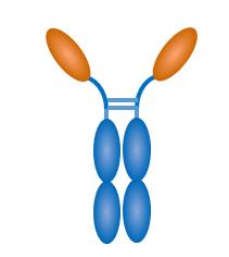
Luspatercept (mIgG2a) (RAP-536) is a fusion protein, consisting of a modified extracellular domain of human ActRIIB linked to the murine IgG2a Fc domain. Luspatercept (mIgG2a) inhibits Smad2/3 signaling, promotes differentiation of late-stage erythroid precursors and mitigates ineffective erythropoiesis (IE) in murine β-thalassemia. Luspatercept (mIgG2a) reduces anemia, α-globin aggregates, hemolysis, and disease complications of IE such as iron overload, splenomegaly, and bone defects.
For research use only. We do not sell to patients.
- Purity : 99.9%
- CAS No.: 1373715-00-4
- Molecular Weight:75.709 kDa
-
Storage:
Please store the product under the recommended conditions in the Certificate of Analysis.


To place orders, for customer services and technical support, please contact: MedChemExpress USA
Tel: 609-228-6898 E-mail: [email protected] [email protected]


-
 Biological Activity
-
Chemical Information
- Protocols
- Purity & Documentation
- References
-
 Help & FAQs




Biological Activity
Description
Species Reactivity
Human
In Vivo
Luspatercept (mIgG2a) (30 mg/kg, i.p., once for 12 h) inhibits overactivation of Smad2/3 in splenic erythroid precursors in murine β-thalassemia mice model[1].
Luspatercept (mIgG2a) (1-10 mg/kg, s.c., twice weekly for 2 months or once for 72 h) promotes terminal erythroid differentiation in the bone marrow and spleen and increases peripheral red cells in EPO-pretreated wild-type mice [1].
MedChemExpress (MCE) has not independently confirmed the accuracy of these methods. They are for reference only.
-
Animal Model:Hbbth1/th1 mice of the B6.D2-Hbbd3th/BrkJ strain (3-4 months old) with hydrogen peroxidenot (50 μM) or not[1].
-
Dosage:1 mg/kg
-
Administration:s.c., twice weekly for 2 months and then collected bone marrow, spleen and liver tissues.
-
Result:Significantly increased 29% RBC number, 16% hemoglobin concentration, and 19% hematocrit.
Decreased 8% mean cell volume, 10% mean cell hemoglobin, and 2% mean cell hemoglobin concentration.
Reduced 33% reticulocytes and a 19% red cell distribution width area.
Induced an overall increase in the proportion of late-stage erythroid precursors, significantly decreased the cell population of R1 while an increase of R3 in bone marrow, and reduced R1 and R2 with a significant increase in R3 in the spleen.
Reduced ∼60% serum EPO level, 36% splenomegaly, serum concentrations of ferritin (15%) and iron (28%), 29% transferrin saturation.
Reduced iron levels in liver (1464 μg/mg dry weight) and kidney (648 μg/mg dry weight) and altered the iron distribution with restoration of architecture in spleen.
Increased Hamp mRNA levels approximately 2 times without the expression changes of Bmp6 in liver and Gdf15 or Twsg1 in spleen.
Reduced 40% mean concentrations of total bilirubin and occurrence of abnormal erythrocyte morphology and hemolytic debris, but increased mean erythrocyte life span (28 vs 20 days).
Markedly reduced heinz bodies and 38% mean membrane-associated α-globin aggregates in peripheral erythrocytes.
Reduced ROS levels significantly in immature (CD71+Ter119+) and mature (CD71−Ter119+) erythroblasts as well as in peripheral red cells.
Reduced peroxide-stimulated ROS levels and significantly increased 17% bone mineral density, 100% trabecular bone volume (100%), number (14%), and thickness (14%).
Increased cortical thickness with a decrease in marrow space.
-
Animal Model:Hbbth1/th1 mice of the B6.D2-Hbbd3th/BrkJ strain (3-4 months old)[1].
-
Dosage:30 mg/kg
-
Administration:i.p., once for 12 h, and then collected bone marrow, spleen and liver tissues.
-
Result:Reduced phosphorylated Smad2/3 level in erythroid precursors of spleens within 12 h.
Inhibited phospho-Smad2 protein expression in erythroid precursors of spleens within 12 h.
-
Animal Model:C57BL/6 wild-type mice were injected intraperitoneally with EPO (1500 units/kg) or not[1].
-
Dosage:1, 10 mg/kg
-
Administration:s.c., (1 mg/kg) twice weekly for 2 months, (10 mg/kg) once for 72 h, and then collected bone marrow, spleen and liver tissues.
-
Result:Promoted terminal erythroid differentiation in the bone marrow and spleen and increased peripheral red cells in EPO-pretreated wild-type mice at 10 mg/kg with EPO stimulation in C57BL/6 wild-type mice.
Did not affect iron parameters and Hamp expression in C57BL/6 wild-type mice.
Conjugated
Unconjugated
Reconsititution
The product can be reconstituted/diluted with sterile PBS or saline.
Format
-
[ACVR2B (activin A receptor type 2B, ActR-IIB, ActRIIB)]2-Mouse IgG2A
Application
ELISA, FACS, Functional assay


Chemical Information
-
CAS No. 1373715-00-4
-
Appearance Liquid
-
Molecular Weight 75.709 kDa
-
Color Colorless to light yellow
-
SMILES
[Luspatercept (mIgG2a)]
-
Synonyms
RAP-536
-
Shipping
Shipping with dry ice.
-
Formulation
Please refer to the lot-specific COA for specific buffer information.
-
Storage
Please store the product under the recommended conditions in the Certificate of Analysis.

Protocols
-
Perls' Prussian Blue Iron Staining
Perls' Prussian blue staining is a histochemical method used to detect non-heme ferric iron (Fe3+) in biological tissues by exploiting an acid-mediated release of loosely bound iron from storage complexes such as ferritin or hemosiderin, followed by its reaction with potassium ferrocyanide to form an insoluble blue ferric ferrocyanide (Prussian blue) precipitate that marks iron localization under light microscopy. The reaction is classically performed under acidic conditions, which liberate Fe3+ ions that subsequently bind ferrocyanide to generate the visible chromogen, enabling spatial visualization of iron deposits in tissues such as brain, liver, and spleen. Histochemical interpretations are limited to a reactive iron pool rather than total iron content, reflecting only histologically accessible iron species rather than tightly protein-bound iron.


Purity & Documentation
-
Data Sheet (263 KB)
-
SDS (252 KB)
- English - EN (252 KB)
- Français - FR (252 KB)
- Deutsch - DE (252 KB)
- Norwegian - NO (252 KB)
- Español - ES (252 KB)
- Swedish - SV (252 KB)
- Italian - IT (252 KB)
- Korean - KR (252 KB)
- Portuguese - PT (252 KB)
-
Inhibitory Antibodies User Guide (603 KB)
References

Calculators
Concentration (start) × Volume (start) = Concentration (final) × Volume (final)


Luspatercept (mIgG2a) Related Classifications
HY-P99720A Related Classifications
 Powered by Bioz