



Lyso-PAF C-18

Based on 1 Customer Validation
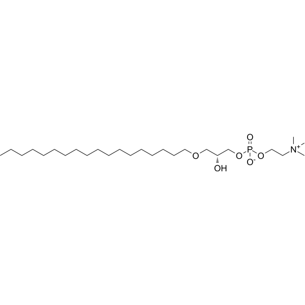
Lyso-PAF C-18 is a single-chain ether phospholipid with membrane regulatory activity, and Lyso-PAF is the deacetylated inactive precursor of PAF. Lyso-PAF C-18 regulates the activity of plasma membrane Ca2+-ATPase, and its mechanism of action may involve altering the lipid microenvironment of this enzyme. Lyso-PAF C-18 activates the calcium ATPase in rat synaptosomal membranes and inhibits the calcium ATPase in rat leukocyte membranes. Lyso-PAF C-18 can be used in cell membrane-related research.
For research use only. We do not sell to patients.
- Purity : 99.74%
- CAS No.: 74430-89-0
- Formula: C26H56NO6P
- Molecular Weight:509.70
-
Storage:Powder -20°C, 3 years ; In solvent -80°C, 6 months , -20°C, 1 month

To place orders, for customer services and technical support, please contact: MedChemExpress USA
Tel: 609-228-6898 E-mail: [email protected] [email protected]


-
 Biological Activity
-
Chemical Information
-
Solvent & Solubility
- Protocols
- Purity & Documentation
- References
-
 Help & FAQs





Biological Activity
Description
Cellular Effect
|
Cell Line

|
Type | Value | Description | References |
|---|---|---|---|---|
| HT-29 | IC50 |
18 μM
Compound: 7b
|
Cytotoxicity against human HT-29 cells assessed as cell viability after 48 hrs by MTT assay
Cytotoxicity against human HT-29 cells assessed as cell viability after 48 hrs by MTT assay
|
[PMID: 19402667] |
In Vitro
Lyso-PAF C-18 forms highly stable Langmuir monolayers at 20 °C under 20 mN/m, whereas its monolayer stability declines at 35 mN/m[1].
MedChemExpress (MCE) has not independently confirmed the accuracy of these methods. They are for reference only.


Chemical Information
-
CAS No. 74430-89-0
-
Appearance Solid
-
Molecular Weight 509.70
-
Formula C26H56NO6P
-
Color White to off-white
-
SMILES
CCCCCCCCCCCCCCCCCCOC[C@@H](O)COP(OCC[N+](C)(C)C)([O-])=O
-
Structure Classification
-
Initial Source
-
Shipping
Room temperature in continental US; may vary elsewhere.
-
Storage
Powder -20°C 3 years In solvent -80°C 6 months -20°C 1 month


Solvent & Solubility
In Vitro:
Ethanol : 12.5 mg/mL (24.52 mM; Need ultrasonic)
Please refer to the solubility information to select the appropriate solvent. Once prepared, please aliquot and store the solution to prevent product inactivation from repeated freeze-thaw cycles.
Storage method and period of stock solution: -80°C, 6 months; -20°C, 1 month. When stored at -80°C, please use it within 6 months. When stored at -20°C, please use it within 1 month.
Please refer to the solubility information to select the appropriate solvent. Once prepared, please aliquot and store the solution to prevent product inactivation from repeated freeze-thaw cycles.
Storage method and period of stock solution: -80°C, 6 months; -20°C, 1 month. When stored at -80°C, please use it within 6 months. When stored at -20°C, please use it within 1 month.
Concentration (start) × Volume (start) = Concentration (final) × Volume (final)


Purity & Documentation
-
Data Sheet (269 KB)
-
SDS (251 KB)
- English - EN (251 KB)
- Français - FR (251 KB)
- Deutsch - DE (251 KB)
- Norwegian - NO (251 KB)
- Español - ES (251 KB)
- Swedish - SV (251 KB)
- Italian - IT (251 KB)
- Korean - KR (251 KB)
- Portuguese - PT (251 KB)
-
Handling Instructions (2659 KB)
References


Complete Stock Solution Preparation Table
Please refer to the solubility information to select the appropriate solvent. Once prepared, please aliquot and store the solution to prevent product inactivation from repeated freeze-thaw cycles.
Storage method and period of stock solution: -80°C, 6 months; -20°C, 1 month. When stored at -80°C, please use it within 6 months. When stored at -20°C, please use it within 1 month.
| Optional Solvent | Concentration Solvent Mass | 1 mg | 5 mg | 10 mg | 25 mg |
|---|---|---|---|---|---|
| Ethanol | 1 mM | 1.9619 mL | 9.8097 mL | 19.6194 mL | 49.0485 mL |
| 5 mM | 0.3924 mL | 1.9619 mL | 3.9239 mL | 9.8097 mL | |
| 10 mM | 0.1962 mL | 0.9810 mL | 1.9619 mL | 4.9048 mL | |
| 15 mM | 0.1308 mL | 0.6540 mL | 1.3080 mL | 3.2699 mL | |
| 20 mM | 0.0981 mL | 0.4905 mL | 0.9810 mL | 2.4524 mL |


Lyso-PAF C-18 Related Classifications
HY-141581 Related Classifications
 Powered by Bioz