



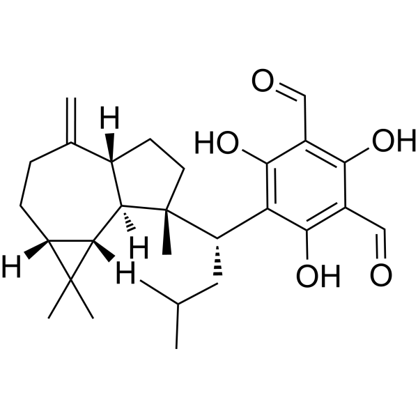
Macrocarpal C

Based on 1 Customer Validation
Macrocarpal C can be isolated from the 95 % ethanol extract of fresh leaves of E. globulus. Macrocarpal C inhibits the growth of T. mentagrophytes via an increase in the permeability of the fungal membrane. Macrocarpal C increases the production of intracellular ROS and? induces apoptosis as a consequence of DNA fragmentation.
Para uso exclusivo en investigación. No vendemos a pacientes.
- Pureza : 95.0%
- No. CAS: 142628-53-3
- Fòrmula: C28H38O5
- Peso molecular:454.60
-
Almacenamiento:
-20°C, sealed storage, away from moisture and light
* In solvent : -80°C, 6 months; -20°C, 1 month (sealed storage, away from moisture and light)

To place orders, for customer services and technical support, please contact: MedChemExpress USA
Tel: 609-228-6898 E-mail: [email protected] [email protected]


-
 Actividad biológica
-
Chemical Information
- Protocolo
- Pureza y Documentación
- Referencias
-
 Help & FAQs




Actividad biológica
Descripciòn
Cellular Effect
|
Cell Line

|
Type | Value | Description | References |
|---|---|---|---|---|
| A549 | IC50 |
<10 μM
Compound: Macrocarpal C
|
Cytotoxicity against human A549 cells assessed as reduction in cell viability incubated for 72 hrs by SRB assay
Cytotoxicity against human A549 cells assessed as reduction in cell viability incubated for 72 hrs by SRB assay
|
[PMID: 27142786] |
| HL-60 | IC50 |
<10 μM
Compound: Macrocarpal C
|
Cytotoxicity against human HL60 cells assessed as reduction in cell viability after 72 hrs by MTT assay
Cytotoxicity against human HL60 cells assessed as reduction in cell viability after 72 hrs by MTT assay
|
[PMID: 27142786] |


Chemical Information
-
No. CAS 142628-53-3
-
Appearance Solid
-
Peso molecular 454.60
-
Fòrmula C28H38O5
-
Color White to light yellow
-
SMILES
CC(C)C[C@@H](C1=C(C(C=O)=C(C(C=O)=C1O)O)O)[C@@]2([C@@]3([H])[C@@]4([H])[C@](CCC([C@]3([H])CC2)=C)([H])C4(C)C)C
-
Structure Classification
-
Initial Source
-
Envío
Room temperature in continental US; may vary elsewhere.
-
Almacenamiento
-20°C, sealed storage, away from moisture and light
* In solvent : -80°C, 6 months; -20°C, 1 month (sealed storage, away from moisture and light)

Protocolo
-
Apoptosis
Apoptosis, also called programmed cell death, is generally characterized by distinct morphological characteristics.
-
TUNEL staining for apoptotic DNA fragmentation
TUNEL staining detects DNA strand breaks by using terminal deoxynucleotidyl transferase to add labeled nucleotides to exposed 3′-OH DNA termini, generating either microscopic staining in fixed cells or tissue sections, or fluorescence/cytometric signal in cell suspensions. TUNEL positivity reflects DNA fragmentation but should not be interpreted alone as definitive apoptosis, because TUNEL can also label necrotic, autolytic, mechanically damaged, or DNA-repair-associated DNA breaks.
-
Annexin V plus membrane-impermeant dye apoptosis staining
Annexin V-based apoptosis assays rely on the detection of phosphatidylserine (PS) externalization from the inner leaflet of the plasma membrane to the outer leaflet, an early biochemical hallmark of apoptosis. Fluorescently labeled Annexin V binds PS in a calcium-dependent manner, enabling identification of early apoptotic cells by flow cytometry or fluorescence microscopy. When combined with a membrane-impermeant DNA-binding dye (e. g. , propidium iodide), this approach allows discrimination between viable (Annexin V−/dye−), early apoptotic (Annexin V+/dye−), and late apoptotic or necrotic (Annexin V+/dye+) cell populations by assessing membrane integrity and PS exposure.
-
ROS/oxidative-stress fluorescent staining
ROS/oxidative-stress fluorescent staining uses cell-permeant fluorogenic probes that become fluorescent after oxidation inside cells or tissues; commonly used examples include DCFH-DA/DCFDA for broad cellular oxidant detection, DHE for superoxide-related signal detection, MitoSOX for mitochondrial superoxide-related signal detection, and CellROX probes for oxidative-stress-associated fluorescence readouts. The assay detects probe oxidation rather than a single ROS species unless the probe and analysis method have been chemically validated for that species. DCFH-DA enters cells, is deacetylated by intracellular esterases to DCFH, and produces fluorescent DCF after oxidation, so the readout is used as an operational measure of total cellular oxidative stress rather than a species-specific ROS measurement. DHE and MitoSOX can report superoxide-related oxidation, but red fluorescence alone can include non-specific ethidium-like oxidation products; HPLC or optimized spectral approaches are
-
Transepithelial/transendothelial electrical resistance assay
TEER measures electrical resistance across epithelial or endothelial monolayers cultured on permeable supports, and the readout reflects ionic conductance through the cell barrier, especially the paracellular pathway regulated by junctional integrity. TEER can be measured without destroying the monolayer and is commonly used before or during transport, permeability, barrier-disruption, and barrier-maturation experiments. TEER values are influenced by biological maturation and technical conditions; reported factors include temperature, medium formulation, passage number, electrode geometry, membrane properties, and junctional length during early monolayer maturation. Therefore, TEER should be interpreted with blank-insert subtraction, area normalization, repeated readings, and, when possible, orthogonal barrier readouts such as FITC-dextran flux or tight-junction staining.
-
Apoptosis Solutions
Apoptosis is a regulated, generally non-lytic cell-death pathway that removes unwanted, damaged, infected, or abnormal cells through coordinated morphological changes, caspase activation, DNA fragmentation, and membrane remodeling. The intrinsic apoptosis pathway is controlled mainly by mitochondrial outer membrane permeabilization, BCL-2 family proteins, cytochrome c release, apoptosome formation, caspase-9 activation, and downstream executioner caspase-3/7 activation. The extrinsic apoptosis pathway is initiated by death receptors such as Fas, TNFR, and TRAIL receptors, which recruit adaptor proteins and activate caspase-8 before engaging executioner caspases or mitochondrial amplification through BID cleavage. Apoptosis is linked to many phenotypes, including cancer cell killing, tissue homeostasis, immune regulation, neurodegeneration, infection response, and treatment-induced cytotoxicity; unresolved questions include how apoptosis interacts with necroptosis, pyroptosis, ferroptos


Pureza y Documentación
-
Ficha de datos (274 KB)
-
SDS (251 KB)
- English - EN (251 KB)
- Français - FR (251 KB)
- Deutsch - DE (251 KB)
- Norwegian - NO (251 KB)
- Español - ES (251 KB)
- Swedish - SV (251 KB)
- Italian - IT (251 KB)
- Korean - KR (251 KB)
- Portuguese - PT (251 KB)
-
Instrucciones de manejo (2659 KB)
Referencias

Calculators
Concentration (start) × Volume (start) = Concentration (final) × Volume (final)


Macrocarpal C Related Classifications
HY-N3344 Related Classifications
 Powered by Bioz