



Tarextumab

Based on 1 Customer Validation
Tarextumab (OMP-59R5) is a cross-reactive, fully human IgG2 antibody that selectively inhibits Notch2 and Notch3 signaling. Tarextumab demonstrates broad-spectrum antitumor efficacy in xenograft models of epithelial tumors. Tarextumab can be used for the study of pancreatic cancer.
For research use only. We do not sell to patients.
- Purity : 99.83%
- CAS No.: 1359940-55-8
- Molecular Weight:142.94 kDa
-
Storage:
Please store the product under the recommended conditions in the Certificate of Analysis.


To place orders, for customer services and technical support, please contact: MedChemExpress USA
Tel: 609-228-6898 E-mail: [email protected] [email protected]


-
 Biological Activity
-
Chemical Information
- Protocols
- Purity & Documentation
- References
-
 Help & FAQs


-

HY-P99258
BrontictuzumabNotch1 Inhibitor -

HY-P99261
Demcizumabanti-DLL4 mAb -
HY-15860
FLI-06Notch Inhibitor -
HY-D0861
EGTACalcium Ion Chelator -

HY-P9904
AtezolizumabAnti-PD-L1 Antibody -

HY-P99041
PanitumumabAnti-EGFR Monoclonal Antibody -
HY-K0207
Anti-Flag Magnetic Beads -

HY-P99756
NirsevimabAnti-RSV Antibody -

HY-P99333
VolociximabIntegrin α5β1 mAb -

HY-P99575
Tarlatamabanti-DLL3/CD3 BiTE antibody -
HY-P99002
Human IgG2 kappa, Isotype ControlIgG2κ Isotype Control -

HY-P99854
DisitamabAnti-HER2 Antibody


Biological Activity
Description
Isotype
Human IgG2 kappa
Recommend Isotype Controls
Species Reactivity
Human
IC50 & Target
NOTCH3
In Vitro
Tarextumab inhibits human Notch2 and Notch3 signaling activity (reduces RLU values) without affecting Notch1 in HeLa cells using a Notch-responsive luciferase reporter assay[1].
Tarextumab downregulates mRNA expression of human Notch2, Notch3, HES1, NANOG, and OCT4, and reverses Gemcitabine (HY-17026)-induced upregulation of EMT-related genes (CDH2, VIM, FN1) in OMP-PN17 pancreatic tumor cells[1].
Tarextumab reduces protein levels of Notch3 extracellular domain (ECD) and active intracellular domain (ICD) in OMP-PN17 pancreatic tumor cells, and decreases the number of cells with high Notch3 ECD staining[1].
MedChemExpress (MCE) has not independently confirmed the accuracy of these methods. They are for reference only.
In Vivo
Tarextumab (40 mg/kg, i.p., once weekly, 4 weeks) does not induce marked tumor growth inhibition in MDA-MB-468 and HCC70 TNBC cell line-derived xenograft models in BALB/c nude mice[2].
MedChemExpress (MCE) has not independently confirmed the accuracy of these methods. They are for reference only.
-
Animal Model:Human pancreatic cancer cells (from PDX models including OMP-PN4, OMP-PN8, OMP-PN17) were subcutaneously implanted into NOD/SCID mice[1]
-
Dosage:40 mg/kg combined with Gemcitabine (HY-17026)
-
Administration:i.p., every other week
-
Result:Achieved enhanced antitumor efficacy.
Showed striking tumor regression.
Reduced cancer stem cell (CSC) frequency.
Delayed tumor recurrence.
Decreased tumor cell proliferation.
Increased pericyte maturation (enhanced desmin-positive pericyte association with CD31-positive endothelial cells) and tumor vascular perfusion and reduced intratumoral hypoxia.
-
Animal Model:Triple-negative breast cancer (TNBC) cells (from cell lines including MDA-MB-468, HCC70) were subcutaneously implanted into BALB/c nude mice[2]
-
Dosage:40 mg/kg
-
Administration:i.p., once weekly, 4 weeks
-
Result:Reduced tumor-initiating cell (TIC) frequency.
Clinical Trial
| NCT Number | Sponsor | Condition | Start Date |
Phase

|
|---|---|---|---|---|
| NCT01329991 | Plexxikon| | 2011-05 | PHASE1 |
Gene ID
Accession
Conjugated
Unconjugated
Reconsititution
The product can be reconstituted/diluted with sterile PBS or saline.
Format
-
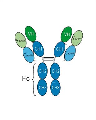
Human IgG2 kappa
Application
ELISA, FACS, Functional assay
Verified Bioactivity
-
 Immobilized NOTCH2 Protein (His Tag), Human can bind Tarextumab. The EC50 for this effect is 1.22 ng/mL.
 -
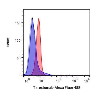 Flow cytometric analysis of 1X10⁶ MCF-7 cells with Tarextumab (HY-P99320, red). Cells were fixed with 4% paraformaldehyde. Then stained with the primary antibody at 1/200 dilution for an hour at 4℃. Alexa Fluor 488-conjugated AffiniPure Goat Anti-Human IgG H&L (AF488) (HY-P83776) was used as the secondary antibody at 1/1,000 dilution for 30 minutes at 4℃. Human IgG2 kappa (HY-P99002, blue) was used as the isotype control, cells without incubation with primary antibody were used as the unlabeled control (black).


Chemical Information
-
CAS No. 1359940-55-8
-
Appearance Liquid
-
Molecular Weight 142.94 kDa
-
Color Colorless to light yellow
-
SMILES
[Tarextumab]
-
Synonyms
OMP-59R5; Anti-Human NOTCH2 Recombinant Antibody
-
Shipping
Shipping with dry ice.
-
Formulation
Please refer to the lot-specific COA for specific buffer information.
-
Storage
Please store the product under the recommended conditions in the Certificate of Analysis.


Purity & Documentation
-
Data Sheet (278 KB)
-
SDS (251 KB)
- English - EN (251 KB)
- Français - FR (251 KB)
- Deutsch - DE (251 KB)
- Norwegian - NO (251 KB)
- Español - ES (251 KB)
- Swedish - SV (251 KB)
- Italian - IT (251 KB)
- Korean - KR (251 KB)
- Portuguese - PT (251 KB)
-
Inhibitory Antibodies User Guide (603 KB)
References
[1]. Yen WC, et al. Targeting Notch signaling with a Notch2/Notch3 antagonist (tarextumab) inhibits tumor growth and decreases tumor-initiating cell frequency. Clin Cancer Res. 2015 May 1;21(9):2084-95. [Content Brief]
[2]. Fu W, et al. EGFR/Notch Antagonists Enhance the Response to Inhibitors of the PI3K-Akt Pathway by Decreasing Tumor-Initiating Cell Frequency. Clin Cancer Res. 2019 May 1;25(9):2835-2847. [Content Brief]

Calculators
Concentration (start) × Volume (start) = Concentration (final) × Volume (final)


Tarextumab Related Classifications
HY-P99320 Related Classifications
 Powered by Bioz