



P62-mediated mitophagy inducer
Based on 6 publication(s) in Google Scholar
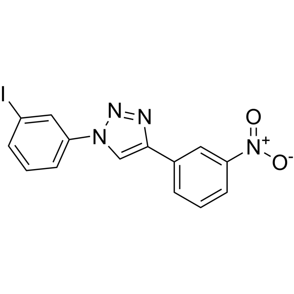
P62-mediated mitophagy inducer (PMI) is a P62-mediated mitophagy activator. P62-mediated mitophagy inducer activates mitochondrial autophagy without recruitment of Parkin or collapse of the mitochondrial membrane potential and remains active in cells lacking a fully functional PINK1/Parkin pathway. P62-mediated mitophagy inducer serves as a pharmacological tool to study the molecular mechanisms of mitosis, avoiding toxicity and some of the non-specific effects associated with the sudden dissipation of mitochondria lacking membrane potential.
For research use only. We do not sell to patients.
- Purity : 99.54%
- CAS No.: 1809031-84-2
- Formula: C14H9IN4O2
- Molecular Weight:392.15
-
Storage:Powder -20°C, 3 years ; In solvent -80°C, 2 years , -20°C, 1 year

To place orders, for customer services and technical support, please contact: MedChemExpress USA
Tel: 609-228-6898 E-mail: [email protected] [email protected]


-
 Biological Activity
-
Chemical Information
-
Solvent & Solubility
- Protocols
- Purity & Documentation
- References
-
 Help & FAQs


-
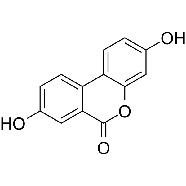
HY-100599
Urolithin AMitophagy Activator -
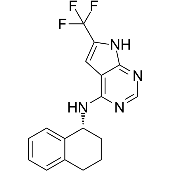
HY-152943
MTK458Mitophagy Activator -
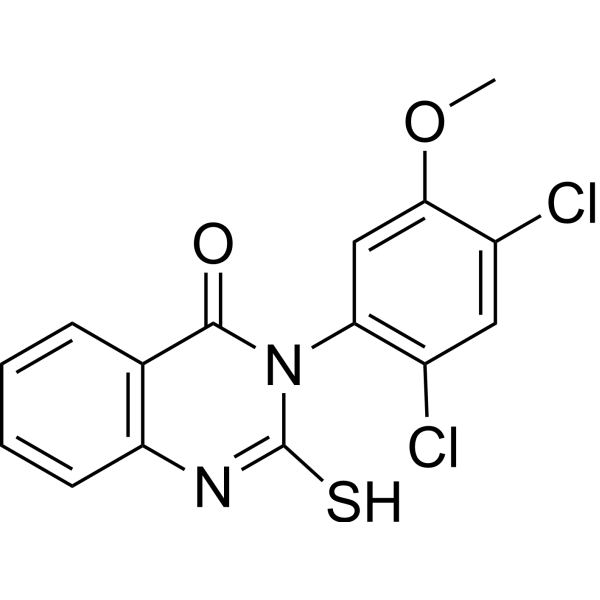
HY-15886
Mdivi-1Drp1 Inhibitor -
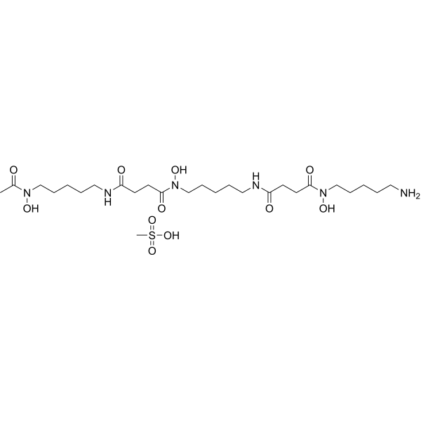
HY-B0988
Deferoxamine mesylateIron Chelator -

HY-10219
RapamycinmTOR Inhibitor -
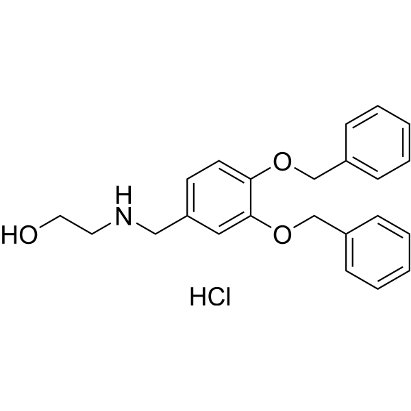
HY-W688687
XIE62-1004-AAutophagy Inducer -
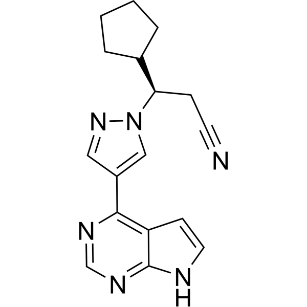
HY-50856
RuxolitinibJAK1/2 Inhibitor -
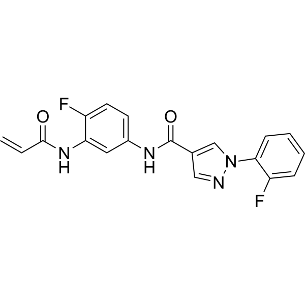
HY-128892
EN6Autophagy Activator -
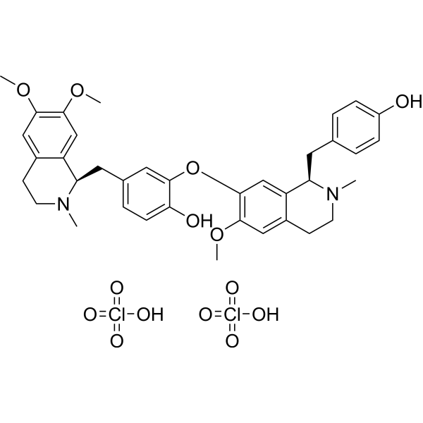
HY-N0485
Liensinine DiperchlorateAutophagy Inhibitor -
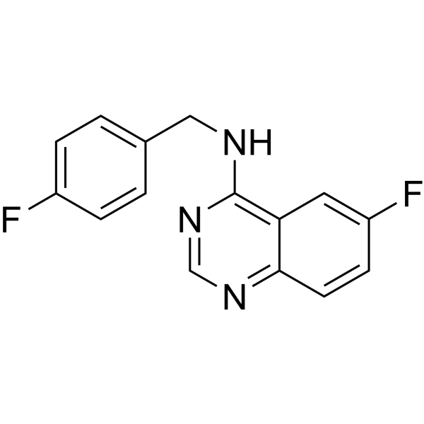
HY-12990
Spautin-1Autophagy Inhibitor -

HY-13259
MG-132Proteasome Inhibitor -

HY-100558
Bafilomycin A1V-ATPase Inhibitor
Publications Citing Use of MedChemExpress (MCE) P62-mediated mitophagy inducer
More- Autophagy. 2025 Jan;21(1):80-101. [Abstract]
- Genes Dis. 2023 Sep 2;11(6):101074. [Abstract]
- Free Radic Biol Med. 2026 Jun 27:254:391-404. [Abstract]
- Radiother Oncol. 2024 Jan:190:110028. [Abstract]
- Cancer Res Commun. 2023 Feb 21;3(2):297-308. [Abstract]
- bioRxiv. 2025 May 13:2025.05.12.653519. [Abstract]
 Customer Validation & Images

-
Cell Proliferation/Viability Assay

-
WB

-
IF

-
WB

-
IF


Biological Activity
Description
IC50 & Target
mitophagy[1].
In Vitro
P62-mediated mitophagy inducer (10 μM; 0, 1, 3, 6, 24 h) stabilizes Nrf2 and (10 μM; 9 h) upregulates P62 expression activating mitophagy[1].
P62-mediated mitophagy (10 μM; 24 h) acts downstream of the PINK1/Parkin signaling pathway in MEFs[1].
P62-mediated mitophagy inducer positively affects mitochondrial poly-ubiquitination and coupling[1].
MedChemExpress (MCE) has not independently confirmed the accuracy of these methods. They are for reference only. Further protocols information, click here.
-
Cell Line:MEFs
-
Concentration:10 µM
-
Incubation Time:9 h
-
Result:Significantly increased p62 mRNA levels.
-
Cell Line:MEFs
-
Concentration:10 µM
-
Incubation Time:24 h
-
Result:Demonstrated Parkin-independent induction of mitochondrial recruitment of P62.
-
Cell Line:MEFs
-
Concentration:10 µM
-
Incubation Time:0, 1, 3, 6, 24 h
-
Result:Exhibited maximum Nrf2 levels after 6 h and remained elevated at 24 h.
Clinical Trial
| NCT Number | Sponsor | Condition | Start Date |
Phase

|
|---|---|---|---|---|
| NCT01329991 | Plexxikon| | 2011-05 | PHASE1 |


Chemical Information
-
CAS No. 1809031-84-2
-
Appearance Solid
-
Molecular Weight 392.15
-
Formula C14H9IN4O2
-
Color Light yellow to yellow
-
SMILES
O=[N+](C1=CC(C2=CN(C3=CC=CC(I)=C3)N=N2)=CC=C1)[O-]
-
Shipping
Room temperature in continental US; may vary elsewhere.
-
Storage
Powder -20°C 3 years In solvent -80°C 2 years -20°C 1 year


Publications (6)
-
Journal Impact Factor
-
Most Recent
-
Autophagy
MANF facilitates breast cancer cell survival under glucose-starvation conditions via prkn-mediated mitophagy regulation. [Abstract]2025 Jan;21(1):80-101. PMID: 39147386
P62-mediated mitophagy inducer purchased from MedChemExpress. Usage Cited in: Autophagy. 2025 Jan;21(1):80-101. [Abstract]
Both pharmacological mitophagy inhibition (baf A1 and mdivi-1) and genetic mitophagy inhibition (DNM1L-KD) reduced and mitophagy agonist PMI (P62-mediated mitophagy inducer, 0.1-10 μM) increased BC cell viability under glucose-starvation conditions (n = 3).
-
Genes Dis
Mitophagy deficiency activates stimulator of interferon genes activation and aggravates pathogenetic cardiac remodeling. [Abstract]2023 Sep 2;11(6):101074. PMID: 39281830
P62-mediated mitophagy inducer purchased from MedChemExpress. Usage Cited in: Genes Dis. 2023 Sep 2;11(6):101074. [Abstract]
Protein levels of Parkin in CFs exposed to Ang II (100 nM) for 24 h with siRNA-Parkin or PMI (P62-mediated mitophagy inducer).
P62-mediated mitophagy inducer purchased from MedChemExpress. Usage Cited in: Genes Dis. 2023 Sep 2;11(6):101074. [Abstract]
CF mitophagy was confirmed by the co-location of mito-tracker green and lyso-tracker red after exposure to Ang II for 24 h with siRNA-Parkin or PMI (P62-mediated mitophagy inducer).
P62-mediated mitophagy inducer purchased from MedChemExpress. Usage Cited in: Genes Dis. 2023 Sep 2;11(6):101074. [Abstract]
Protein levels and quantification of STING, p-IRF3, and IRF3 in CFs exposed to Ang II with siRNA-Parkin or PMI (P62-mediated mitophagy inducer).
P62-mediated mitophagy inducer purchased from MedChemExpress. Usage Cited in: Genes Dis. 2023 Sep 2;11(6):101074. [Abstract]
Representative immunofluorescence staining and quantitative analysis of α-SMA in CFs exposed to Ang II with siRNA-Parkin or PMI (P62-mediated mitophagy inducer).

P62-mediated mitophagy inducer purchased from MedChemExpress. Usage Cited in: Genes Dis. 2023 Sep 2;11(6):101074. [Abstract]
CF cytosolic mtDNA levels were measured with RT-PCR after exposure to Ang II for 24 h with siRNA-Parkin or PMI (P62-mediated mitophagy inducer).
-
Free Radic Biol Med
DRP1-mediated mitophagy facilitates oral squamous cell carcinoma tumor cell survival under glucose restriction. [Abstract]2026 Jun 27:254:391-404. PMID: 42364552 -
Radiother Oncol
Mitophagy induction improves salivary gland stem/progenitor cell function by reducing senescence after irradiation. [Abstract]2024 Jan:190:110028. PMID: 38007043 -
Cancer Res Commun
Combination of a New Oral Demethylating Agent, OR2100, and Venetoclax for Treatment of Acute Myeloid Leukemia. [Abstract]2023 Feb 21;3(2):297-308. PMID: 36860654 -
bioRxiv
LOXL2 Deletion Triggers TMJ Osteoarthritis While Overexpression Protects Against NF-κβ-Induced Chondrocyte Apoptosis. [Abstract]2025 May 13:2025.05.12.653519. PMID: 40463059


Solvent & Solubility
In Vitro:
DMSO : 8.33 mg/mL (21.24 mM; Need ultrasonic; Hygroscopic DMSO has a significant impact on the solubility of product, please use newly opened DMSO)
Please refer to the solubility information to select the appropriate solvent. Once prepared, please aliquot and store the solution to prevent product inactivation from repeated freeze-thaw cycles.
Storage method and period of stock solution: -80°C, 2 years; -20°C, 1 year. When stored at -80°C, please use it within 2 years. When stored at -20°C, please use it within 1 year.
Please refer to the solubility information to select the appropriate solvent. Once prepared, please aliquot and store the solution to prevent product inactivation from repeated freeze-thaw cycles.
Storage method and period of stock solution: -80°C, 2 years; -20°C, 1 year. When stored at -80°C, please use it within 2 years. When stored at -20°C, please use it within 1 year.
Concentration (start) × Volume (start) = Concentration (final) × Volume (final)
In Vivo:
Select the appropriate dissolution method based on your experimental animal and administration route.
- For the following dissolution methods, please ensure to first prepare a clear stock solution using an In Vitro approach and then sequentially add co-solvents:
- To ensure reliable experimental results, the clarified stock solution can be appropriately stored based on storage conditions. As for the working solution for In Vivo experiments, it is recommended to prepare freshly and use it on the same day.
- The percentages shown for the solvents indicate their volumetric ratio in the final prepared solution. If precipitation or phase separation occurs during preparation, heat and/or sonication can be used to aid dissolution.
Add each solvent one by one: 10% DMSO 40% PEG300 5% Tween-80 45% Saline
Solubility: ≥ 1.25 mg/mL (3.19 mM); Clear solution
This protocol yields a clear solution of ≥ 1.25 mg/mL (saturation unknown).
Taking 1 mL working solution as an example, add 100 μL DMSO stock solution (12.5 mg/mL) to 400 μL PEG300, and mix evenly; then add 50 μL Tween-80 and mix evenly; then add 450 μL Saline to adjust the volume to 1 mL.
Preparation of Saline: Dissolve 0.9 g sodium chloride in ddH₂O and dilute to 100 mL to obtain a clear Saline solution.
In Vivo Dissolution Calculator
Please enter the basic information of animal experiments:
-
-
-
-
Recommended: Prepare an additional quantity of animals to account for potential losses during experiments.
Please enter your animal formula composition:
-
%DMSO +
Recommended: Keep the proportion of DMSO in working solution below 2% if your animal is weak.
-
%+
-
+%Tween-80 + +
-
%Saline +
The co-solvents required include: DMSO, . All of co-solvents are available by MedChemExpress (MCE). , Tween 80. All of co-solvents are available by MedChemExpress (MCE).
Working solution concentration: 0.22 mg/mL
Method for preparing stock solution: mg drug dissolved in μL DMSO. Stock solution concentration: mg/mL.
1. Take μL DMSO stock solution;
2. Add μL .
μL , mix evenly;
3. Then add μL Tween 80, mix evenly;
4. Then add μL
Please ensure that the stock solution in the first step is dissolved to a clear state, and add co-solvents in sequence. You can use ultrasonic heating (ultrasonic cleaner, recommended frequency 20-40 kHz), vortexing, etc. to assist dissolution.

Protocols
-
Mitochondrial membrane-potential fluorescent assay
Mitochondrial membrane potential fluorescent assays estimate ΔΨm in living cells using lipophilic cationic dyes such as TMRM, TMRE, rhodamine 123, and JC-1, which accumulate in mitochondria according to membrane polarization; loss of signal after FCCP or CCCP treatment is interpreted as mitochondrial depolarization. TMRM/TMRE and rhodamine 123 are commonly used for semi-quantitative live-cell microscopy or flow cytometry, while JC-1 can report a shift from red aggregate fluorescence to green monomer fluorescence during depolarization; interpretation requires controls because dye concentration, quenching mode, cell type, dye efflux, and mitochondrial mass can affect fluorescence independently of ΔΨm.
-
Fluorescent plasma-membrane potential dye assay
Fluorescent plasma-membrane potential dye assays measure changes in cell membrane potential using voltage-sensitive dyes whose fluorescence changes when cells depolarize or hyperpolarize. Anionic bis-oxonol dyes such as DiBAC4(3) enter depolarized cells more readily and show increased fluorescence after intracellular binding, while hyperpolarization reduces dye accumulation and fluorescence. FMP/FLIPR membrane-potential dyes are used for faster, homogeneous microplate assays of ion-channel or receptor-mediated membrane-potential changes.
-
Autophagy
Autophagy is a process in which eukaryotic cells use lysosomes to degrade their own cytoplasmic proteins and damaged organelles under the regulation of autophagy related gene (Atg). Microtubule-associated proteins light chain 3 (LC3) is recognized as autophagy marker, which transfers from cytoplasmic LC3 (LC3-I) to membrane type (LC3-II). LC3-II/I ratio could be detected by Western Blot and fluorescence microscopy.
-
Mitochondrial membrane-potential and mitochondrial mass staining
Mitochondrial membrane potential staining measures the electrochemical polarization across the mitochondrial inner membrane in live cells using lipophilic cationic fluorescent probes; early rhodamine-based work showed that selective mitochondrial dye accumulation is lost when the mitochondrial transmembrane potential is dissipated. JC-1 reports mitochondrial polarization by shifting from green monomer fluorescence to red J-aggregate fluorescence as dye concentration increases within energized mitochondria; therefore, the red/green fluorescence ratio is used as a relative readout of mitochondrial membrane potential. TMRE or TMRM staining provides a single-channel relative readout because these cationic rhodamine esters accumulate in polarized mitochondria, and lower fluorescence indicates reduced mitochondrial polarization when acquisition and dye-loading conditions are controlled. Mitochondrial mass staining is commonly performed with MitoTracker Green FM or related MitoTracker dyes as
-
Lysosome and acidic-vesicle live-cell staining
Lysosome and acidic-vesicle live-cell staining detects acidic intracellular compartments by using membrane-permeant acidotropic probes that accumulate in low-pH vesicles, including lysosomes, late endosomes, autolysosomes, and acidic phagosomes. LysoTracker staining is commonly used as an intensity-based readout of acidic lysosomal compartment abundance or enlargement, while acridine orange produces green fluorescence in less concentrated compartments and red fluorescence after concentration-dependent accumulation in acidic vesicular organelles. Loss or reduction of acridine-orange red signal can be used as a readout of lysosomal membrane permeabilization or reduced acidic-vesicle integrity. This protocol is designed for live cultured cells and can be adapted for fluorescence microscopy, high-content imaging, plate-reader readout, or flow cytometry when the selected literature supports the readout. Because these dyes report acidotropic accumulation rather than lysosome identity alone,
-
Mitophagy Solutions
Mitophagy is the selective autophagic degradation of mitochondria and functions as a mitochondrial quality-control pathway that removes damaged, depolarized, excess, or developmentally programmed mitochondria. The pathway links mitochondrial damage recognition, autophagosome recruitment, lysosomal delivery, and mitochondrial turnover to phenotypes such as mitochondrial homeostasis, oxidative-stress control, metabolic remodeling, differentiation, and neurodegeneration-related mitochondrial fidelity. The best-characterized damage-induced pathway is the PINK1-Parkin axis. Parkin is recruited selectively to impaired mitochondria and promotes their autophagic elimination, while mitochondrial depolarization stabilizes PINK1 on damaged mitochondria, recruits Parkin, and activates Parkin-dependent mitophagy. PINK1 also phosphorylates ubiquitin to activate Parkin E3 ubiquitin ligase activity, and PINK1-driven ubiquitin phosphorylation creates a feed-forward signal for recruiting autophagy machi
-
Macroautophagy Solutions
Macroautophagy is a conserved lysosome-dependent degradation pathway in which cytoplasmic material is sequestered into double-membrane autophagosomes and delivered to lysosomes for degradation and recycling. The pathway supports cellular homeostasis during nutrient limitation, organelle stress, protein-aggregate accumulation, infection, differentiation, and tissue remodeling by coupling cargo sequestration, autophagosome maturation, lysosomal fusion, and degradation of cargo-derived macromolecules. The core molecular sequence includes initiation by nutrient- and stress-regulated autophagy machinery, autophagosome nucleation, LC3/ATG8-family conjugation to autophagosomal membranes, cargo selection through receptors such as SQSTM1/p62, autophagosome-lysosome fusion, and lysosomal degradation. LC3 was identified as a mammalian homolog of yeast Atg8 that localizes to autophagosomal membranes after processing, and p62/SQSTM1 was shown to connect ubiquitinated cargo with autophagic degradati


Purity & Documentation
-
Data Sheet (275 KB)
-
SDS (393 KB)
- English - EN (393 KB)
- Français - FR (393 KB)
- Deutsch - DE (393 KB)
- Norwegian - NO (393 KB)
- Español - ES (393 KB)
- Swedish - SV (393 KB)
- Italian - IT (393 KB)
- Korean - KR (393 KB)
- Portuguese - PT (393 KB)
-
Handling Instructions (2659 KB)
References


Complete Stock Solution Preparation Table
Please refer to the solubility information to select the appropriate solvent. Once prepared, please aliquot and store the solution to prevent product inactivation from repeated freeze-thaw cycles.
Storage method and period of stock solution: -80°C, 2 years; -20°C, 1 year. When stored at -80°C, please use it within 2 years. When stored at -20°C, please use it within 1 year.
| Optional Solvent | Concentration Solvent Mass | 1 mg | 5 mg | 10 mg | 25 mg |
|---|---|---|---|---|---|
| DMSO | 1 mM | 2.5500 mL | 12.7502 mL | 25.5004 mL | 63.7511 mL |
| 5 mM | 0.5100 mL | 2.5500 mL | 5.1001 mL | 12.7502 mL | |
| 10 mM | 0.2550 mL | 1.2750 mL | 2.5500 mL | 6.3751 mL | |
| 15 mM | 0.1700 mL | 0.8500 mL | 1.7000 mL | 4.2501 mL | |
| 20 mM | 0.1275 mL | 0.6375 mL | 1.2750 mL | 3.1876 mL |


P62-mediated mitophagy inducer Related Classifications
HY-115576 Related Classifications
 Powered by Bioz