



PACMA 31
Based on 6 publication(s) in Google Scholar
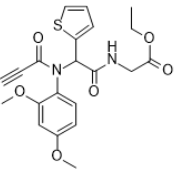
PACMA 31 is an irreversible, orally active protein disulfide isomerase (PDI) inhibitor with an IC50 of 10 μM. PACMA 31 forms a covalent bond with the active site cysteines of PDI. PACMA 31 shows tumor targeting ability and significantly suppresses ovarian tumor growth without causing toxicity to normal tissues. PACMA 31 is a click chemistry reagent, it contains an Alkyne group and can undergo copper-catalyzed azide-alkyne cycloaddition (CuAAc) with molecules containing Azide groups.
For research use only. We do not sell to patients.
- Purity : 99.32%
- CAS No.: 1401089-31-3
- Formula: C21H22N2O6S
- Molecular Weight:430.47
-
Storage:Powder -20°C, 3 years , 4°C, 2 years ; In solvent -80°C, 6 months , -20°C, 1 month

To place orders, for customer services and technical support, please contact: MedChemExpress USA
Tel: 609-228-6898 E-mail: [email protected] [email protected]


-
 Biological Activity
-
Chemical Information
-
Solvent & Solubility
- Protocols
- Purity & Documentation
- References
-
 Help & FAQs


-
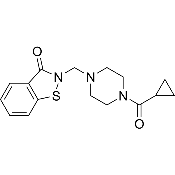
HY-100432
LOC14PDI Inhibitor -
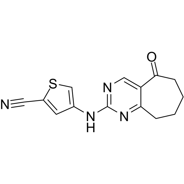
HY-W107464
G6PDi-1G6PD Inhibitor -
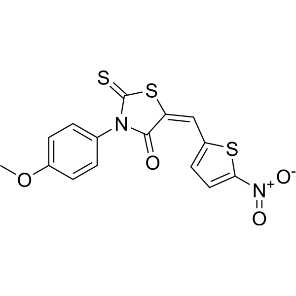
HY-100430
CCF642PDI Inhibitor -
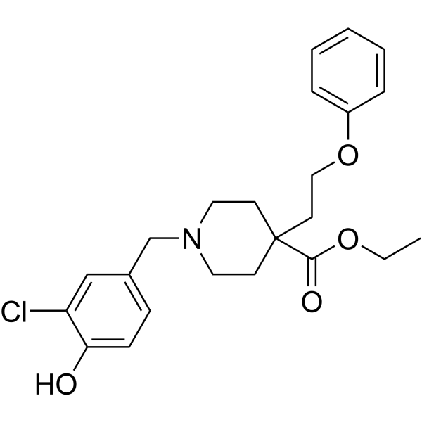
HY-114086
ML359PDI Inhibitor -
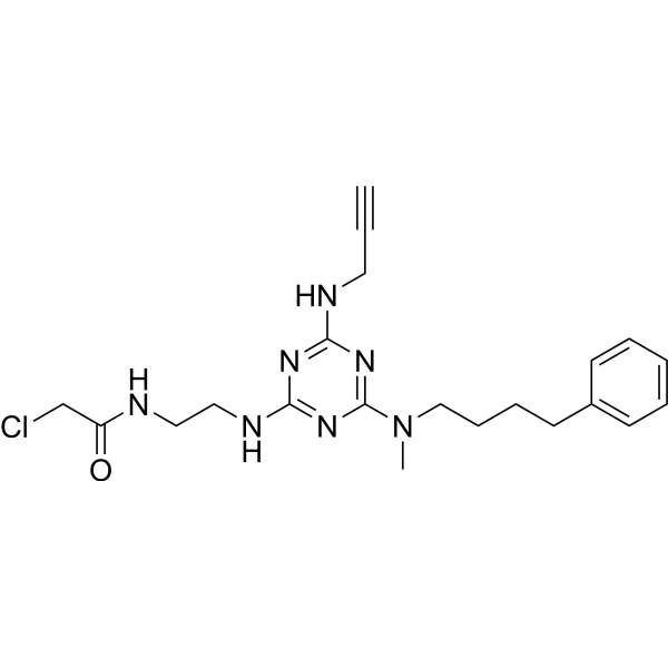
HY-117570
KSC-34PDIA1 Inhibitor -
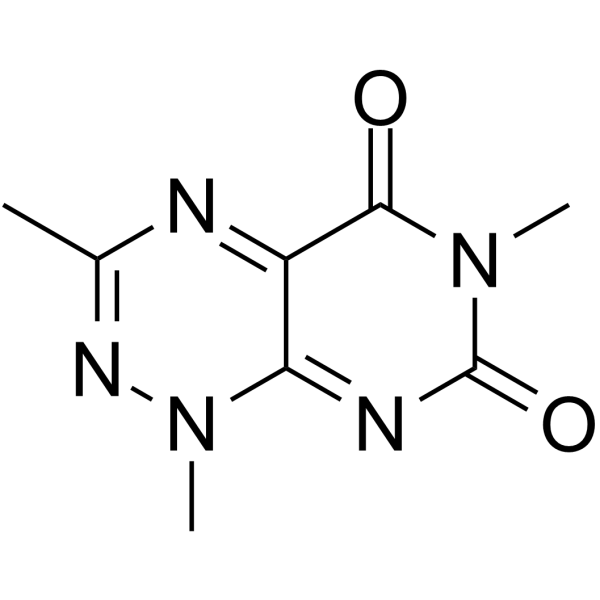
HY-111117
3-MethyltoxoflavinPDI Inhibitor -
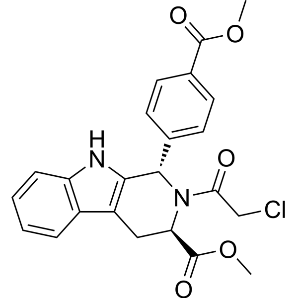
HY-100218A
RSL3GPX4 Inhibitor -
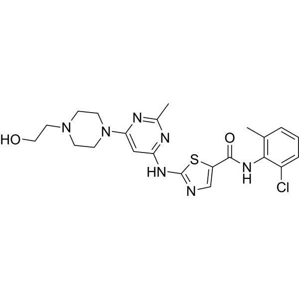
HY-10181
DasatinibSrc/Bcr-Abl Inhibitor -
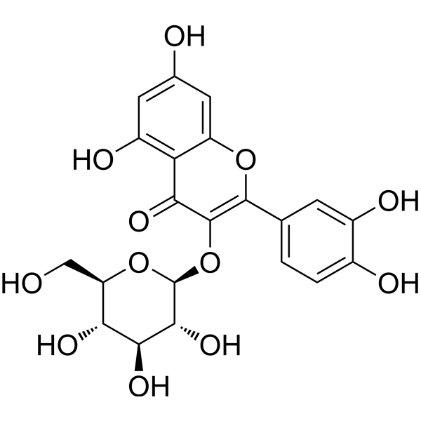
HY-N1445
IsoquercitrinNO Synthase Inhibitor -

HY-B0015
PaclitaxelAntitumor Agent -
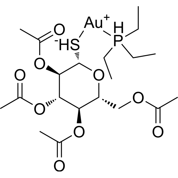
HY-B1123
AuranofinBacterial Inhibitor -
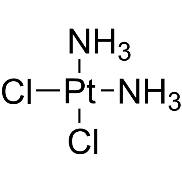
HY-17394
CisplatinDNA Alkylator
Publications Citing Use of MedChemExpress (MCE) PACMA 31
More Customer Validation & Images

-
WB

-
RT-PCR

-
Cell Proliferation/Viability Assay

-
Flow Cytometry

-
IP


Biological Activity
Description
IC50 & Target
|
PDIA1 |
In Vitro
PACMA 31 (0-10 μM; 24 hours) significantly inhibits colony formation in OVCAR-8 cells in a dose-dependent manner[1].
MedChemExpress (MCE) has not independently confirmed the accuracy of these methods. They are for reference only. Further protocols information, click here.
In Vivo
MedChemExpress (MCE) has not independently confirmed the accuracy of these methods. They are for reference only.
-
Animal Model:Athymic mice (bearing OVCAR-8 cells)[1]
-
Dosage:20-200 mg/kg
-
Administration:I.p., per day for the first 3 wk with 5-d on and 2-d off treatment cycles, and dose was escalated to 40 mg/kg per day for the next 7 d; p.o., the initial dose of 20 mg/kg per day was gradually increased by 20 mg/kg per day with each dose for 3 d before it was orally dosed at 200 mg/kg per day for an additional 32 d, increasing the dose from 20 to 200 mg/kg
-
Result:Compared with the control group, i.p. or per os administration of PACMA 31 significantly inhibited tumor growth by 85% and 65% at day 62, respectively.


Chemical Information
-
CAS No. 1401089-31-3
-
Appearance Solid
-
Molecular Weight 430.47
-
Formula C21H22N2O6S
-
Color White to off-white
-
SMILES
COC1=CC=C(N(C(C2=CC=CS2)C(NCC(OCC)=O)=O)C(C#C)=O)C(OC)=C1
-
Shipping
Room temperature in continental US; may vary elsewhere.
-
Storage
Powder -20°C 3 years 4°C 2 years In solvent -80°C 6 months -20°C 1 month


Publications (6)
-
Journal Impact Factor
-
Most Recent
-
J Transl Med
Single-cell transcriptomics identifies PDIA4 as a marker of progression and therapeutic vulnerability in multiple myeloma. [Abstract]2025 Oct 21;23(1):1136. PMID: 41121130
PACMA 31 purchased from MedChemExpress. Usage Cited in: J Transl Med. 2025 Oct 21;23(1):1136. [Abstract]
Flow cytometry was used to quantify the apoptosis rate of RPMI-8226 cells after treatment with untreated (no treatment), bortezomib, PACMA 31 (20 μM), or a combination of both for 48 hours. Annexin V/7-AAD staining was used to assess apoptosis.
-
Cell Chem Biol
2025 Oct 16;32(10):1235-1248.e34. PMID: 41056949 -
Diabetes Metab J
Serpina3c Mitigates Adipose Tissue Inflammation by Inhibiting the HIF1α-Mediated Endoplasmic Reticulum Overoxidation in Adipocytes. [Abstract]2026 Jan;50(1):62-76. PMID: 40403760
PACMA 31 purchased from MedChemExpress. Usage Cited in: Diabetes Metab J. 2026 Jan;50(1):62-76. [Abstract]
PACMA31 (0.4 μM). The protein levels of MAPK signaling pathway in 3cKD 3T3-L1 adipocytes and quantification of the relative protein band density.
PACMA 31 purchased from MedChemExpress. Usage Cited in: Diabetes Metab J. 2026 Jan;50(1):62-76. [Abstract]
3cKD 3T3-L1 adipocytes were pretreated with 10 μM Ero1α inhibitor EN460 or 0.4 μM PDI inhibitor propynoylcarbamoylmethylamide 31 (PACMA31) alone or in combination for 2 hours, followed by stimulation with 500 μM PA for 48 hours. The mRNA levels of specified genes in 3T3-L1 adipocytes were detected.
-
Int J Antimicrob Agents
Targeting the human protein disulfide isomerase A3 as a broad-spectrum antiviral approach against human respiratory viruses. [Abstract]2026 Apr 2:107802. PMID: 41935586
PACMA 31 purchased from MedChemExpress. Usage Cited in: Int J Antimicrob Agents. 2026 Apr 2:107802. [Abstract]
Antiviral activity of LOC14, 16F16, and PACMA31. VRAs were performed in HCT-8 cells infected with hCoV-OC43 and treated with increasing concentrations of the PDIA3 inhibitors 1 h before, during, and post-infection (full treatment).
-
-
PACMA 31 purchased from MedChemExpress. Usage Cited in: bioRxiv. 2025 March 21.
Immunoblot of input and streptavidin-affinity purified P1 modified proteins in THP1 null cells primed for 16 h with LPS (1 µg/mL) treated with the indicated concentration of P1 and/or PACMA31 for 6 h. P1 modified proteins were modified with biotin through click-dependent addition of a biotinazide to the alkyne moiety of P1.


Solvent & Solubility
In Vitro:
DMSO : 100 mg/mL (232.30 mM; Need ultrasonic; Hygroscopic DMSO has a significant impact on the solubility of product, please use newly opened DMSO)
Please refer to the solubility information to select the appropriate solvent. Once prepared, please aliquot and store the solution to prevent product inactivation from repeated freeze-thaw cycles.
Storage method and period of stock solution: -80°C, 6 months; -20°C, 1 month. When stored at -80°C, please use it within 6 months. When stored at -20°C, please use it within 1 month.
Please refer to the solubility information to select the appropriate solvent. Once prepared, please aliquot and store the solution to prevent product inactivation from repeated freeze-thaw cycles.
Storage method and period of stock solution: -80°C, 6 months; -20°C, 1 month. When stored at -80°C, please use it within 6 months. When stored at -20°C, please use it within 1 month.
Concentration (start) × Volume (start) = Concentration (final) × Volume (final)
In Vivo:
Select the appropriate dissolution method based on your experimental animal and administration route.
- For the following dissolution methods, please ensure to first prepare a clear stock solution using an In Vitro approach and then sequentially add co-solvents:
- To ensure reliable experimental results, the clarified stock solution can be appropriately stored based on storage conditions. As for the working solution for In Vivo experiments, it is recommended to prepare freshly and use it on the same day.
- The percentages shown for the solvents indicate their volumetric ratio in the final prepared solution. If precipitation or phase separation occurs during preparation, heat and/or sonication can be used to aid dissolution.
Add each solvent one by one: 10% DMSO 40% PEG300 5% Tween-80 45% Saline
Solubility: ≥ 2.5 mg/mL (5.81 mM); Clear solution
This protocol yields a clear solution of ≥ 2.5 mg/mL (saturation unknown).
Taking 1 mL working solution as an example, add 100 μL DMSO stock solution (25.0 mg/mL) to 400 μL PEG300, and mix evenly; then add 50 μL Tween-80 and mix evenly; then add 450 μL Saline to adjust the volume to 1 mL.
Preparation of Saline: Dissolve 0.9 g sodium chloride in ddH₂O and dilute to 100 mL to obtain a clear Saline solution.
In Vivo Dissolution Calculator
Please enter the basic information of animal experiments:
-
-
-
-
Recommended: Prepare an additional quantity of animals to account for potential losses during experiments.
Please enter your animal formula composition:
-
%DMSO +
Recommended: Keep the proportion of DMSO in working solution below 2% if your animal is weak.
-
%+
-
+%Tween-80 + +
-
%Saline +
The co-solvents required include: DMSO, . All of co-solvents are available by MedChemExpress (MCE). , Tween 80. All of co-solvents are available by MedChemExpress (MCE).
Working solution concentration: 0.22 mg/mL
Method for preparing stock solution: mg drug dissolved in μL DMSO. Stock solution concentration: mg/mL.
1. Take μL DMSO stock solution;
2. Add μL .
μL , mix evenly;
3. Then add μL Tween 80, mix evenly;
4. Then add μL
Please ensure that the stock solution in the first step is dissolved to a clear state, and add co-solvents in sequence. You can use ultrasonic heating (ultrasonic cleaner, recommended frequency 20-40 kHz), vortexing, etc. to assist dissolution.

Protocols
-
EdU Incorporation Assay (Click Chemistry-Based DNA Synthesis Measurement)
The EdU incorporation assay measures DNA synthesis by adding the thymidine analog 5-ethynyl-2′-deoxyuridine to cells or tissues, where it is incorporated into newly synthesized DNA during S phase. Incorporated EdU is detected by copper-catalyzed azide-alkyne cycloaddition, in which a fluorescent azide covalently reacts with the ethynyl group on EdU, allowing S-phase cells to be detected by fluorescence microscopy, flow cytometry, or high-content imaging. EdU detection does not require DNA denaturation or anti-BrdU antibody access, which preserves sample structure and improves compatibility with immunostaining and multiparameter cytometry compared with BrdU-based detection. EdU can be cytotoxic in a cell-type- and exposure-dependent manner, so pulse duration, concentration, and continuous-labeling designs should be validated for each cell type.
-
Cell Cytotoxicity Assay
Cytotoxicity assays are usually based on the assessment of cell membrane damage, which can also be indirectly detected by measuring cell viability. Detection methods include MTT assay, CKK-8 assay, LDH assay and ATP assay, etc.
-
Patient-Derived Xenograft (PDX)
Patient-derived xenograft (PDX) models are generated by engrafting primary human tumor tissue directly into immunodeficient mice, allowing in vivo propagation of patient tumor biology without initial in vitro adaptation. These models are used to preserve key histopathological and molecular characteristics of the original tumor and enable assessment of tumor growth dynamics and therapeutic response in a living organism. The biological readout is tumor engraftment and subsequent growth in the murine host, which reflects the ability of human tumor cells to survive, vascularize, and expand in an immunocompromised microenvironment.
-
Patient-Derived Orthotopic Xenograft (PDOX)
Patient-derived orthotopic xenograft (PDOX) modeling implants fresh patient tumor tissue or patient-derived tumor cells into the anatomically corresponding organ or tissue site of immunodeficient mice, usually by surgical orthotopic implantation, to preserve patient tumor histology, local microenvironmental context, invasion, metastatic behavior, and treatment-response features better than subcutaneous implantation. PDOX readouts include tumor engraftment, orthotopic tumor growth, local invasion, metastasis, recurrence after resection, histologic similarity to the donor tumor, biomarker retention, molecular concordance, survival, and response or resistance to therapy. PDOX models are used for preclinical drug testing and individualized therapy evaluation, but engraftment success varies by tumor type and specimen quality.
-
Subcutaneous Cell-Line-Derived Xenograft
Subcutaneous cell-line-derived xenograft (CDX) models are established by implanting cultured human cancer cell lines into immunodeficient mice, where the injected cells form localized tumors that can be monitored in vivo as a measure of tumorigenic potential, growth kinetics, and treatment response. These models are widely used in oncology research because they allow reproducible tumor formation and enable comparative assessment of tumor growth between different cell lines or genetic manipulations in a controlled in vivo microenvironment. Subcutaneous implantation of cancer cells in immunodeficient mice is a standard approach for evaluating tumor growth behavior and therapeutic response across multiple cancer types, including prostate, esophageal, pancreatic, and colon cancer models.
-
Orthotopic Cell-Line Xenograft
Orthotopic cell-line xenograft models involve implantation of human cancer cell lines into the anatomically corresponding organ of immunodeficient mice to reproduce tumor growth within a native microenvironment, enabling more clinically relevant tumor behavior compared with subcutaneous models. These models are widely used because orthotopic placement better recapitulates tumor progression, including invasion and metastatic spread, which are often underrepresented in heterotopic implantation systems. Compared with conventional xenografts, orthotopic implantation is described as more technically complex but provides improved simulation of tumor-microenvironment interactions and metastatic behavior, making it particularly valuable for translational oncology research. Surgical orthotopic implantation approaches have been emphasized as enabling faithful reproduction of clinical cancer features, including metastasis and disease progression patterns that align with the tumor’s organ of origi


Purity & Documentation
-
Data Sheet (280 KB)
-
SDS (480 KB)
- English - EN (480 KB)
- Français - FR (480 KB)
- Deutsch - DE (480 KB)
- Norwegian - NO (480 KB)
- Español - ES (480 KB)
- Swedish - SV (480 KB)
- Italian - IT (480 KB)
- Korean - KR (480 KB)
- Portuguese - PT (480 KB)
-
Handling Instructions (2659 KB)
References


Complete Stock Solution Preparation Table
Please refer to the solubility information to select the appropriate solvent. Once prepared, please aliquot and store the solution to prevent product inactivation from repeated freeze-thaw cycles.
Storage method and period of stock solution: -80°C, 6 months; -20°C, 1 month. When stored at -80°C, please use it within 6 months. When stored at -20°C, please use it within 1 month.
| Optional Solvent | Concentration Solvent Mass | 1 mg | 5 mg | 10 mg | 25 mg |
|---|---|---|---|---|---|
| DMSO | 1 mM | 2.3230 mL | 11.6152 mL | 23.2304 mL | 58.0761 mL |
| 5 mM | 0.4646 mL | 2.3230 mL | 4.6461 mL | 11.6152 mL | |
| 10 mM | 0.2323 mL | 1.1615 mL | 2.3230 mL | 5.8076 mL | |
| 15 mM | 0.1549 mL | 0.7743 mL | 1.5487 mL | 3.8717 mL | |
| 20 mM | 0.1162 mL | 0.5808 mL | 1.1615 mL | 2.9038 mL | |
| 25 mM | 0.0929 mL | 0.4646 mL | 0.9292 mL | 2.3230 mL | |
| 30 mM | 0.0774 mL | 0.3872 mL | 0.7743 mL | 1.9359 mL | |
| 40 mM | 0.0581 mL | 0.2904 mL | 0.5808 mL | 1.4519 mL | |
| 50 mM | 0.0465 mL | 0.2323 mL | 0.4646 mL | 1.1615 mL | |
| 60 mM | 0.0387 mL | 0.1936 mL | 0.3872 mL | 0.9679 mL | |
| 80 mM | 0.0290 mL | 0.1452 mL | 0.2904 mL | 0.7260 mL | |
| 100 mM | 0.0232 mL | 0.1162 mL | 0.2323 mL | 0.5808 mL |


PACMA 31 Related Classifications
HY-100433 Related Classifications
 Powered by Bioz