



PB28 dihydrochloride

Based on 1 Customer Validation
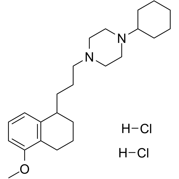
PB28 dihydrochloride, a cyclohexylpiperazine derivative, is a high affinity and selective sigma 2 (σ2) receptor agonist with a Ki of 0.68 nM. PB28 dihydrochloride is also a σ1 antagonist with a Ki of 0.38 nM. PB28 dihydrochloride is less affinity for other receptors. PB28 dihydrochloride inhibits electrically evoked twitch in guinea pig bladder and ileum with EC50 values of 2.62 μM and 3.96 μM, respectively. PB28 dihydrochloride can modulate SARS-CoV-2-human protein-protein interaction. PB28 dihydrochloride induces caspase-independent apoptosis and has antitumor activity.
For research use only. We do not sell to patients.
- Purity : 99.63%
- CAS No.: 172907-03-8
- Formula: C24H40Cl2N2O
- Molecular Weight:443.49
-
Storage:
4°C, sealed storage, away from moisture
* In solvent : -80°C, 6 months; -20°C, 1 month (sealed storage, away from moisture)

To place orders, for customer services and technical support, please contact: MedChemExpress USA
Tel: 609-228-6898 E-mail: [email protected] [email protected]


-
 Biological Activity
-
Chemical Information
-
Solvent & Solubility
- Protocols
- Purity & Documentation
- References
-
 Help & FAQs


-
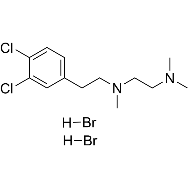
HY-16996A
BD-1047 dihydrobromideSigma-1 Receptor Antagonist -
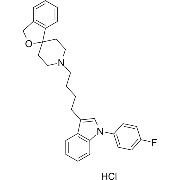
HY-14221A
Siramesine hydrochlorideSigma-2 Receptor Agonist -
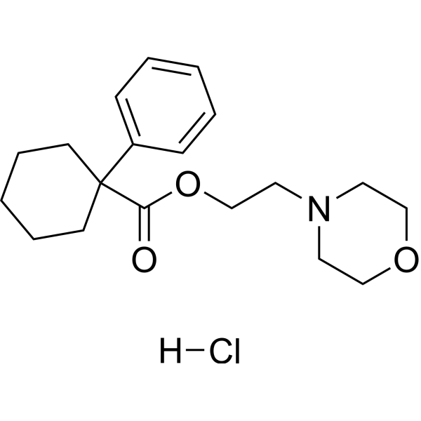
HY-18100A
PRE-084 hydrochlorideS1R Agonist -

HY-108511R
PB28 dihydrochloride (Standard)Reference Standard -
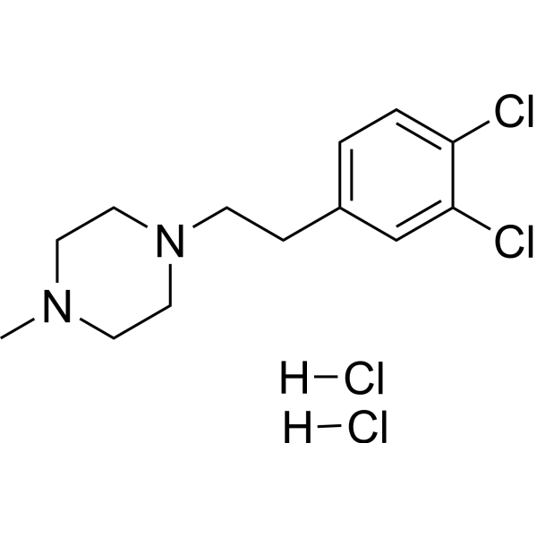
HY-18101A
BD-1063 dihydrochlorideσ-1R Antagonist -
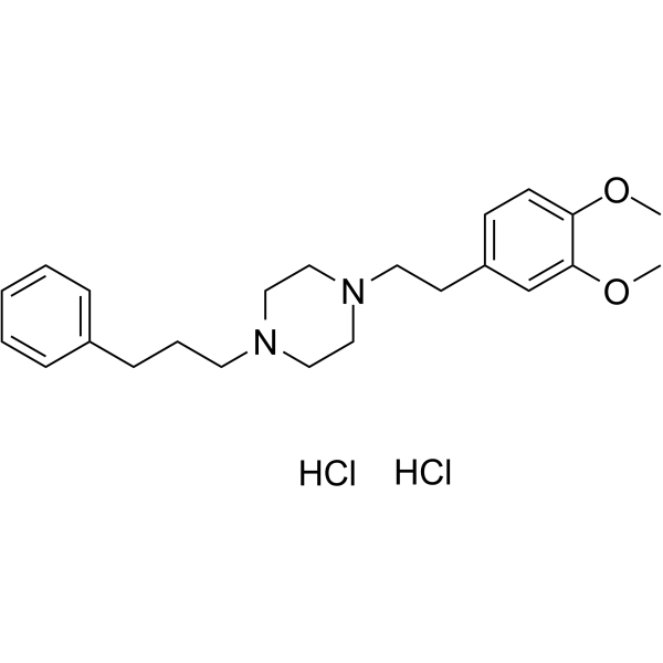
HY-13510
Cutamesine dihydrochlorideσ1R Agonist -
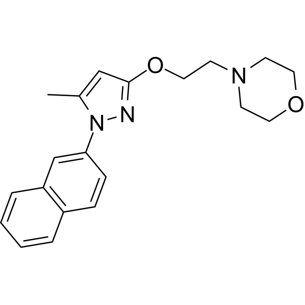
HY-18099
S1RAσ1R Antagonist -
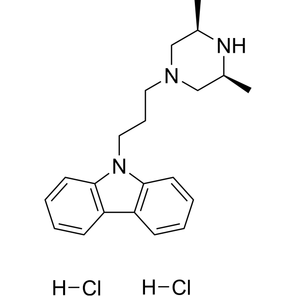
HY-108510
Rimcazole dihydrochlorideσ Receptor Antagonist -
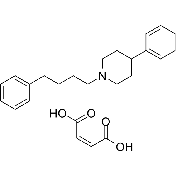
HY-101043
4-PPBP maleateσ 1 Receptor Ligand/Agonist -
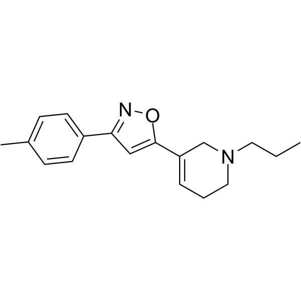
HY-108512
PD 144418σ1 Receptor Ligand -
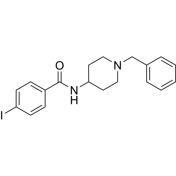
HY-100155
4-IBPσ1 Receptor Agonist -
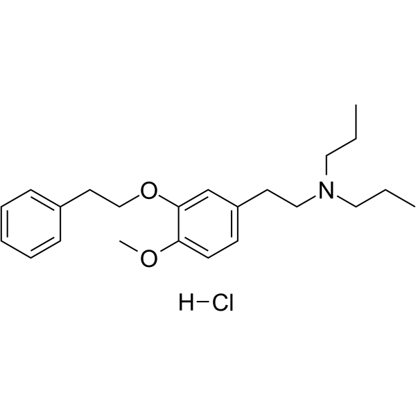
HY-101484A
NE-100 hydrochlorideSigma Receptor Antagonist
All Sigma Receptor Isoforms
More

Biological Activity
Description
In Vitro
PB28 (15-25 nM; 24-48 hours; MCF7 and MCF7 ADR cells) dihydrochloride treatment shows an accumulation in the G0-G1 phase for MCF7 and MCF7 ADR cells that are time and concentration independent[1].
?
PB28 dihydrochloride has a higher σ2 receptor affinity expressed as Ki (0.28 nM and 0.17 nM in MCF7 and MCF7 ADR cells, respectively) than σ1 receptor affinity (13.0 nMand 10.0 nM, respectively)[1].
?
PB28 dihydrochloride inhibits cell growth of MCF7 and MCF7 ADR cells with IC50s of 25 nM and 15 nM, respectively after 2-day treatment[1].
?
PB28 dihydrochloride induces apoptosis through a caspase-independent pathway[1].
?
PB28 dihydrochloride also reduces P-gp expression in a concentration- and time-dependent manner (approximately 60% in MCF7 and 90% in MCF7 ADR)[1].
?
PB28 dihydrochloride displays antiproliferative and cytotoxic effects in both C6 rat glioma and SK-N-SH human neuroblastoma cell lines[1].
MedChemExpress (MCE) has not independently confirmed the accuracy of these methods. They are for reference only. Further protocols information, click here.
-
Cell Line:MCF7 and MCF7 ADR cells
-
Concentration:25 nM and 15 nM
-
Incubation Time:24 hours, 48 hours
-
Result:Showed an accumulation in the G0-G1 phase for MCF7 and MCF7 ADR cells that were time and concentration independent.
In Vivo
MedChemExpress (MCE) has not independently confirmed the accuracy of these methods. They are for reference only.
-
Animal Model:C57BL/6 female mice (10 weeks old) injected with Panc02 cells[2]
-
Dosage:10.7 mg/mL
-
Administration:Intraperitoneal injection; daily; for two weeks
-
Result:Inhibited tumor growth in Panc02 tumor burden mice.


Chemical Information
-
CAS No. 172907-03-8
-
Appearance Solid
-
Molecular Weight 443.49
-
Formula C24H40Cl2N2O
-
Color White to off-white
-
SMILES
[H]Cl.[H]Cl.COC1=CC=CC2=C1CCCC2CCCN3CCN(C4CCCCC4)CC3
-
Shipping
Room temperature in continental US; may vary elsewhere.
-
Storage
4°C, sealed storage, away from moisture
* In solvent : -80°C, 6 months; -20°C, 1 month (sealed storage, away from moisture)


Solvent & Solubility
In Vitro:
H2O : 9 mg/mL (20.29 mM; Need ultrasonic and warming)
DMSO : 5 mg/mL (11.27 mM; ultrasonic and warming and heat to 60°C; Hygroscopic DMSO has a significant impact on the solubility of product, please use newly opened DMSO)
Please refer to the solubility information to select the appropriate solvent. Once prepared, please aliquot and store the solution to prevent product inactivation from repeated freeze-thaw cycles.
Storage method and period of stock solution: -80°C, 6 months; -20°C, 1 month (sealed storage, away from moisture). When stored at -80°C, please use it within 6 months. When stored at -20°C, please use it within 1 month.
* Note: If you choose water as the stock solution, please dilute it to the working solution, then filter and sterilize it with a 0.22 μm filter before use.
Please refer to the solubility information to select the appropriate solvent. Once prepared, please aliquot and store the solution to prevent product inactivation from repeated freeze-thaw cycles.
Storage method and period of stock solution: -80°C, 6 months; -20°C, 1 month (sealed storage, away from moisture). When stored at -80°C, please use it within 6 months. When stored at -20°C, please use it within 1 month.
* Note: If you choose water as the stock solution, please dilute it to the working solution, then filter and sterilize it with a 0.22 μm filter before use.
Concentration (start) × Volume (start) = Concentration (final) × Volume (final)
In Vivo:
Select the appropriate dissolution method based on your experimental animal and administration route.
- For the following dissolution methods, please ensure to first prepare a clear stock solution using an In Vitro approach and then sequentially add co-solvents:
- To ensure reliable experimental results, the clarified stock solution can be appropriately stored based on storage conditions. As for the working solution for In Vivo experiments, it is recommended to prepare freshly and use it on the same day.
- The percentages shown for the solvents indicate their volumetric ratio in the final prepared solution. If precipitation or phase separation occurs during preparation, heat and/or sonication can be used to aid dissolution.
Add each solvent one by one: 10% DMSO 40% PEG300 5% Tween-80 45% Saline
Solubility: ≥ 1 mg/mL (2.25 mM); Clear solution
This protocol yields a clear solution of ≥ 1 mg/mL (saturation unknown).
Taking 1 mL working solution as an example, add 100 μL DMSO stock solution (10.0 mg/mL) to 400 μL PEG300, and mix evenly; then add 50 μL Tween-80 and mix evenly; then add 450 μL Saline to adjust the volume to 1 mL.
Preparation of Saline: Dissolve 0.9 g sodium chloride in ddH₂O and dilute to 100 mL to obtain a clear Saline solution.
Add each solvent one by one: 10% DMSO 90% (20% SBE-β-CD in Saline)
Solubility: ≥ 1 mg/mL (2.25 mM); Clear solution
This protocol yields a clear solution of ≥ 1 mg/mL (saturation unknown).
Taking 1 mL working solution as an example, add 100 μL DMSO stock solution (10.0 mg/mL) to 900 μL 20% SBE-β-CD in Saline, and mix evenly.
Preparation of 20% SBE-β-CD in Saline (4°C, storage for one week): 2 g SBE-β-CD powder is dissolved in 10 mL Saline, completely dissolve until clear.
In Vivo Dissolution Calculator
Please enter the basic information of animal experiments:
-
-
-
-
Recommended: Prepare an additional quantity of animals to account for potential losses during experiments.
Please enter your animal formula composition:
-
%DMSO +
Recommended: Keep the proportion of DMSO in working solution below 2% if your animal is weak.
-
%+
-
+%Tween-80 + +
-
%Saline +
The co-solvents required include: DMSO, . All of co-solvents are available by MedChemExpress (MCE). , Tween 80. All of co-solvents are available by MedChemExpress (MCE).
Working solution concentration: 0.22 mg/mL
Method for preparing stock solution: mg drug dissolved in μL DMSO. Stock solution concentration: mg/mL. * In solvent : -80°C, 6 months; -20°C, 1 month (sealed storage, away from moisture)
1. Take μL DMSO stock solution;
2. Add μL .
μL , mix evenly;
3. Then add μL Tween 80, mix evenly;
4. Then add μL
Please ensure that the stock solution in the first step is dissolved to a clear state, and add co-solvents in sequence. You can use ultrasonic heating (ultrasonic cleaner, recommended frequency 20-40 kHz), vortexing, etc. to assist dissolution.

Protocols
-
Apoptosis
Apoptosis, also called programmed cell death, is generally characterized by distinct morphological characteristics.
-
TUNEL staining for apoptotic DNA fragmentation
TUNEL staining detects DNA strand breaks by using terminal deoxynucleotidyl transferase to add labeled nucleotides to exposed 3′-OH DNA termini, generating either microscopic staining in fixed cells or tissue sections, or fluorescence/cytometric signal in cell suspensions. TUNEL positivity reflects DNA fragmentation but should not be interpreted alone as definitive apoptosis, because TUNEL can also label necrotic, autolytic, mechanically damaged, or DNA-repair-associated DNA breaks.
-
Annexin V plus membrane-impermeant dye apoptosis staining
Annexin V-based apoptosis assays rely on the detection of phosphatidylserine (PS) externalization from the inner leaflet of the plasma membrane to the outer leaflet, an early biochemical hallmark of apoptosis. Fluorescently labeled Annexin V binds PS in a calcium-dependent manner, enabling identification of early apoptotic cells by flow cytometry or fluorescence microscopy. When combined with a membrane-impermeant DNA-binding dye (e. g. , propidium iodide), this approach allows discrimination between viable (Annexin V−/dye−), early apoptotic (Annexin V+/dye−), and late apoptotic or necrotic (Annexin V+/dye+) cell populations by assessing membrane integrity and PS exposure.
-
Pull-down
The pull-down assay is an in vitro technique used to detect physical interactions between two or more proteins and an invaluable tool for confirming a predicted protein-protein interaction or identifying novel interacting partners. This method typically involves the use of affinity purification with various wash and elution steps.
-
Immunoprecipitation
Immunoprecipitation (IP) is an experimental method that uses the principle of antibody specific binding to purify and enrich target proteins.
-
Protocol for Bimolecular Fluorescence Complementation (BiFC) Assay
Bimolecular fluorescence complementation detects protein-protein proximity in living or fixed cells by fusing two candidate interaction partners to nonfluorescent N- and C-terminal fragments of a fluorescent protein; when the partners interact or remain close enough, the fluorescent fragments complement, mature, and generate a fluorescent signal at the site of the protein complex. The BiFC readout is fluorescence intensity and subcellular localization of the reconstituted fluorophore, which reflects formation or stabilization of a protein complex rather than direct biochemical binding kinetics; BiFC is therefore useful for mapping where interactions occur in cancer cells, neurons, macrophages, organoid-derived cells, or drug-screening systems, but results should be validated by independent assays such as co-IP or Western blot. BiFC signal formation is delayed by fluorophore maturation and can stabilize otherwise transient complexes, so it is not a real-time reversible interaction assay
-
Apoptosis Solutions
Apoptosis is a regulated, generally non-lytic cell-death pathway that removes unwanted, damaged, infected, or abnormal cells through coordinated morphological changes, caspase activation, DNA fragmentation, and membrane remodeling. The intrinsic apoptosis pathway is controlled mainly by mitochondrial outer membrane permeabilization, BCL-2 family proteins, cytochrome c release, apoptosome formation, caspase-9 activation, and downstream executioner caspase-3/7 activation. The extrinsic apoptosis pathway is initiated by death receptors such as Fas, TNFR, and TRAIL receptors, which recruit adaptor proteins and activate caspase-8 before engaging executioner caspases or mitochondrial amplification through BID cleavage. Apoptosis is linked to many phenotypes, including cancer cell killing, tissue homeostasis, immune regulation, neurodegeneration, infection response, and treatment-induced cytotoxicity; unresolved questions include how apoptosis interacts with necroptosis, pyroptosis, ferroptos
-
Subcutaneous Cell-Line-Derived Xenograft
Subcutaneous cell-line-derived xenograft (CDX) models are established by implanting cultured human cancer cell lines into immunodeficient mice, where the injected cells form localized tumors that can be monitored in vivo as a measure of tumorigenic potential, growth kinetics, and treatment response. These models are widely used in oncology research because they allow reproducible tumor formation and enable comparative assessment of tumor growth between different cell lines or genetic manipulations in a controlled in vivo microenvironment. Subcutaneous implantation of cancer cells in immunodeficient mice is a standard approach for evaluating tumor growth behavior and therapeutic response across multiple cancer types, including prostate, esophageal, pancreatic, and colon cancer models.
-
Co-Immunoprecipitation
Co-immunoprecipitation technology can verify protein interaction based on the specific immune reaction between antibodies and antigens.
-
Protocol for Yeast Two-Hybrid (Y2H) Assay
The yeast two-hybrid assay detects binary protein-protein interactions by separating a transcription factor into a DNA-binding domain fused to a "bait" protein and a transcriptional activation domain fused to a "prey" protein; if bait and prey interact in yeast, the transcription factor is reconstituted and activates reporter genes such as HIS3, ADE2, lacZ, MEL1, or other selectable/readable reporters. The readout is yeast growth on selective medium and/or reporter activity, which reflects proximity-dependent transcriptional activation in the yeast nucleus rather than direct biochemical binding in the original mammalian, tumor, neuronal, macrophage, or organoid context. Because yeast two-hybrid can generate false positives and false negatives, interaction claims should be validated using independent assays such as co-immunoprecipitation, Western blot, immunofluorescence colocalization, BiFC, pull-down, or mammalian two-hybrid assays.


Purity & Documentation
-
Data Sheet (279 KB)
-
SDS (393 KB)
- English - EN (393 KB)
- Français - FR (393 KB)
- Deutsch - DE (393 KB)
- Norwegian - NO (393 KB)
- Español - ES (393 KB)
- Swedish - SV (393 KB)
- Italian - IT (393 KB)
- Korean - KR (393 KB)
- Portuguese - PT (393 KB)
-
Handling Instructions (2659 KB)
References
[1]. Amalia Azzariti, et al. Cyclohexylpiperazine Derivative PB28, a sigma2 Agonist and sigma1 Antagonist Receptor, Inhibits Cell Growth, Modulates P-glycoprotein, and Synergizes With Anthracyclines in Breast Cancer. Mol Cancer Ther. 2006 Jul;5(7):1807-16. [Content Brief]
[2]. Maria Laura Pati, et al. Sigma-2 Receptor Agonist Derivatives of 1-Cyclohexyl-4-[3-(5-methoxy-1,2,3,4-tetrahydronaphthalen-1-yl)propyl]piperazine (PB28) Induce Cell Death via Mitochondrial Superoxide Production and Caspase Activation in Pancreatic Cancer. [Content Brief]
[3]. Nicola A Colabufo, et al. A New Method for Evaluating sigma(2) Ligand Activity in the Isolated Guinea-Pig Bladder. Naunyn Schmiedebergs Arch Pharmacol. 2003 Aug;368(2):106-12. [Content Brief]
[4]. Francesco Berardi, et al. Exploring the Importance of Piperazine N-atoms for sigma(2) Receptor Affinity and Activity in a Series of Analogs of 1-cyclohexyl-4-[3-(5-methoxy-1,2,3,4-tetrahydronaphthalen-1-yl)propyl]piperazine (PB28). J Med Chem. 2009 Dec 10 [Content Brief]
[5]. David E Gordon, et al. A SARS-CoV-2-Human Protein-Protein Interaction Map Reveals Drug Targets and Potential Drug-Repurposing. bioRxiv. 2020 Mar 22;2020.03.22.002386. [Content Brief]


Complete Stock Solution Preparation Table
Please refer to the solubility information to select the appropriate solvent. Once prepared, please aliquot and store the solution to prevent product inactivation from repeated freeze-thaw cycles.
Storage method and period of stock solution: -80°C, 6 months; -20°C, 1 month (sealed storage, away from moisture). When stored at -80°C, please use it within 6 months. When stored at -20°C, please use it within 1 month.
| Optional Solvent | Concentration Solvent Mass | 1 mg | 5 mg | 10 mg | 25 mg |
|---|---|---|---|---|---|
| DMSO / H2O | 1 mM | 2.2548 mL | 11.2742 mL | 22.5484 mL | 56.3711 mL |
| 5 mM | 0.4510 mL | 2.2548 mL | 4.5097 mL | 11.2742 mL | |
| 10 mM | 0.2255 mL | 1.1274 mL | 2.2548 mL | 5.6371 mL | |
| H2O | 15 mM | 0.1503 mL | 0.7516 mL | 1.5032 mL | 3.7581 mL |
| 20 mM | 0.1127 mL | 0.5637 mL | 1.1274 mL | 2.8186 mL |
* Note: If you choose water as the stock solution, please dilute it to the working solution, then filter and sterilize it with a 0.22 μm filter before use.


PB28 dihydrochloride Related Classifications
HY-108511 Related Classifications
 Powered by Bioz