



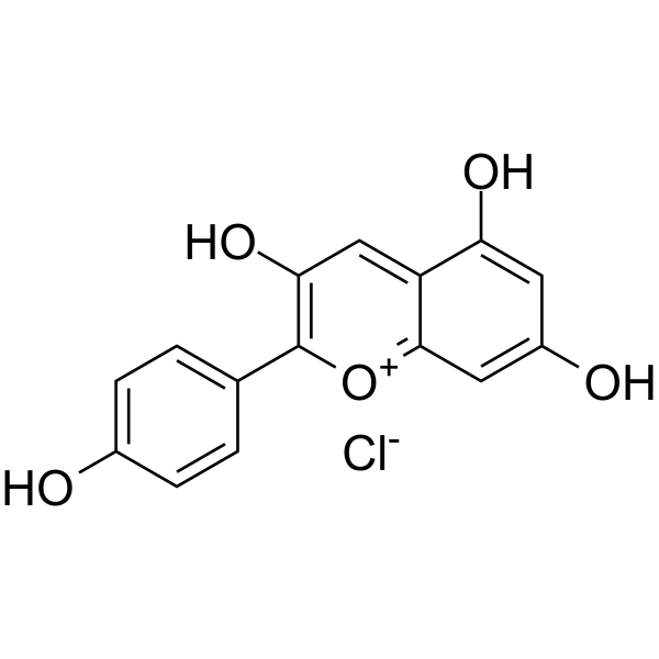
Pelargonidin chloride
Based on 1 publication(s) in Google Scholar
Pelargonidin chloride is an anthocyanidin and also is a scavenger of nitric oxide radical and has antioxidant activities. Pelargonidin inhibits cell viability and induces cell cycle arrest at sub-G1 phase. Pelargonidin chloride increases the mRNA and protein expression of HO-1, NQO1, Nrf2. Pelargonidin chloride improves Aβ-induced memory and learning impairment.
For research use only. We do not sell to patients.
- Purity : 98.52%
- CAS No.: 134-04-3
- Formula: C15H11ClO5
- Molecular Weight:306.70
-
Storage:
-20°C, sealed storage, away from moisture
* In solvent : -80°C, 6 months; -20°C, 1 month (sealed storage, away from moisture)

To place orders, for customer services and technical support, please contact: MedChemExpress USA
Tel: 609-228-6898 E-mail: [email protected] [email protected]


-
 Biological Activity
-
Chemical Information
-
Solvent & Solubility
- Protocols
- Purity & Documentation
- References
-
 Help & FAQs


-
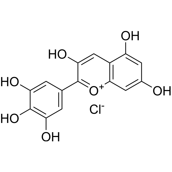
HY-N2409
Delphinidin chlorideAnthocyanidin -
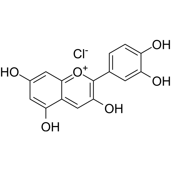
HY-N0499
Cyanidin ChlorideAntioxidant -
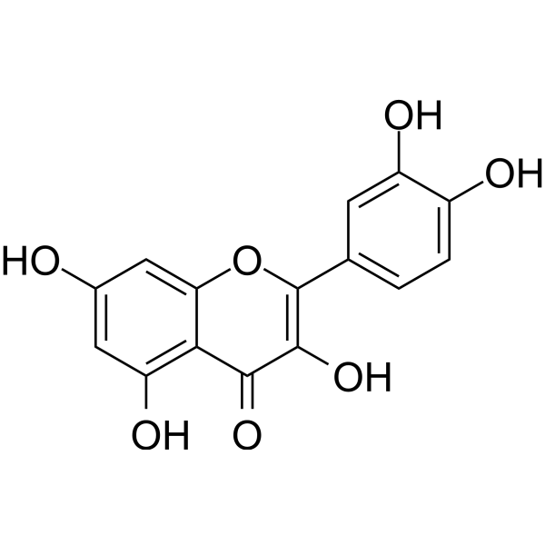
HY-18085
QuercetinFlavonoid Antioxidant -
HY-B0166
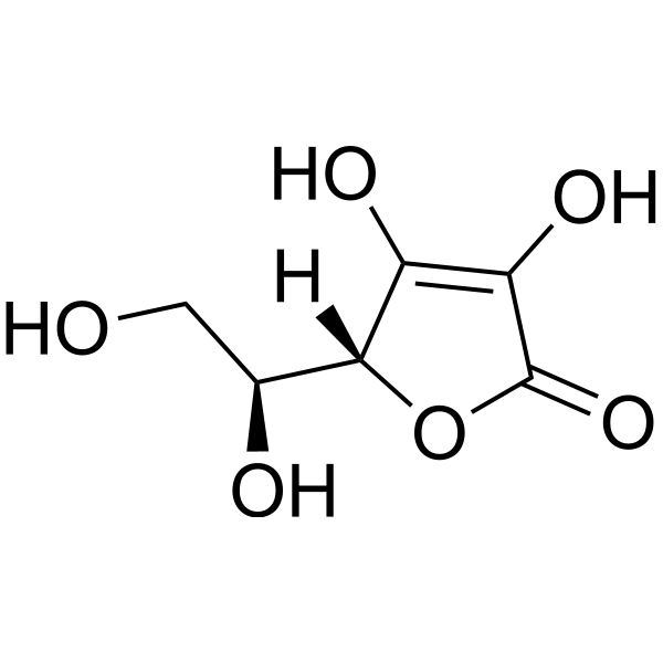
L-Ascorbic acidAntioxidant -

HY-15371
ForskolinAdenylate Cyclase Activator -
HY-N0162
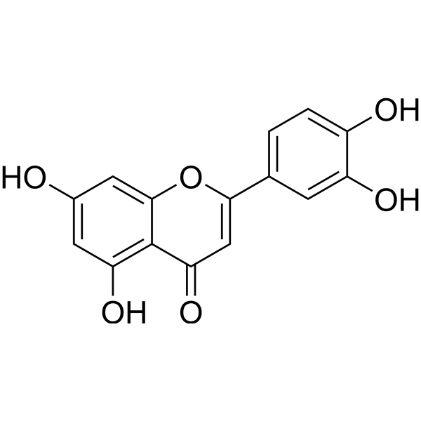
LuteolinNrf2 Inhibitor -
HY-126410
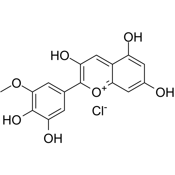
Petunidin chloridePEN Inhibitor -
HY-14596
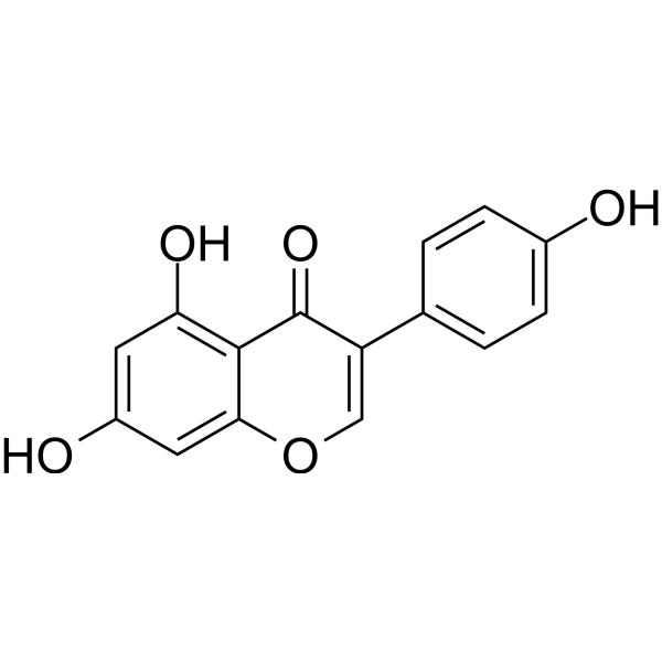
GenisteinAutophagy Inducer -
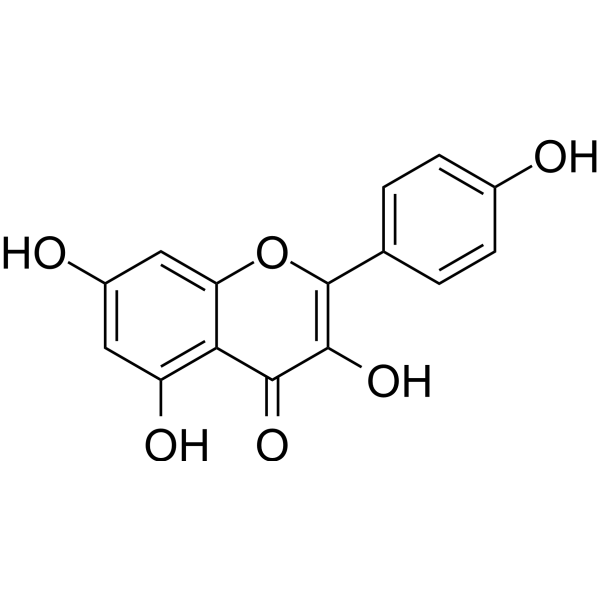
HY-14590
KaempferolEstrogen Receptor α Inhibitor -
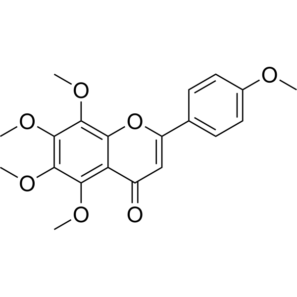
HY-N0133
TangeretinApoptosis Inducer -
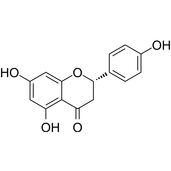
HY-N0100
NaringeninFlavonoid -
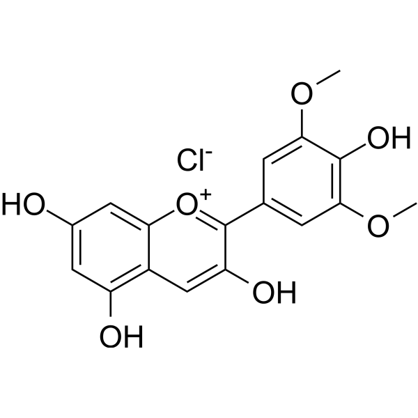
HY-122496
Malvidin chlorideApoptosis
Publications Citing Use of MedChemExpress (MCE) Pelargonidin chloride
More

Biological Activity
Description
In Vitro
Pelargonidin chloride (10-100 µM; 1, 3, 5 days) inhibits cell viability in a time- and dose-dependent manner in JB6 P+ cells[2].
Pelargonidin chloride (10, 30, 50 µM; 3 days) induces cell cycle arrest at sub-G1 phase in a dose-dependent manner[2].
Pelargonidin chloride (0,10, 30, 50 µM; 5 days) increases the mRNA and protein expression of HO-1, NQO1, Nrf2 in a dose-dependent manner[2].
MedChemExpress (MCE) has not independently confirmed the accuracy of these methods. They are for reference only. Further protocols information, click here.
-
Cell Line:JB6 P+ cells
-
Concentration:10, 30, 50, 70, 100 μM
-
Incubation Time:1, 3, 5 days
-
Result:Showed cytotoxicity in a time- and dose-dependent manner.
-
Cell Line:JB6 P+ cells
-
Concentration:10, 30, 50 µM
-
Incubation Time:3 days
-
Result:Induced cell cycle arrest at sub-G1 phase in a dose-dependent manner.
-
Cell Line:JB6 P+ cells
-
Concentration:0,10, 30, 50 µM
-
Incubation Time:6h, 5 days
-
Result:Promoted the mRNA expression of HO-1 and NQO1 after 6 hours and 5 days.
In Vivo
MedChemExpress (MCE) has not independently confirmed the accuracy of these methods. They are for reference only.
-
Animal Model:250-300 g, 12 weeks Wistar rats (Aβ 25-35 (1 µg/µL for 5 μL, injectes bilaterally into the hippocampi)[3]
-
Dosage:3 mg/kg
-
Administration:IP; once
-
Result:Significant decreased in escape latency and traveled distance in comparison to the Aβ-treated group, increased the antioxidant power of hippocampus homogenate.


Chemical Information
-
CAS No. 134-04-3
-
Appearance Solid
-
Molecular Weight 306.70
-
Formula C15H11ClO5
-
Color Brown to reddish brown
-
SMILES
OC1=CC=C(C2=C(O)C=C3C(C=C(O)C=C3O)=[O+]2)C=C1.[Cl-]
-
Structure Classification
-
Initial Source
-
Shipping
Room temperature in continental US; may vary elsewhere.
-
Storage
-20°C, sealed storage, away from moisture
* In solvent : -80°C, 6 months; -20°C, 1 month (sealed storage, away from moisture)


Publications (1)
-
Journal Impact Factor
-
Most Recent
-
Food Chem
Flavonoid-mediated metabolic underpinning quality variation in red bud-sport pear mutants. [Abstract]2025 Oct 15:489:144992. PMID: 40466530


Solvent & Solubility
In Vitro:
DMSO : 50 mg/mL (163.03 mM; Need ultrasonic; Hygroscopic DMSO has a significant impact on the solubility of product, please use newly opened DMSO)
Please refer to the solubility information to select the appropriate solvent. Once prepared, please aliquot and store the solution to prevent product inactivation from repeated freeze-thaw cycles.
Storage method and period of stock solution: -80°C, 6 months; -20°C, 1 month (sealed storage, away from moisture). When stored at -80°C, please use it within 6 months. When stored at -20°C, please use it within 1 month.
Please refer to the solubility information to select the appropriate solvent. Once prepared, please aliquot and store the solution to prevent product inactivation from repeated freeze-thaw cycles.
Storage method and period of stock solution: -80°C, 6 months; -20°C, 1 month (sealed storage, away from moisture). When stored at -80°C, please use it within 6 months. When stored at -20°C, please use it within 1 month.
Concentration (start) × Volume (start) = Concentration (final) × Volume (final)

Protocols
-
RNA extraction experimental
By lysing cells, releasing RNA, and removing impurities such as proteins and DNA, high-purity RNA products are finally obtained. The commonly used traditional method is the guanidine isothiocyanate/phenol/chloroform method (Trizol), which is suitable for a variety of animal materials including animal tissues, microorganisms, cultured cells, etc., and most plant materials.
-
Western Blot
Western blotting (WB) is a commonly used experimental method in molecular biology, biochemistry, and immunogenetics for identifying and quantifying target proteins. It combines gel electrophoresis with immunoassay, enabling researchers to analyze protein expression, post-translational modifications, and molecular weight.
-
Cell Cytotoxicity Assay
Cytotoxicity assays are usually based on the assessment of cell membrane damage, which can also be indirectly detected by measuring cell viability. Detection methods include MTT assay, CKK-8 assay, LDH assay and ATP assay, etc.
-
Mammalian live/dead viability and cytotoxicity staining
Live/dead viability and cytotoxicity staining assays are based on the simultaneous detection of intracellular esterase activity in metabolically active (viable) cells and membrane integrity loss in non-viable cells. In commonly used dual-staining approaches, membrane-permeant fluorogenic substrates are converted by intracellular esterases into fluorescent products in live cells, while impermeant DNA-binding dyes selectively enter cells with compromised plasma membranes and label nucleic acids in dead or dying cells, enabling discrimination between viable and non-viable populations by fluorescence microscopy or flow cytometry.
-
Flow cytometric DNA-content cell-cycle staining
Flow cytometric DNA-content cell-cycle staining measures the fluorescence intensity of DNA-bound fluorochromes in single cells or nuclei to estimate DNA content distributions, allowing assignment of populations to G0/G1, S, and G2/M phases by DNA histogram deconvolution. Propidium iodide (PI) intercalates into DNA, and PI fluorescence is proportional to cellular DNA content when staining is performed under conditions that make DNA accessible and minimize non-DNA signal. Cells with G2/M DNA content are expected to show approximately twice the fluorescence intensity of G0/G1 cells, while S-phase cells occupy intermediate fluorescence values. PI-based DNA-content analysis can also detect cells with fractional DNA content, often reported as sub-G1, when DNA fragmentation and extraction during staining reduce retained DNA signal in apoptotic cells. DAPI is an alternative DNA fluorochrome for univariate DNA-content analysis, while bivariate approaches combining DNA content with proliferation
-
BrdU Incorporation Assay
Bromodeoxyuridine (BrdU) incorporation assay is based on the principle that BrdU, a thymidine analog, is incorporated into newly synthesized DNA during the S phase of the cell cycle, thereby serving as a marker of DNA replication and cellular proliferation. Incorporated BrdU can be detected using anti-BrdU antibodies following DNA denaturation, enabling visualization or quantification of proliferating cells through immunochemical detection methods such as immunofluorescence or immunohistochemistry.
-
CCK-8/WST-8 Cell Proliferation Assay
The CCK-8/WST-8 assay is based on the reduction of the water-soluble tetrazolium salt WST-8 to a water-soluble formazan product by cellular dehydrogenases in metabolically active cells, where the generated formazan amount is proportional to the number of living cells and is quantified by measuring absorbance in the visible range, providing a colorimetric readout for cell viability and proliferation assessment. This class of tetrazolium-based assays improves upon earlier MTT-based systems by producing a water-soluble formazan, eliminating the need for organic solubilization steps and enabling direct spectrophotometric measurement in culture medium.
-
Protocol for Cell Cycle
Cell-cycle analysis by flow cytometry measures DNA content in single cells to estimate the fraction of cells in G0/G1, S, and G2/M phases. Propidium iodide intercalates into DNA, and after RNA removal with RNase, fluorescence intensity reflects cellular DNA content: 2N cells are assigned to G0/G1, cells between 2N and 4N to S phase, and 4N cells to G2/M. DNA-content analysis alone cannot reliably separate G0 from G1 or G2 from M. Ki-67 can distinguish quiescent G0 cells from cycling cells, EdU or BrdU incorporation marks active DNA synthesis in S phase, and phospho-histone H3 staining identifies mitotic cells within the 4N population.
-
MTT Cell Proliferation Assay
The MTT assay is a colorimetric endpoint assay for estimating viable cell number, cell growth, cytotoxicity, or cell activation in cultured mammalian cells. Living cells reduce the yellow tetrazolium salt MTT into purple/blue formazan, while dead cells do not generate the same signal; the resulting color can be quantified with a multiwell spectrophotometer. MTT reduction is commonly interpreted as a readout of metabolic activity that often correlates with viable cell number, but it should not be treated as a direct cell-counting method unless the assay is optimized for the cell type and experimental condition. Studies show that MTT reduction can involve mitochondrial and non-mitochondrial reducing systems, and formazan may accumulate in intracellular lipid droplets rather than simply marking mitochondria.
-
Cell Viability Determination by MTT Colorimetric Assay
The following protocol uses the MTT colorimetric assay as a classic literature-established method for assessing cell viability/metabolic activity in cultured mammalian cells. MTT[3-(4,5-dimethylthiazol-2-yl)-2,5-diphenyltetrazolium bromide] is reduced by metabolically active cells to a colored formazan product; the amount of formazan is quantified spectrophotometrically and provides an indirect measure of metabolically active viable cells. Importantly, MTT reduction reflects cellular oxidoreductase/metabolic activity rather than an absolute direct count of living cells, so changes in cellular metabolism can alter the signal independently of cell number.
-
Protocol For Protein Expression And Purification
Recombinant protein expression in Escherichia coli followed by purification of a His-tagged soluble protein by immobilized metal affinity chromatography (IMAC), with optional MBP fusion and TEV tag removal when the construct includes these elements. The biological readout is production of the encoded target protein, detected as an inducible band at the expected molecular mass by SDS-PAGE and quantified by total protein assay or chromatographic absorbance; the purification readout is enrichment of the target protein in elution fractions after selective binding of polyhistidine residues to immobilized Ni2+/metal-chelate resin and elution by imidazole-containing buffer. Expression is driven by an inducible bacterial expression system, commonly T7/lac-based, in which IPTG or lactose/auto-induction activates transcription and translation of the cloned gene; lower induction temperature, lower inducer concentration, induction timing, and solubility-enhancing fusion tags can influence the frac
-
Alzheimer’s Disease Modeling
Alzheimer’s Disease (AD) is a neurodegenerative disorder characterized by a progressive decline in cognitive functions and loss of specific types of neurons and synapses. Alzheimer's symptoms can be simulated in mice by injecting drugs (such as Aβ) or genetically modified.


Purity & Documentation
-
Data Sheet (287 KB)
-
SDS (393 KB)
- English - EN (393 KB)
- Français - FR (393 KB)
- Deutsch - DE (393 KB)
- Norwegian - NO (393 KB)
- Español - ES (393 KB)
- Swedish - SV (393 KB)
- Italian - IT (393 KB)
- Korean - KR (393 KB)
- Portuguese - PT (393 KB)
-
Handling Instructions (2659 KB)
References
[1]. S A van Acke, et al. Flavonoids as Scavengers of Nitric Oxide Radical.Biochem Biophys Res Commun. 1995 Sep 25;214(3):755-9. [Content Brief]
[2]. Soleimani Asl S, et al. Pelargonidin exhibits restoring effects against amyloid β-induced deficits in the hippocampus of male rats. Med J Islam Repub Iran. 2019 Dec 12;33:135. [Content Brief]
[3]. Li S, et al. Pelargonidin reduces the TPA induced transformation of mouse epidermal cells -potential involvement of Nrf2 promoter demethylation. Chem Biol Interact. 2019 Aug 25;309:108701. [Content Brief]


Complete Stock Solution Preparation Table
Please refer to the solubility information to select the appropriate solvent. Once prepared, please aliquot and store the solution to prevent product inactivation from repeated freeze-thaw cycles.
Storage method and period of stock solution: -80°C, 6 months; -20°C, 1 month (sealed storage, away from moisture). When stored at -80°C, please use it within 6 months. When stored at -20°C, please use it within 1 month.
| Optional Solvent | Concentration Solvent Mass | 1 mg | 5 mg | 10 mg | 25 mg |
|---|---|---|---|---|---|
| DMSO | 1 mM | 3.2605 mL | 16.3026 mL | 32.6052 mL | 81.5129 mL |
| 5 mM | 0.6521 mL | 3.2605 mL | 6.5210 mL | 16.3026 mL | |
| 10 mM | 0.3261 mL | 1.6303 mL | 3.2605 mL | 8.1513 mL | |
| 15 mM | 0.2174 mL | 1.0868 mL | 2.1737 mL | 5.4342 mL | |
| 20 mM | 0.1630 mL | 0.8151 mL | 1.6303 mL | 4.0756 mL | |
| 25 mM | 0.1304 mL | 0.6521 mL | 1.3042 mL | 3.2605 mL | |
| 30 mM | 0.1087 mL | 0.5434 mL | 1.0868 mL | 2.7171 mL | |
| 40 mM | 0.0815 mL | 0.4076 mL | 0.8151 mL | 2.0378 mL | |
| 50 mM | 0.0652 mL | 0.3261 mL | 0.6521 mL | 1.6303 mL | |
| 60 mM | 0.0543 mL | 0.2717 mL | 0.5434 mL | 1.3585 mL | |
| 80 mM | 0.0408 mL | 0.2038 mL | 0.4076 mL | 1.0189 mL | |
| 100 mM | 0.0326 mL | 0.1630 mL | 0.3261 mL | 0.8151 mL |


Pelargonidin chloride Related Classifications
HY-W011370 Related Classifications
 Powered by Bioz