



PFI-90
Based on 1 publication(s) in Google Scholar
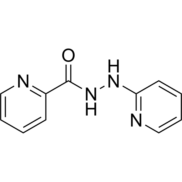
PFI-90 is a selective inhibitor of histone demethylase (KDM3B) that inhibits PAX3-FOXO1 action. PFI-90 induces apoptosis and myogenic differentiation, resulting in the cell death increased. PFI-90 has the potential for the antitumor activity. (patent WO2021101929A1).
For research use only. We do not sell to patients.
- Purity : 99.86%
- CAS No.: 53995-62-3
- Formula: C11H10N4O
- Molecular Weight:214.22
-
Storage:Powder -20°C, 3 years , 4°C, 2 years ; In solvent -80°C, 6 months , -20°C, 1 month

To place orders, for customer services and technical support, please contact: MedChemExpress USA
Tel: 609-228-6898 E-mail: [email protected] [email protected]


-
 Biological Activity
-
Chemical Information
-
Solvent & Solubility
- Protocols
- Purity & Documentation
- References
-
 Help & FAQs


-
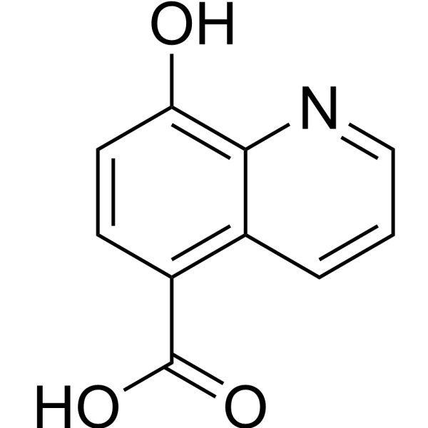
HY-12304
IOX12OG Oxygenases Inhibitor -
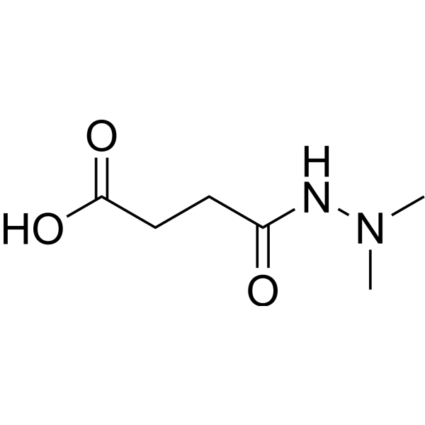
HY-13643
DaminozideKDM2/7 Inhibitor -
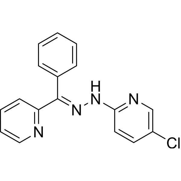
HY-13953
JIB-04Histone Demethylase Inhibitor -
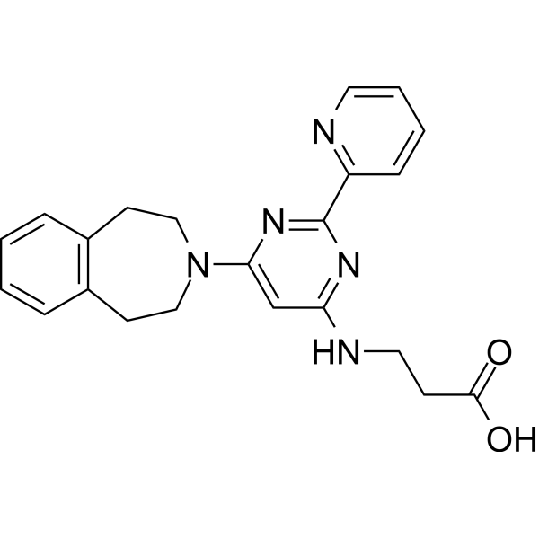
HY-15648
GSK-J1JMJD3/KDM6B Inhibitor -
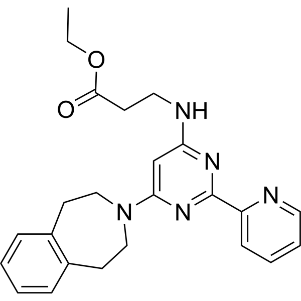
HY-15648B
GSK-J4JMJD3/UTX Inhibitor -
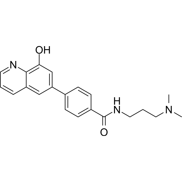
HY-12725
ML324JMJD2 Inhibitor -
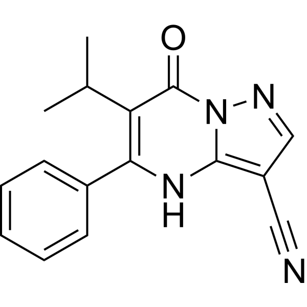
HY-100421
CPI-455KDM5 Inhibitor -
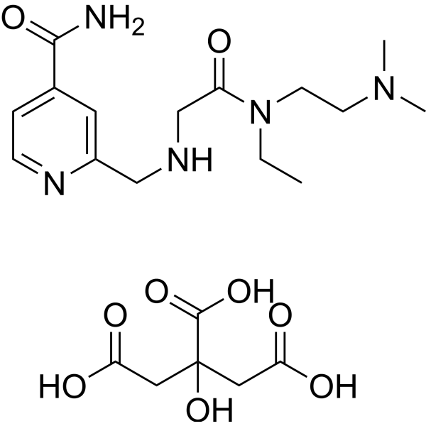
HY-102047B
KDOAM-25 citrateKDM5 Inhibitor -
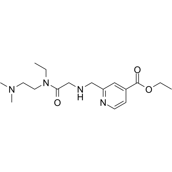
HY-120400
KDM5-C70KDM5 Inhibitor -
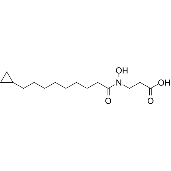
HY-107573
KDM2/7-IN-1KDM2/7 Inhibitor -
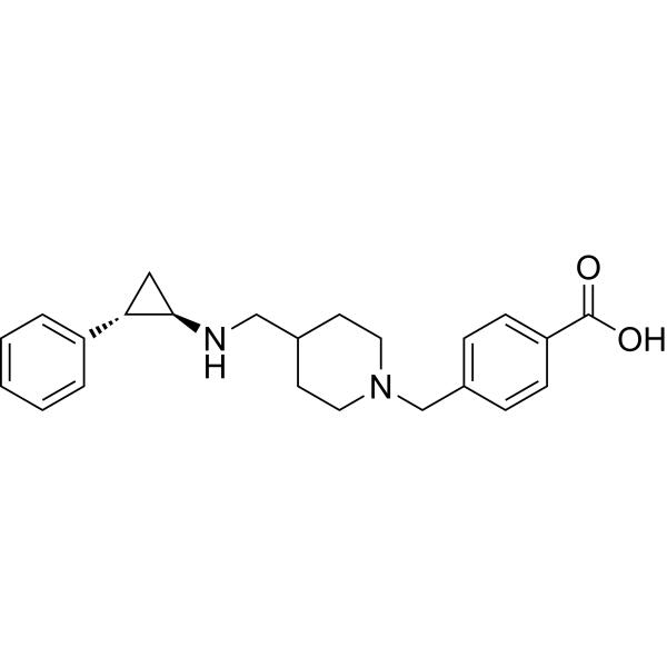
HY-18632
GSK2879552LSD1 Inhibitor -
HY-12635
SP2509LSD1 Antagonist
Publications Citing Use of MedChemExpress (MCE) PFI-90
More

Biological Activity
Description
IC50 & Target
|
KDM3 |
In Vitro
PFI-90 shows dose response in RH4, RH30, OSA-CL and TC-32 cells, with IC50s of 812, 3200, 1895 and 1113 nM, respectively[1].
?
PFI-90 (3 μΜ; 24 hours) increases apoptosis in RH4 and SCMC cells[1].
MedChemExpress (MCE) has not independently confirmed the accuracy of these methods. They are for reference only. Further protocols information, click here.
-
Cell Line:RH4 and SCMC cells
-
Concentration:3 μΜ
-
Incubation Time:24 hours
-
Result:Apoptosis increased in RH4 and SCMC cells.


Chemical Information
-
CAS No. 53995-62-3
-
Appearance Solid
-
Molecular Weight 214.22
-
Formula C11H10N4O
-
Color Light yellow to yellow
-
SMILES
O=C(C1=NC=CC=C1)NNC2=NC=CC=C2
-
Shipping
Room temperature in continental US; may vary elsewhere.
-
Storage
Powder -20°C 3 years 4°C 2 years In solvent -80°C 6 months -20°C 1 month


Publications (1)
-
Journal Impact Factor
-
Most Recent
-
Clin Exp Med
Corin: a dual inhibitor for KDM1A/HDAC1, suppresses hepatocellular carcinoma by triggering cuproptosis. [Abstract]2025 Nov 25;26(1):33. PMID: 41286164


Solvent & Solubility
In Vitro:
DMSO : 100 mg/mL (466.81 mM; Need ultrasonic; Hygroscopic DMSO has a significant impact on the solubility of product, please use newly opened DMSO)
Please refer to the solubility information to select the appropriate solvent. Once prepared, please aliquot and store the solution to prevent product inactivation from repeated freeze-thaw cycles.
Storage method and period of stock solution: -80°C, 6 months; -20°C, 1 month. When stored at -80°C, please use it within 6 months. When stored at -20°C, please use it within 1 month.
Please refer to the solubility information to select the appropriate solvent. Once prepared, please aliquot and store the solution to prevent product inactivation from repeated freeze-thaw cycles.
Storage method and period of stock solution: -80°C, 6 months; -20°C, 1 month. When stored at -80°C, please use it within 6 months. When stored at -20°C, please use it within 1 month.
Concentration (start) × Volume (start) = Concentration (final) × Volume (final)
In Vivo:
Select the appropriate dissolution method based on your experimental animal and administration route.
- For the following dissolution methods, please ensure to first prepare a clear stock solution using an In Vitro approach and then sequentially add co-solvents:
- To ensure reliable experimental results, the clarified stock solution can be appropriately stored based on storage conditions. As for the working solution for In Vivo experiments, it is recommended to prepare freshly and use it on the same day.
- The percentages shown for the solvents indicate their volumetric ratio in the final prepared solution. If precipitation or phase separation occurs during preparation, heat and/or sonication can be used to aid dissolution.
Add each solvent one by one: 10% DMSO 90% (20% SBE-β-CD in Saline)
Solubility: 2.5 mg/mL (11.67 mM); Clear solution; Need ultrasonic
This protocol yields a clear solution of 2.5 mg/mL.
Taking 1 mL working solution as an example, add 100 μL DMSO stock solution (25.0 mg/mL) to 900 μL 20% SBE-β-CD in Saline, and mix evenly.
Preparation of 20% SBE-β-CD in Saline (4°C, storage for one week): 2 g SBE-β-CD powder is dissolved in 10 mL Saline, completely dissolve until clear.
In Vivo Dissolution Calculator
Please enter the basic information of animal experiments:
-
-
-
-
Recommended: Prepare an additional quantity of animals to account for potential losses during experiments.
Please enter your animal formula composition:
-
%DMSO +
Recommended: Keep the proportion of DMSO in working solution below 2% if your animal is weak.
-
%+
-
+%Tween-80 + +
-
%Saline +
The co-solvents required include: DMSO, . All of co-solvents are available by MedChemExpress (MCE). , Tween 80. All of co-solvents are available by MedChemExpress (MCE).
Working solution concentration: 0.22 mg/mL
Method for preparing stock solution: mg drug dissolved in μL DMSO. Stock solution concentration: mg/mL.
1. Take μL DMSO stock solution;
2. Add μL .
μL , mix evenly;
3. Then add μL Tween 80, mix evenly;
4. Then add μL
Please ensure that the stock solution in the first step is dissolved to a clear state, and add co-solvents in sequence. You can use ultrasonic heating (ultrasonic cleaner, recommended frequency 20-40 kHz), vortexing, etc. to assist dissolution.

Protocols
-
Apoptosis
Apoptosis, also called programmed cell death, is generally characterized by distinct morphological characteristics.
-
TUNEL staining for apoptotic DNA fragmentation
TUNEL staining detects DNA strand breaks by using terminal deoxynucleotidyl transferase to add labeled nucleotides to exposed 3′-OH DNA termini, generating either microscopic staining in fixed cells or tissue sections, or fluorescence/cytometric signal in cell suspensions. TUNEL positivity reflects DNA fragmentation but should not be interpreted alone as definitive apoptosis, because TUNEL can also label necrotic, autolytic, mechanically damaged, or DNA-repair-associated DNA breaks.
-
Cell Cytotoxicity Assay
Cytotoxicity assays are usually based on the assessment of cell membrane damage, which can also be indirectly detected by measuring cell viability. Detection methods include MTT assay, CKK-8 assay, LDH assay and ATP assay, etc.
-
Mammalian live/dead viability and cytotoxicity staining
Live/dead viability and cytotoxicity staining assays are based on the simultaneous detection of intracellular esterase activity in metabolically active (viable) cells and membrane integrity loss in non-viable cells. In commonly used dual-staining approaches, membrane-permeant fluorogenic substrates are converted by intracellular esterases into fluorescent products in live cells, while impermeant DNA-binding dyes selectively enter cells with compromised plasma membranes and label nucleic acids in dead or dying cells, enabling discrimination between viable and non-viable populations by fluorescence microscopy or flow cytometry.
-
Annexin V plus membrane-impermeant dye apoptosis staining
Annexin V-based apoptosis assays rely on the detection of phosphatidylserine (PS) externalization from the inner leaflet of the plasma membrane to the outer leaflet, an early biochemical hallmark of apoptosis. Fluorescently labeled Annexin V binds PS in a calcium-dependent manner, enabling identification of early apoptotic cells by flow cytometry or fluorescence microscopy. When combined with a membrane-impermeant DNA-binding dye (e. g. , propidium iodide), this approach allows discrimination between viable (Annexin V−/dye−), early apoptotic (Annexin V+/dye−), and late apoptotic or necrotic (Annexin V+/dye+) cell populations by assessing membrane integrity and PS exposure.
-
Apoptosis Solutions
Apoptosis is a regulated, generally non-lytic cell-death pathway that removes unwanted, damaged, infected, or abnormal cells through coordinated morphological changes, caspase activation, DNA fragmentation, and membrane remodeling. The intrinsic apoptosis pathway is controlled mainly by mitochondrial outer membrane permeabilization, BCL-2 family proteins, cytochrome c release, apoptosome formation, caspase-9 activation, and downstream executioner caspase-3/7 activation. The extrinsic apoptosis pathway is initiated by death receptors such as Fas, TNFR, and TRAIL receptors, which recruit adaptor proteins and activate caspase-8 before engaging executioner caspases or mitochondrial amplification through BID cleavage. Apoptosis is linked to many phenotypes, including cancer cell killing, tissue homeostasis, immune regulation, neurodegeneration, infection response, and treatment-induced cytotoxicity; unresolved questions include how apoptosis interacts with necroptosis, pyroptosis, ferroptos
-
Subcutaneous Cell-Line-Derived Xenograft
Subcutaneous cell-line-derived xenograft (CDX) models are established by implanting cultured human cancer cell lines into immunodeficient mice, where the injected cells form localized tumors that can be monitored in vivo as a measure of tumorigenic potential, growth kinetics, and treatment response. These models are widely used in oncology research because they allow reproducible tumor formation and enable comparative assessment of tumor growth between different cell lines or genetic manipulations in a controlled in vivo microenvironment. Subcutaneous implantation of cancer cells in immunodeficient mice is a standard approach for evaluating tumor growth behavior and therapeutic response across multiple cancer types, including prostate, esophageal, pancreatic, and colon cancer models.


Purity & Documentation
-
Data Sheet (272 KB)
-
SDS (393 KB)
- English - EN (393 KB)
- Français - FR (393 KB)
- Deutsch - DE (393 KB)
- Norwegian - NO (393 KB)
- Español - ES (393 KB)
- Swedish - SV (393 KB)
- Italian - IT (393 KB)
- Korean - KR (393 KB)
- Portuguese - PT (393 KB)
-
Handling Instructions (2659 KB)
References


Complete Stock Solution Preparation Table
Please refer to the solubility information to select the appropriate solvent. Once prepared, please aliquot and store the solution to prevent product inactivation from repeated freeze-thaw cycles.
Storage method and period of stock solution: -80°C, 6 months; -20°C, 1 month. When stored at -80°C, please use it within 6 months. When stored at -20°C, please use it within 1 month.
| Optional Solvent | Concentration Solvent Mass | 1 mg | 5 mg | 10 mg | 25 mg |
|---|---|---|---|---|---|
| DMSO | 1 mM | 4.6681 mL | 23.3405 mL | 46.6810 mL | 116.7025 mL |
| 5 mM | 0.9336 mL | 4.6681 mL | 9.3362 mL | 23.3405 mL | |
| 10 mM | 0.4668 mL | 2.3340 mL | 4.6681 mL | 11.6702 mL | |
| 15 mM | 0.3112 mL | 1.5560 mL | 3.1121 mL | 7.7802 mL | |
| 20 mM | 0.2334 mL | 1.1670 mL | 2.3340 mL | 5.8351 mL | |
| 25 mM | 0.1867 mL | 0.9336 mL | 1.8672 mL | 4.6681 mL | |
| 30 mM | 0.1556 mL | 0.7780 mL | 1.5560 mL | 3.8901 mL | |
| 40 mM | 0.1167 mL | 0.5835 mL | 1.1670 mL | 2.9176 mL | |
| 50 mM | 0.0934 mL | 0.4668 mL | 0.9336 mL | 2.3340 mL | |
| 60 mM | 0.0778 mL | 0.3890 mL | 0.7780 mL | 1.9450 mL | |
| 80 mM | 0.0584 mL | 0.2918 mL | 0.5835 mL | 1.4588 mL | |
| 100 mM | 0.0467 mL | 0.2334 mL | 0.4668 mL | 1.1670 mL |


PFI-90 Related Classifications
HY-139348 Related Classifications
 Powered by Bioz