



Poly(ethylene glycol) diacrylate (MW 700)

Based on 1 Customer Validation
Poly(ethylene glycol) diacrylate (PEGDA) (MW 700) is a versatile polymer material that is widely used as a building material for hydrogel scaffolds, especially in the fields of tissue engineering and regenerative medicine. Poly(ethylene glycol) diacrylate (MW 700) mimics the extracellular matrix (ECM), providing support and growth space for cells.
For research use only. We do not sell to patients.
- Purity : 99.79%
- CAS No.: 26570-48-9
- Formula: (C2H4O)nC6H6O3
- Molecular Weight:700.00 (Average)
-
Storage:Pure form -20°C, 3 years ; In solvent -80°C, 6 months , -20°C, 1 month


To place orders, for customer services and technical support, please contact: MedChemExpress USA
Tel: 609-228-6898 E-mail: [email protected] [email protected]


-
 Biological Activity
-
Chemical Information
-
Solvent & Solubility
- Protocols
- Purity & Documentation
- References
-
 Help & FAQs


-

HY-W110542L
Poly(ethylene glycol) diacrylate (MW 3500)Polyethylene Glycol Derivative -

HY-W110542G
Poly(ethylene glycol) diacrylate (MW 6000)Polyethylene Glycol Derivative -

HY-W110542O
Poly(ethylene glycol) diacrylate (MW 575),MEHQ as inhibitorPolyethylene Glycol Derivative -

HY-W110542N
Poly(ethylene glycol) diacrylate (MW 250),MEHQ as inhibitorPolyethylene Glycol Derivative -

HY-W110542P
Poly(ethylene glycol) diacrylate (MW 4000), MEHQ as inhibitorPolyethylene Glycol Derivative -

HY-W110542D
Poly(ethylene glycol) diacrylate (MW 1000)Polyethylene Glycol Derivative -
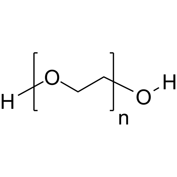
HY-Y0873
PEG300Neutral Polymer -
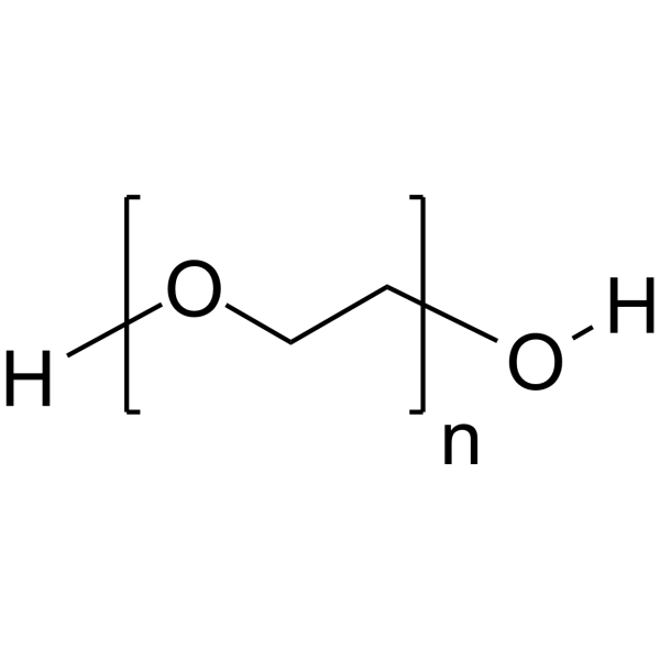
HY-Y0873A
PEG400 -

HY-W110542M
Poly(ethylene glycol) diacrylate (MW 20000)Polyethylene Glycol Derivative -
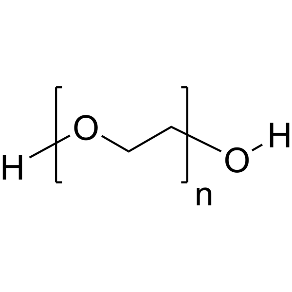
HY-Y0873J
PEG8000Biochemical reagent -

HY-W110542H
Poly(ethylene glycol) diacrylate (MW 10000)Polyethylene Glycol Derivative -
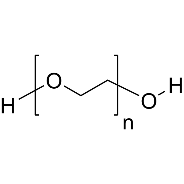
HY-Y0873C
PEG3350Neutral Polymer


Biological Activity
Description
In Vitro
Poly(ethylene glycol) diacrylate (MW 700) and cellulose nanofibers (CNF) jointly form a hydrogel scaffold. When the thickness of the scaffold reaches 0.4 mm, its surface is rich in micropores and the pore diameters are mostly between 20 and 50 μm, which is suitable for cell attachment[2].
Product Properties:
This product is readily soluble in water and can be prepared into hydrogel precursor solutions of varying concentrations. Adding the photoinitiator LAP (HY-44076) to this precursor solution triggers crosslinking upon illumination, yielding a hydrogel with a certain mechanical strength.
Instructions:
1. Dissolve this product at a concentration of 10-20 (w/w)% in pure water or phosphate buffer at room temperature, depending on the intended use. Add 0.1-0.3% (w/w)% LAP photoinitiator to prepare a gel precursor solution.
2. Add the above solution to the mold and refrigerate. Irradiation under 405 nm light for 30-60 seconds will form PEGDA hydrogel.
MedChemExpress (MCE) has not independently confirmed the accuracy of these methods. They are for reference only. Further protocols information, click here.


Chemical Information
-
CAS No. 26570-48-9
-
Appearance Liquid
-
Molecular Weight 700.00 (Average)
-
Formula (C2H4O)nC6H6O3
-
Color Colorless to light yellow
-
SMILES
O=C(C=C)OCCOC(C=C)=O.[n]
-
Synonyms
PEGDA (MW 700)
-
Shipping
Room temperature in continental US; may vary elsewhere.
-
Storage
Pure form -20°C 3 years In solvent -80°C 6 months -20°C 1 month


Solvent & Solubility
In Vitro:
DMSO : 200 mg/mL (Need ultrasonic; Hygroscopic DMSO has a significant impact on the solubility of product, please use newly opened DMSO)

Protocols
-
3D Hydrogel Synthetic Scaffold Culture
3D hydrogel synthetic scaffold culture embeds cells, spheroids, organoids, or tissue fragments inside a hydrated crosslinked polymer network so that cells receive matrix and cell-cell cues in three dimensions rather than from a flat plastic surface. A literature-supported model protocol is PEG-4MAL hydrogel culture, in which four-arm maleimide-terminated PEG is functionalized with cysteine-containing adhesive peptides such as RGD and crosslinked with protease-degradable peptides such as GPQ-W; this creates a defined, modular scaffold that supports human organoid generation and culture. The readouts are scaffold-supported growth, morphology, lumen formation, budding, viability, proliferation, lineage-marker expression, and matrix-dependent expansion or differentiation; reported assays include transmitted-light imaging, immunofluorescence, in situ hybridization, qRT-PCR, and rheological characterization.
-
Fibrosis/Collagen Morphometry
Fibrosis and collagen morphometry is based on the quantitative visualization of fibrillar collagen deposition in tissue sections using histochemical stains such as Sirius Red (Picrosirius Red) or Masson's trichrome, followed by image-based or polarization-enhanced analysis to estimate collagen proportional area as a surrogate of extracellular matrix accumulation during fibrotic remodeling. Sirius Red combined with polarized light microscopy enhances detection of collagen fibers due to birefringence properties, enabling more specific visualization of collagen type I and III fibrils compared to conventional bright-field histology, while whole-section or region-restricted digital morphometry reduces field-selection bias in fibrosis assessment. Alternative quantitative approaches include second harmonic generation (SHG) and two-photon excited fluorescence microscopy, which enable label-free detection of fibrillar collagen and have been validated against histological staining and biochemica
-
Collagen: Sirius Red Staining
Sirius Red or picrosirius red staining is a histochemical method for visualizing collagen-rich extracellular matrix in tissue sections, and collagen fibers are detected as red-stained structures under bright-field microscopy with enhanced birefringence under polarized light. Picrosirius red is useful for assessing total collagen organization, distribution, and fibrosis burden, but polarized color should not be interpreted as a definitive collagen type I versus type III readout because color is affected by fiber orientation, thickness, and packing.
-
ECM-Embedded Organoid (Matrigel/Dome) Culture
ECM-embedded organoid dome culture embeds epithelial stem cells, crypts, organoid fragments, or tumor-derived epithelial cells in a basement-membrane-like hydrogel such as Matrigel, allowing 3D growth, self-organization, lumen formation, budding or cystic morphogenesis, and lineage maintenance under defined niche-factor-containing medium. The primary readouts are organoid establishment efficiency, growth, morphology, passaging capacity, lineage-marker expression, and, when fluorescently labeled lines are used, microscopy- or flow-cytometry-based quantification of population behavior in 3D culture.
-
Matrigel/ECM Transwell Invasion Assay
The Matrigel/ECM Transwell invasion assay measures the ability of cells to move toward a chemoattractant while crossing an extracellular-matrix barrier placed on a porous membrane; therefore, the readout reflects both chemotactic motility and matrix invasion rather than migration alone. Matrigel is a basement-membrane-rich matrix derived from Engelbreth-Holm-Swarm mouse sarcoma and has been used as a reconstituted basement membrane barrier in chemoinvasion assays. The assay readout is generated by quantifying cells that reach the underside of the insert membrane or lower compartment after incubation, commonly by staining and counting invaded cells or by fluorescence-based quantification.
-
Matrigel Transwell/Boyden Chamber Invasion Assay
Matrigel Transwell/Boyden chamber invasion assay measures the ability of cells to degrade or traverse an extracellular matrix-coated porous membrane and move from an upper chamber toward a chemoattractant in a lower chamber. Invasion is distinguished from migration by coating the membrane with Matrigel or basement membrane matrix; uncoated inserts measure migration, while coated inserts require cells to cross an ECM barrier before reaching the underside of the membrane.


Purity & Documentation
-
Data Sheet (270 KB)
-
SDS (614 KB)
- English - EN (614 KB)
- Français - FR (614 KB)
- Deutsch - DE (614 KB)
- Norwegian - NO (614 KB)
- Español - ES (614 KB)
- Swedish - SV (614 KB)
- Italian - IT (614 KB)
- Korean - KR (614 KB)
- Portuguese - PT (614 KB)
-
Handling Instructions (2659 KB)
References
[2]. Huang L, et al. Antibacterial poly (ethylene glycol) diacrylate/chitosan hydrogels enhance mechanical adhesiveness and promote skin regeneration. Carbohydr Polym. 2019 Dec 1;225:115110. [Content Brief]
[3]. Sun D, et al. Tuning the Poisson's ratio of poly(ethylene glycol) diacrylate/cellulose nanofibril aerogel scaffold precisely for cultivation of bone marrow mesenchymal stem cell. J Biomed Mater Res A. 2023 Apr;111(4):502-513. [Content Brief]

Calculators
Concentration (start) × Volume (start) = Concentration (final) × Volume (final)


Poly(ethylene glycol) diacrylate (MW 700) Related Classifications
HY-W110542J Related Classifications
 Powered by Bioz