



PRDX1-IN-1
Based on 3 publication(s) in Google Scholar
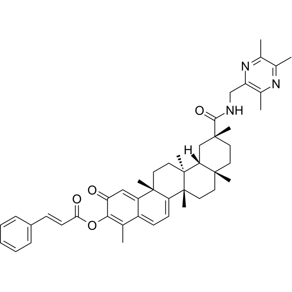
PRDX1-IN-1 is a selective inhibtor of PRDX1 with an IC50 value of 0.164 μM. PRDX1-IN-1 can be used in researches related to cancer.PRDX1-IN-1 promots intracellular ROS accumulation, and inhibits the proliferation, invasion and migration of cancer cells besides inducing apoptosis. PRDX1-IN-1 could be used in cancer research.
For research use only. We do not sell to patients.
- Purity : 98.04%
- CAS No.: 2996096-63-8
- Formula: C46H55N3O4
- Molecular Weight:713.95
-
Storage:Powder -20°C, 3 years , 4°C, 2 years ; In solvent -80°C, 6 months , -20°C, 1 month

To place orders, for customer services and technical support, please contact: MedChemExpress USA
Tel: 609-228-6898 E-mail: [email protected] [email protected]


-
 Biological Activity
-
Chemical Information
-
Solvent & Solubility
- Protocols
- Purity & Documentation
- References
-
 Help & FAQs


Publications Citing Use of MedChemExpress (MCE) PRDX1-IN-1
More

Biological Activity
Description
IC50 & Target
IC50:0.164μM(potent peroxiredoxin 1,PRDX1)[1]
Cellular Effect
|
Cell Line

|
Type | Value | Description | References |
|---|---|---|---|---|
| A549 | IC50 |
1.92 μM
Compound: 7e
|
Antiproliferative activity against human A549 cells assessed as inhibition of cell growth incubated for 48 hrs by MTT assay
Antiproliferative activity against human A549 cells assessed as inhibition of cell growth incubated for 48 hrs by MTT assay
|
[PMID: 37499289] |
| MDA-MB-231 | IC50 |
2.67 μM
Compound: 7e
|
Antiproliferative activity against human MDA-MB-231 cells assessed as inhibition of cell growth incubated for 48 hrs by MTT assay
Antiproliferative activity against human MDA-MB-231 cells assessed as inhibition of cell growth incubated for 48 hrs by MTT assay
|
[PMID: 37499289] |
| NCI-H1975 | IC50 |
1.99 μM
Compound: 7e
|
Antiproliferative activity against human NCI-H1975 cells assessed as inhibition of cell growth incubated for 48 hrs by MTT assay
Antiproliferative activity against human NCI-H1975 cells assessed as inhibition of cell growth incubated for 48 hrs by MTT assay
|
[PMID: 37499289] |
| SK-HEP1 | IC50 |
2.42 μM
Compound: 7e
|
Antiproliferative activity against human SK-HEP1 cells assessed as inhibition of cell growth incubated for 48 hrs by MTT assay
Antiproliferative activity against human SK-HEP1 cells assessed as inhibition of cell growth incubated for 48 hrs by MTT assay
|
[PMID: 37499289] |
In Vitro
PRDX1-IN-1 inhibites the proliferation activities of the human lung cancer cells A549, lung cancer cell lines (LTEP-a-2 and H1975), human breast cancer cell line (MDA-MB-231), human hepatoma cell line (SK-Hep-1) with the IC50 values of 1.92 μM, 2.93 μM, 1.99 μM, 2.67 μM, 2.42μM, respectively[1].
PRDX1-IN-1 (compound 7e)(2 μM or 4 μM, 24 h) promotes intracellular ROS accumulation, and inhibits the invasion and migration of human lung cancer cells A549[1].
PRDX1-IN-1 (2 μM or 4 μM, 24 h) induces the apoptosis of A549 cells[1].
PRDX1-IN-1 (2 μM or 4 μM, 6 h) suppresses the key signaling pathways (AKT and ERK) and promotes the expression of apoptosis-related proteins (cleaved caspase-3/8 and cleaved PARP) in A549 cells[1].
MedChemExpress (MCE) has not independently confirmed the accuracy of these methods. They are for reference only. Further protocols information, click here.
-
Cell Line:lung cancer cell lines (LTEP-a-2 and H1975), human breast cancer cell line (MDA-MB-231), human hepatoma cell line (SK-Hep-1)
-
Concentration:0.5–10 μM
-
Incubation Time:48 h
-
Result:Inhibited the proliferation activities of cancer cells A549, LTEP-a-2, H1975, MDA-MB-231, SK-Hep-1.
-
Cell Line:human lung cancer cells A549
-
Concentration:2 μM or 4 μM
-
Incubation Time:24 h
-
Result:Increased the ratio of the total number of early (annexin-V+/PI− ) and late (annexin-V+/PI+) apoptotic cells significantly.
-
Cell Line:A549 cell
-
Concentration:2 μM or 4 μM
-
Incubation Time:6 h
-
Result:Decreased the phosphorylation levels of PI3K, AKT, C-RAF and ERK.
-
Cell Line:A549 cell
-
Concentration:2 μM or 4 μM
-
Incubation Time:24 - 48 h
-
Result:Inhibted the cell matrigel and invasion.
In Vivo
MedChemExpress (MCE) has not independently confirmed the accuracy of these methods. They are for reference only.
-
Animal Model:C57BL/6J male mice injected with Lewis cell (lung cancer)[1]
-
Dosage:0.5 or 1 mg/kg
-
Administration:intraperitoneal injection (i.p.), every day for 19 days.
-
Result:Inhibited tumor growth, with the tumor growth inhibition (TGI) values of 77.47% and 69.89% in the groups of 1 mg/kg and 0.5 mg/kg, respectively.
Induced the changes in morphological characteristics of tumor cells, including cell agglutination, contraction, and nuclear chromatin marginalization.


Chemical Information
-
CAS No. 2996096-63-8
-
Appearance Solid
-
Molecular Weight 713.95
-
Formula C46H55N3O4
-
Color Light yellow to yellow
-
SMILES
O=C(OC1=C(C)C2=CC=C([C@](CC[C@]3(C)[C@@]4([H])C[C@@](C(NCC5=C(C)N=C(C)C(C)=N5)=O)(C)CC3)(C)[C@@]4(C)CC6)[C@]6(C)C2=CC1=O)/C=C/C7=CC=CC=C7
-
Shipping
Room temperature in continental US; may vary elsewhere.
-
Storage
Powder -20°C 3 years 4°C 2 years In solvent -80°C 6 months -20°C 1 month


Publications (3)
-
Journal Impact Factor
-
Most Recent
-
-
Free Radic Biol Med
Peroxiredoxin-1 aggravates hypoxia-induced renal injury by promoting inflammation through the TLR4/MAPK/NF-κB signaling pathway. [Abstract]2025 Aug 16:236:176-187. PMID: 40398686 -
Exp Cell Res
Exosomes derived from diabetic microenvironment-preconditioned mesenchymal stem cells ameliorate nonalcoholic fatty liver disease and inhibit pyroptosis of hepatocytes. [Abstract]2024 Nov 7;443(2):114325. PMID: 39521106


Solvent & Solubility
In Vitro:
DMSO : 100 mg/mL (140.07 mM; Need ultrasonic; Hygroscopic DMSO has a significant impact on the solubility of product, please use newly opened DMSO)
Please refer to the solubility information to select the appropriate solvent. Once prepared, please aliquot and store the solution to prevent product inactivation from repeated freeze-thaw cycles.
Storage method and period of stock solution: -80°C, 6 months; -20°C, 1 month. When stored at -80°C, please use it within 6 months. When stored at -20°C, please use it within 1 month.
Please refer to the solubility information to select the appropriate solvent. Once prepared, please aliquot and store the solution to prevent product inactivation from repeated freeze-thaw cycles.
Storage method and period of stock solution: -80°C, 6 months; -20°C, 1 month. When stored at -80°C, please use it within 6 months. When stored at -20°C, please use it within 1 month.
Concentration (start) × Volume (start) = Concentration (final) × Volume (final)
In Vivo:
Select the appropriate dissolution method based on your experimental animal and administration route.
- For the following dissolution methods, please ensure to first prepare a clear stock solution using an In Vitro approach and then sequentially add co-solvents:
- To ensure reliable experimental results, the clarified stock solution can be appropriately stored based on storage conditions. As for the working solution for In Vivo experiments, it is recommended to prepare freshly and use it on the same day.
- The percentages shown for the solvents indicate their volumetric ratio in the final prepared solution. If precipitation or phase separation occurs during preparation, heat and/or sonication can be used to aid dissolution.
Add each solvent one by one: 10% DMSO 40% PEG300 5% Tween-80 45% Saline
Solubility: 2.5 mg/mL (3.50 mM); Clear solution; Need ultrasonic
This protocol yields a clear solution of 2.5 mg/mL.
Taking 1 mL working solution as an example, add 100 μL DMSO stock solution (25.0 mg/mL) to 400 μL PEG300, and mix evenly; then add 50 μL Tween-80 and mix evenly; then add 450 μL Saline to adjust the volume to 1 mL.
Preparation of Saline: Dissolve 0.9 g sodium chloride in ddH₂O and dilute to 100 mL to obtain a clear Saline solution.
In Vivo Dissolution Calculator
Please enter the basic information of animal experiments:
-
-
-
-
Recommended: Prepare an additional quantity of animals to account for potential losses during experiments.
Please enter your animal formula composition:
-
%DMSO +
Recommended: Keep the proportion of DMSO in working solution below 2% if your animal is weak.
-
%+
-
+%Tween-80 + +
-
%Saline +
The co-solvents required include: DMSO, . All of co-solvents are available by MedChemExpress (MCE). , Tween 80. All of co-solvents are available by MedChemExpress (MCE).
Working solution concentration: 0.22 mg/mL
Method for preparing stock solution: mg drug dissolved in μL DMSO. Stock solution concentration: mg/mL.
1. Take μL DMSO stock solution;
2. Add μL .
μL , mix evenly;
3. Then add μL Tween 80, mix evenly;
4. Then add μL
Please ensure that the stock solution in the first step is dissolved to a clear state, and add co-solvents in sequence. You can use ultrasonic heating (ultrasonic cleaner, recommended frequency 20-40 kHz), vortexing, etc. to assist dissolution.

Protocols
-
Apoptosis
Apoptosis, also called programmed cell death, is generally characterized by distinct morphological characteristics.
-
TUNEL staining for apoptotic DNA fragmentation
TUNEL staining detects DNA strand breaks by using terminal deoxynucleotidyl transferase to add labeled nucleotides to exposed 3′-OH DNA termini, generating either microscopic staining in fixed cells or tissue sections, or fluorescence/cytometric signal in cell suspensions. TUNEL positivity reflects DNA fragmentation but should not be interpreted alone as definitive apoptosis, because TUNEL can also label necrotic, autolytic, mechanically damaged, or DNA-repair-associated DNA breaks.
-
Cell Cytotoxicity Assay
Cytotoxicity assays are usually based on the assessment of cell membrane damage, which can also be indirectly detected by measuring cell viability. Detection methods include MTT assay, CKK-8 assay, LDH assay and ATP assay, etc.
-
Annexin V plus membrane-impermeant dye apoptosis staining
Annexin V-based apoptosis assays rely on the detection of phosphatidylserine (PS) externalization from the inner leaflet of the plasma membrane to the outer leaflet, an early biochemical hallmark of apoptosis. Fluorescently labeled Annexin V binds PS in a calcium-dependent manner, enabling identification of early apoptotic cells by flow cytometry or fluorescence microscopy. When combined with a membrane-impermeant DNA-binding dye (e. g. , propidium iodide), this approach allows discrimination between viable (Annexin V−/dye−), early apoptotic (Annexin V+/dye−), and late apoptotic or necrotic (Annexin V+/dye+) cell populations by assessing membrane integrity and PS exposure.
-
ROS/oxidative-stress fluorescent staining
ROS/oxidative-stress fluorescent staining uses cell-permeant fluorogenic probes that become fluorescent after oxidation inside cells or tissues; commonly used examples include DCFH-DA/DCFDA for broad cellular oxidant detection, DHE for superoxide-related signal detection, MitoSOX for mitochondrial superoxide-related signal detection, and CellROX probes for oxidative-stress-associated fluorescence readouts. The assay detects probe oxidation rather than a single ROS species unless the probe and analysis method have been chemically validated for that species. DCFH-DA enters cells, is deacetylated by intracellular esterases to DCFH, and produces fluorescent DCF after oxidation, so the readout is used as an operational measure of total cellular oxidative stress rather than a species-specific ROS measurement. DHE and MitoSOX can report superoxide-related oxidation, but red fluorescence alone can include non-specific ethidium-like oxidation products; HPLC or optimized spectral approaches are
-
Cell migration
Cell migration is a method that plays an important role in wound healing, cell differentiation, embryonic development, etc.
-
Apoptosis Solutions
Apoptosis is a regulated, generally non-lytic cell-death pathway that removes unwanted, damaged, infected, or abnormal cells through coordinated morphological changes, caspase activation, DNA fragmentation, and membrane remodeling. The intrinsic apoptosis pathway is controlled mainly by mitochondrial outer membrane permeabilization, BCL-2 family proteins, cytochrome c release, apoptosome formation, caspase-9 activation, and downstream executioner caspase-3/7 activation. The extrinsic apoptosis pathway is initiated by death receptors such as Fas, TNFR, and TRAIL receptors, which recruit adaptor proteins and activate caspase-8 before engaging executioner caspases or mitochondrial amplification through BID cleavage. Apoptosis is linked to many phenotypes, including cancer cell killing, tissue homeostasis, immune regulation, neurodegeneration, infection response, and treatment-induced cytotoxicity; unresolved questions include how apoptosis interacts with necroptosis, pyroptosis, ferroptos
-
Cell invasion
Cell invasion is the ability of cells to migrate from one area to another via the extracellular matrix. Cell invasion is the response of normal and cancer cells to chemical and mechanical stimuli. Before migrating to a new region, the extracellular matrix is degraded by proteases within the cell. Cell invasion often occurs during wound repair, vascularization and inflammation, abnormal tissue invasion, and tumor cell metastasis.
-
Cell Viability Determination by MTT Colorimetric Assay
The following protocol uses the MTT colorimetric assay as a classic literature-established method for assessing cell viability/metabolic activity in cultured mammalian cells. MTT[3-(4,5-dimethylthiazol-2-yl)-2,5-diphenyltetrazolium bromide] is reduced by metabolically active cells to a colored formazan product; the amount of formazan is quantified spectrophotometrically and provides an indirect measure of metabolically active viable cells. Importantly, MTT reduction reflects cellular oxidoreductase/metabolic activity rather than an absolute direct count of living cells, so changes in cellular metabolism can alter the signal independently of cell number.


Purity & Documentation
-
Data Sheet (278 KB)
-
SDS (251 KB)
- English - EN (251 KB)
- Français - FR (251 KB)
- Deutsch - DE (251 KB)
- Norwegian - NO (251 KB)
- Español - ES (251 KB)
- Swedish - SV (251 KB)
- Italian - IT (251 KB)
- Korean - KR (251 KB)
- Portuguese - PT (251 KB)
-
Handling Instructions (2659 KB)
References


Complete Stock Solution Preparation Table
Please refer to the solubility information to select the appropriate solvent. Once prepared, please aliquot and store the solution to prevent product inactivation from repeated freeze-thaw cycles.
Storage method and period of stock solution: -80°C, 6 months; -20°C, 1 month. When stored at -80°C, please use it within 6 months. When stored at -20°C, please use it within 1 month.
| Optional Solvent | Concentration Solvent Mass | 1 mg | 5 mg | 10 mg | 25 mg |
|---|---|---|---|---|---|
| DMSO | 1 mM | 1.4007 mL | 7.0033 mL | 14.0066 mL | 35.0165 mL |
| 5 mM | 0.2801 mL | 1.4007 mL | 2.8013 mL | 7.0033 mL | |
| 10 mM | 0.1401 mL | 0.7003 mL | 1.4007 mL | 3.5016 mL | |
| 15 mM | 0.0934 mL | 0.4669 mL | 0.9338 mL | 2.3344 mL | |
| 20 mM | 0.0700 mL | 0.3502 mL | 0.7003 mL | 1.7508 mL | |
| 25 mM | 0.0560 mL | 0.2801 mL | 0.5603 mL | 1.4007 mL | |
| 30 mM | 0.0467 mL | 0.2334 mL | 0.4669 mL | 1.1672 mL | |
| 40 mM | 0.0350 mL | 0.1751 mL | 0.3502 mL | 0.8754 mL | |
| 50 mM | 0.0280 mL | 0.1401 mL | 0.2801 mL | 0.7003 mL | |
| 60 mM | 0.0233 mL | 0.1167 mL | 0.2334 mL | 0.5836 mL | |
| 80 mM | 0.0175 mL | 0.0875 mL | 0.1751 mL | 0.4377 mL | |
| 100 mM | 0.0140 mL | 0.0700 mL | 0.1401 mL | 0.3502 mL |


PRDX1-IN-1 Related Classifications
HY-149394 Related Classifications
 Powered by Bioz