



Hematoxylin-eosin staining is a routine histological method that stains nuclei mainly blue-purple with hemalum and stains cytoplasm, extracellular matrix, and many stromal components pink with eosin, allowing tissue architecture, cell morphology, necrosis, inflammation, fibrosis, tumor growth pattern, and treatment-associated injury to be evaluated by light microscopy. In cancer cells, primary neurons, mouse tumor models, intestinal organoids, inflammatory macrophage preparations, and drug-screening tissues, H&E is a morphology assay rather than a molecular assay; it should be interpreted with complementary molecular or immunostaining assays when the biological question concerns specific proteins, RNA levels, ferroptosis, mitophagy, or immune phenotypes.
Hematoxylin-Eosin (H&E) Staining
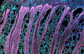
Hematoxylin-eosin (H&E) staining is a commonly used staining method for tissue sections. First, tissue sections are stained with hematoxylin, which stains cell nuclei a dark blue. Next, staining with eosin makes the cytoplasm and collagen fibers appear red. This staining method is widely used in the fields of pathology, histology and anatomy, allowing researchers to more clearly observe and distinguish tissue structures under the microscope, and provides basic histological information for disease diagnosis and research.
Related Experimental Schemes


