



Pyrromethene 597

Based on 1 Customer Validation
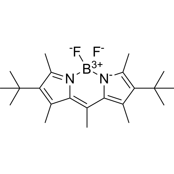
Pyrromethene 597 is a temperature-sensitive BODIPY-class laser fluorescent dye whose fluorescence anisotropy decreases with increasing temperature, Ex/Em =527/557 nm. Pyrromethene 597 can serve as a temperature probe in solution and as a whispering gallery mode (WGM) microlaser gain medium. Pyrromethene 597 can be doped into photoresists for in situ fabrication of microlaser devices within living cells. Pyrromethene 597 can be used in fluorescence imaging and microlaser-related research.
For research use only. We do not sell to patients.
- Purity : 99.67%
- CAS No.: 137829-79-9
- Formula: C22H33BF2N2
- Molecular Weight:374.32
-
Storage:
4°C, protect from light
* In solvent : -80°C, 6 months; -20°C, 1 month (protect from light)

To place orders, for customer services and technical support, please contact: MedChemExpress USA
Tel: 609-228-6898 E-mail: [email protected] [email protected]


-
 Biological Activity
-
Chemical Information
- Protocols
- Purity & Documentation
- References
-
 Help & FAQs


-
HY-D0082
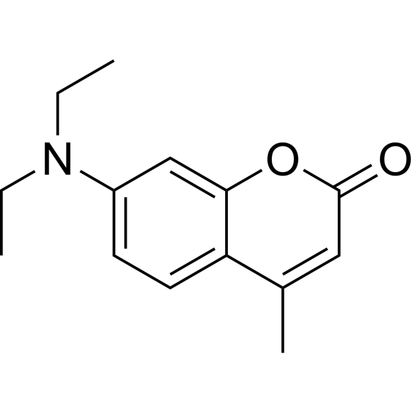
7-Diethylamino-4-methylcoumarinLaser Dye -
HY-W090090
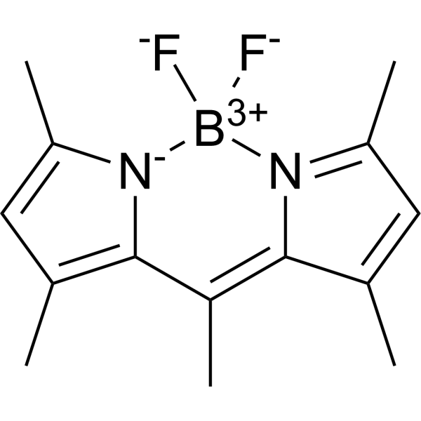
BODIPY 493/503Neutral Lipid Dye -

HY-DY1004
BODIPY 493/503 (solution)Neutral Lipid Dye -

HY-W248121
Pyrromethene 567BODIPY Dye -
HY-W040258
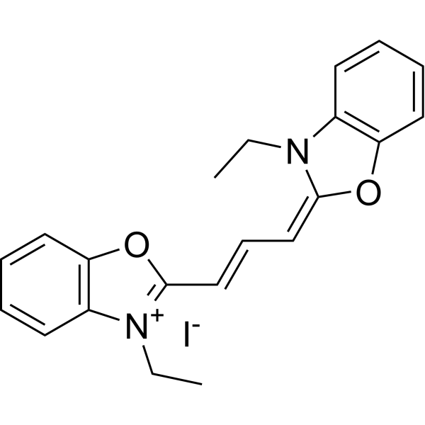
3,3'-Diethyloxacarbocyanine iodideMicroviscosity Fluorescent Probe -

HY-W072992
Iron(II) phthalocyanineMetal-organic Framework -

HY-108362
Iron(III) mesotetraphenylporphine chlorideMetal-organic Framework -

HY-W073017
Copper(II) Phthalocyanine (purified by sublimation)Metal-organic Framework


Biological Activity
Description
In Vitro
Pyrromethene 597 exhibits temperature-dependent fluorescence anisotropy in DMSO/methanol solution, with dr/dT = -1.39 × 10⁻⁴ K-1, indicating that it is a useful temperature probe[1].
Pyrromethene 597-doped IP-n162 photoresist (5 s UV LED polymerization) forms whispering-gallery-mode microlasers as small as 9 μm in diameter inside living HeLa cells[2].
MedChemExpress (MCE) has not independently confirmed the accuracy of these methods. They are for reference only. Further protocols information, click here.


Chemical Information
-
CAS No. 137829-79-9
-
Appearance Solid
-
Molecular Weight 374.32
-
Formula C22H33BF2N2
-
Color Orange to red
-
SMILES
[F-][B+3]1([N]2=C(C)C(C(C)(C)C)=C(C)C2=C(C)C3=C(C)C(C(C)(C)C)=C(C)[N-]31)[F-]
-
Shipping
Room temperature in continental US; may vary elsewhere.
-
Storage
4°C, protect from light
* In solvent : -80°C, 6 months; -20°C, 1 month (protect from light)

Protocols
-
Bioluminescent/Fluorescent Imaging Xenograft
Bioluminescent and fluorescent imaging xenograft models use tumor cells engineered to express optical reporters so tumor engraftment, growth, dissemination, and treatment response can be monitored longitudinally in living animals and validated ex vivo. Bioluminescence imaging usually measures luciferase activity after substrate administration and is commonly used as a surrogate for viable reporter-expressing tumor burden, while fluorescence imaging measures reporter or probe emission and can support tumor localization, ex vivo confirmation, or complementary multimodal analysis.


Purity & Documentation
-
Data Sheet (279 KB)
-
SDS (392 KB)
- English - EN (392 KB)
- Français - FR (392 KB)
- Deutsch - DE (392 KB)
- Norwegian - NO (392 KB)
- Español - ES (392 KB)
- Swedish - SV (392 KB)
- Italian - IT (392 KB)
- Korean - KR (392 KB)
- Portuguese - PT (392 KB)
-
Handling Instructions (2659 KB)
References

Calculators
Concentration (start) × Volume (start) = Concentration (final) × Volume (final)


Pyrromethene 597 Related Classifications
HY-W248115 Related Classifications
 Powered by Bioz