



Quercetin 3-(6″-caffeoylsophoroside)
Quercetin 3-(6″-caffeoylsophoroside) is an orally active α-amylase inhibitor, with an IC50 of 73.66 μg/mL. Quercetin 3-(6″-caffeoylsophoroside) presents in thehydro-methanolic extract of Cardamine hirsuta Linn. Quercetin 3-(6″-caffeoylsophoroside) shows the antidiabetic activities by oxidative stress reduction and α-amylase inhibition. Quercetin 3-(6″-caffeoylsophoroside) can be used for diabetes mellitus research.
For research use only. We do not sell to patients.
- CAS No.: 1032595-77-9
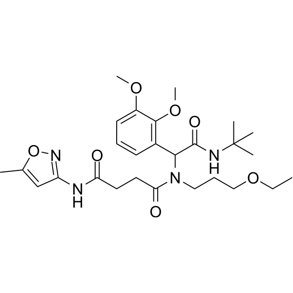
- Formula: C27H40N4O7
- Molecular Weight:532.63
-
Storage:
Please store the product under the recommended conditions in the Certificate of Analysis.

To place orders, for customer services and technical support, please contact: MedChemExpress USA
Tel: 609-228-6898 E-mail: [email protected] [email protected]


-
 Biological Activity
-
Chemical Information
- Protocols
- Purity & Documentation
- References
-
 Help & FAQs




Biological Activity
Description
In Vivo
Quercetin 3-(6″-caffeoylsophoroside) (125, 250, 500 mg/kg, p.o.) improves type 2 diabetes mellitus of SD rats by oxidative stress reduction and α-amylase inhibition[1].
MedChemExpress (MCE) has not independently confirmed the accuracy of these methods. They are for reference only.
-
Animal Model:FED HFD (45% fat) for 30 days to induce type 2 diabetes mellitus in SD rats[1]
-
Dosage:125, 250, 500 mg/kg
-
Administration:Oral gavage (p.o.)
-
Result:Significantly decreased the blood glucose level, improved biochemical parameters as well as oxidative stress by reduction of lipid peroxidation, and increased high-density lipoproteins with dose of 500mg/kg.
Enhanced activities of glutathione-s-transferase, glutathione, superoxide dismutase.
Restored cellular architecture in the histopathological examination.
-
Animal Model:Acute oral toxicity in 8-week male SD rats[1]
-
Dosage:50, 100, 500, 1000, and 2000 mg/kg
-
Administration:Oral gavage (p.o.)
-
Result:Safe for use in anti-diabetic research.


Chemical Information
-
CAS No. 1032595-77-9
-
Molecular Weight 532.63
-
Formula C27H40N4O7
-
SMILES
O=C(CCC(N(C(C1=C(OC)C(OC)=CC=C1)C(NC(C)(C)C)=O)CCCOCC)=O)NC2=NOC(C)=C2
-
Structure Classification
-
Initial Source
-
Shipping
Room temperature in continental US; may vary elsewhere.
-
Storage
Please store the product under the recommended conditions in the Certificate of Analysis.

Protocols
-
Cell Cytotoxicity Assay
Cytotoxicity assays are usually based on the assessment of cell membrane damage, which can also be indirectly detected by measuring cell viability. Detection methods include MTT assay, CKK-8 assay, LDH assay and ATP assay, etc.
-
ROS/oxidative-stress fluorescent staining
ROS/oxidative-stress fluorescent staining uses cell-permeant fluorogenic probes that become fluorescent after oxidation inside cells or tissues; commonly used examples include DCFH-DA/DCFDA for broad cellular oxidant detection, DHE for superoxide-related signal detection, MitoSOX for mitochondrial superoxide-related signal detection, and CellROX probes for oxidative-stress-associated fluorescence readouts. The assay detects probe oxidation rather than a single ROS species unless the probe and analysis method have been chemically validated for that species. DCFH-DA enters cells, is deacetylated by intracellular esterases to DCFH, and produces fluorescent DCF after oxidation, so the readout is used as an operational measure of total cellular oxidative stress rather than a species-specific ROS measurement. DHE and MitoSOX can report superoxide-related oxidation, but red fluorescence alone can include non-specific ethidium-like oxidation products; HPLC or optimized spectral approaches are
-
Research Protocol for Metabolic Diseases
AMP-activated protein kinase, AMPK, is a conserved cellular energy sensor that responds to reduced cellular energy status and coordinates metabolism by increasing ATP-generating catabolic pathways while suppressing ATP-consuming anabolic processes. In metabolic disease research, the AMPK pathway is experimentally relevant because it regulates hepatic lipid synthesis, fatty acid oxidation, glucose production, skeletal-muscle glucose disposal, mTORC1-linked biosynthesis, autophagy, mitochondrial homeostasis, and whole-body energy balance. The central pathway logic is that energy stress, metformin, exercise-like stimulation, or direct AMPK activators increase AMPKα Thr172 phosphorylation and downstream substrate phosphorylation, including ACC and RAPTOR. Phosphorylation of ACC suppresses lipogenesis and supports fatty acid oxidation, whereas phosphorylation of RAPTOR suppresses mTORC1 signaling and links cellular energy status to growth and protein synthesis control. The pathway is linked


Purity & Documentation
References

Calculators
Concentration (start) × Volume (start) = Concentration (final) × Volume (final)


Quercetin 3-(6″-caffeoylsophoroside) Related Classifications
HY-N11844 Related Classifications
 Powered by Bioz