



Queuine
Based on 1 publication(s) in Google Scholar
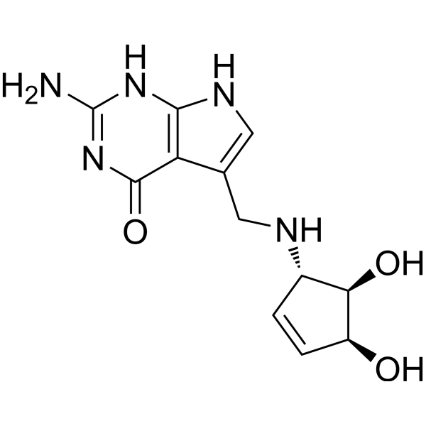
Queuine is a selective substrate for tRNA guanine transglycosylase (TGT) and can be incorporated into eukaryotic tRNA. Queuine promotes tRNA modification, affecting mitochondrial function and Warburg metabolic phenotype. If Queuine is deficient, aerobic glycolysis can be enhanced, oxidative phosphorylation can be inhibited, and Warburg metabolism can be promoted, accompanied by increased ammonia and lactate production and increased lactate dehydrogenase activity. Queuine can be used for autoimmune diseases (such as experimental models of multiple sclerosis) and cancer metabolic regulation, and its deficiency is associated with low tRNA modification in tumor cells.
For research use only. We do not sell to patients.
- Purity : 99.74%
- CAS No.: 72496-59-4
- Formula: C12H15N5O3
- Molecular Weight:277.28
-
Storage:Powder -20°C, 3 years , 4°C, 2 years ; In solvent -80°C, 6 months , -20°C, 1 month

To place orders, for customer services and technical support, please contact: MedChemExpress USA
Tel: 609-228-6898 E-mail: [email protected] [email protected]


-
 Biological Activity
-
Chemical Information
-
Solvent & Solubility
- Protocols
- Purity & Documentation
- References
-
 Help & FAQs



Publications Citing Use of MedChemExpress (MCE) Queuine
More

Biological Activity
Description
In Vitro
Queuine (1 μM; 5 days, supplemented every 24 h) reduces lactate dehydrogenase (LDH) activity in HeLa cells compared to the queuine group and limits glucose consumption and lactate production. In the absence of queuine, cells exhibits Warburg-type metabolic characteristics[2].
Queuine is a natural substrate of TGT and irreversibly replaces guanine (G34) in tRNA by human TGTase[2].
Queuine is a modified pyrrolopyrimidine nucleoside synthesized only by bacteria. It irreversibly replaces guanine (G34) in the tRNA anticodon loop by eukaryotic tRNA guanine transglycosylase (TGT) to form queuosine nucleotides, which specifically modify tRNA isoacceptors corresponding to aspartic acid, asparagine, histidine and tyrosine[3].
Queuine's biological activity is reflected in the following aspects: Queuine deficiency is a characteristic of rapidly proliferating cells (such as tumor cells and activated T cells in autoimmune diseases), and it can regulate cell metabolism by affecting the accuracy of tRNA translation, such as promoting Warburg-type metabolism (enhancing aerobic glycolysis and inhibiting oxidative phosphorylation)[3].
MedChemExpress (MCE) has not independently confirmed the accuracy of these methods. They are for reference only.
In Vivo
Queuine (30 mg/kg, 100 μL; intraperitoneal injection; once a day; 5 days) has no significant effect on the EAE score of the mouse experimental autoimmune encephalomyelitis (EAE) model, but reduces the tRNA low modification level of splenocyte CD4+ T cells[3].
TGT substrate design based on Queuine (such as 6-thioguanine 6TG, novel synthetic substrate NPPDAG) shows significant therapeutic effects in the mouse EAE model. NPPDAG selectively inhibits effector T cell proliferation and central nervous system infiltration through TGT-mediated tRNA modification, reduces the release of proinflammatory cytokines (such as IL-17 and IFN-γ), and completely reverses the clinical symptoms of EAE mice. It is ineffective in TGT-deficient mice, confirming the TGT-dependent therapeutic mechanism[3].
MedChemExpress (MCE) has not independently confirmed the accuracy of these methods. They are for reference only.


Chemical Information
-
CAS No. 72496-59-4
-
Appearance Solid
-
Molecular Weight 277.28
-
Formula C12H15N5O3
-
Color White to off-white
-
SMILES
O[C@@H]([C@H](C=C1)O)[C@H]1NCC2=CNC(NC(N)=N3)=C2C3=O
-
Structure Classification
-
Initial Source
eubacteria
-
Shipping
Room temperature in continental US; may vary elsewhere.
-
Storage
Powder -20°C 3 years 4°C 2 years In solvent -80°C 6 months -20°C 1 month


Publications (1)
-
Journal Impact Factor
-
Most Recent


Solvent & Solubility
In Vitro:
DMSO : 100 mg/mL (360.65 mM; Need ultrasonic; Hygroscopic DMSO has a significant impact on the solubility of product, please use newly opened DMSO)
Please refer to the solubility information to select the appropriate solvent. Once prepared, please aliquot and store the solution to prevent product inactivation from repeated freeze-thaw cycles.
Storage method and period of stock solution: -80°C, 6 months; -20°C, 1 month. When stored at -80°C, please use it within 6 months. When stored at -20°C, please use it within 1 month.
Please refer to the solubility information to select the appropriate solvent. Once prepared, please aliquot and store the solution to prevent product inactivation from repeated freeze-thaw cycles.
Storage method and period of stock solution: -80°C, 6 months; -20°C, 1 month. When stored at -80°C, please use it within 6 months. When stored at -20°C, please use it within 1 month.
Concentration (start) × Volume (start) = Concentration (final) × Volume (final)
In Vivo:
Select the appropriate dissolution method based on your experimental animal and administration route.
- For the following dissolution methods, please ensure to first prepare a clear stock solution using an In Vitro approach and then sequentially add co-solvents:
- To ensure reliable experimental results, the clarified stock solution can be appropriately stored based on storage conditions. As for the working solution for In Vivo experiments, it is recommended to prepare freshly and use it on the same day.
- The percentages shown for the solvents indicate their volumetric ratio in the final prepared solution. If precipitation or phase separation occurs during preparation, heat and/or sonication can be used to aid dissolution.
Add each solvent one by one: 10% DMSO 40% PEG300 5% Tween-80 45% Saline
Solubility: ≥ 2.5 mg/mL (9.02 mM); Clear solution
This protocol yields a clear solution of ≥ 2.5 mg/mL (saturation unknown).
Taking 1 mL working solution as an example, add 100 μL DMSO stock solution (25.0 mg/mL) to 400 μL PEG300, and mix evenly; then add 50 μL Tween-80 and mix evenly; then add 450 μL Saline to adjust the volume to 1 mL.
Preparation of Saline: Dissolve 0.9 g sodium chloride in ddH₂O and dilute to 100 mL to obtain a clear Saline solution.
Add each solvent one by one: 10% DMSO 90% (20% SBE-β-CD in Saline)
Solubility: ≥ 2.5 mg/mL (9.02 mM); Clear solution
This protocol yields a clear solution of ≥ 2.5 mg/mL (saturation unknown).
Taking 1 mL working solution as an example, add 100 μL DMSO stock solution (25.0 mg/mL) to 900 μL 20% SBE-β-CD in Saline, and mix evenly.
Preparation of 20% SBE-β-CD in Saline (4°C, storage for one week): 2 g SBE-β-CD powder is dissolved in 10 mL Saline, completely dissolve until clear.
In Vivo Dissolution Calculator
Please enter the basic information of animal experiments:
-
-
-
-
Recommended: Prepare an additional quantity of animals to account for potential losses during experiments.
Please enter your animal formula composition:
-
%DMSO +
Recommended: Keep the proportion of DMSO in working solution below 2% if your animal is weak.
-
%+
-
+%Tween-80 + +
-
%Saline +
The co-solvents required include: DMSO, . All of co-solvents are available by MedChemExpress (MCE). , Tween 80. All of co-solvents are available by MedChemExpress (MCE).
Working solution concentration: 0.22 mg/mL
Method for preparing stock solution: mg drug dissolved in μL DMSO. Stock solution concentration: mg/mL.
1. Take μL DMSO stock solution;
2. Add μL .
μL , mix evenly;
3. Then add μL Tween 80, mix evenly;
4. Then add μL
Please ensure that the stock solution in the first step is dissolved to a clear state, and add co-solvents in sequence. You can use ultrasonic heating (ultrasonic cleaner, recommended frequency 20-40 kHz), vortexing, etc. to assist dissolution.


Purity & Documentation
-
Data Sheet (278 KB)
-
SDS (252 KB)
- English - EN (252 KB)
- Français - FR (252 KB)
- Deutsch - DE (252 KB)
- Norwegian - NO (252 KB)
- Español - ES (252 KB)
- Swedish - SV (252 KB)
- Italian - IT (252 KB)
- Korean - KR (252 KB)
- Portuguese - PT (252 KB)
-
Handling Instructions (2659 KB)
References
[1]. Yifeng Yuan, et al. Discovery of novel bacterial queuine salvage enzymes and pathways in human pathogens. Proc Natl Acad Sci U S A. 2019 Sep 17;116(38):19126-19135. [Content Brief]
[2]. Hayes P, et al. Queuine Micronutrient Deficiency Promotes Warburg Metabolism and Reversal of the Mitochondrial ATP Synthase in Hela Cells. Nutrients. 2020 Mar 24;12(3):871. [Content Brief]
[3]. Varghese S, et al. In vivo modification of tRNA with an artificial nucleobase leads to full disease remission in an animal model of multiple sclerosis. Nucleic Acids Res. 2017 Feb 28;45(4):2029-2039. [Content Brief]


Complete Stock Solution Preparation Table
Please refer to the solubility information to select the appropriate solvent. Once prepared, please aliquot and store the solution to prevent product inactivation from repeated freeze-thaw cycles.
Storage method and period of stock solution: -80°C, 6 months; -20°C, 1 month. When stored at -80°C, please use it within 6 months. When stored at -20°C, please use it within 1 month.
| Optional Solvent | Concentration Solvent Mass | 1 mg | 5 mg | 10 mg | 25 mg |
|---|---|---|---|---|---|
| DMSO | 1 mM | 3.6065 mL | 18.0323 mL | 36.0646 mL | 90.1616 mL |
| 5 mM | 0.7213 mL | 3.6065 mL | 7.2129 mL | 18.0323 mL | |
| 10 mM | 0.3606 mL | 1.8032 mL | 3.6065 mL | 9.0162 mL | |
| 15 mM | 0.2404 mL | 1.2022 mL | 2.4043 mL | 6.0108 mL | |
| 20 mM | 0.1803 mL | 0.9016 mL | 1.8032 mL | 4.5081 mL | |
| 25 mM | 0.1443 mL | 0.7213 mL | 1.4426 mL | 3.6065 mL | |
| 30 mM | 0.1202 mL | 0.6011 mL | 1.2022 mL | 3.0054 mL | |
| 40 mM | 0.0902 mL | 0.4508 mL | 0.9016 mL | 2.2540 mL | |
| 50 mM | 0.0721 mL | 0.3606 mL | 0.7213 mL | 1.8032 mL | |
| 60 mM | 0.0601 mL | 0.3005 mL | 0.6011 mL | 1.5027 mL | |
| 80 mM | 0.0451 mL | 0.2254 mL | 0.4508 mL | 1.1270 mL | |
| 100 mM | 0.0361 mL | 0.1803 mL | 0.3606 mL | 0.9016 mL |


Queuine Related Classifications
HY-N10574 Related Classifications
 Powered by Bioz