



(Rac)-Upacicalcet

Based on 1 Customer Validation
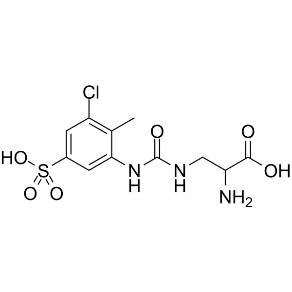
(Rac)-Upacicalcet is the racemate of Upacicalcet (HY-109106). Upacicalcet is a non-peptide calcimimetic that acts as a CaSR agonist (EC50 = 10.8 nM). Upacicalcet reduces serum intact parathyroid hormone (iPTH) and serum Ca2+ levels, reducing hypocalcemia and gastrointestinal complications. Upacicalcet improves vascular calcification and bone disorders in the Adenine (HY-B0152)-induced secondary hyperparathyroidism (SHPT) rat model. Upacicalcet inhibits cortical pore formation and reduces bone fibrosis in rats with chronic kidney disease (CKD). Upacicalcet is useful for studying SHPT.
For research use only. We do not sell to patients.
- Purity : 95.0%
- CAS No.: 2649575-19-7
- Formula: C11H14ClN3O6S
- Molecular Weight:351.76
-
Storage:Powder -20°C, 3 years , 4°C, 2 years ; In solvent -80°C, 6 months , -20°C, 1 month

To place orders, for customer services and technical support, please contact: MedChemExpress USA
Tel: 609-228-6898 E-mail: [email protected] [email protected]


-
 Biological Activity
-
Chemical Information
-
Solvent & Solubility
- Protocols
- Purity & Documentation
- References
-
 Help & FAQs


-
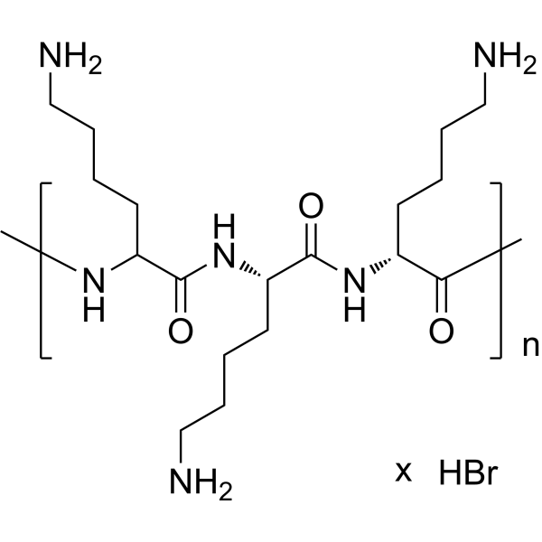
HY-139201A
Poly-D-lysine hydrobromide (MW 70000-150000)CaSR Agonist -
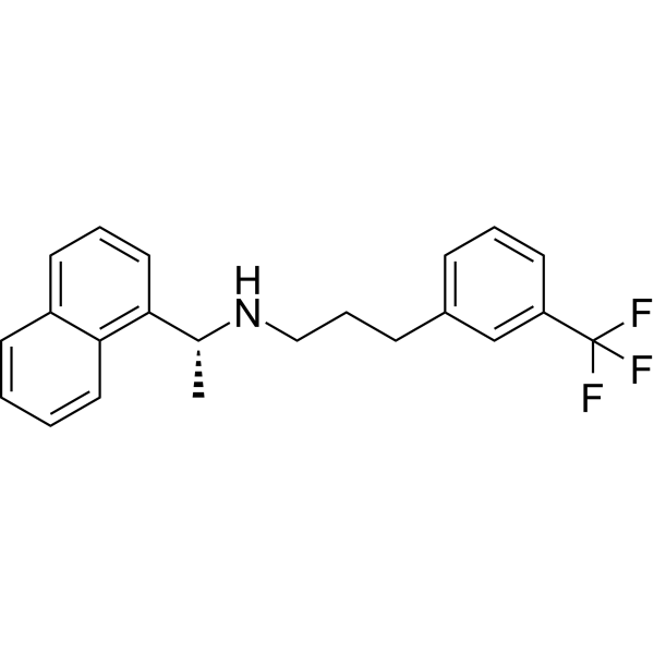
HY-70037
CinacalcetCaSR Agonist -
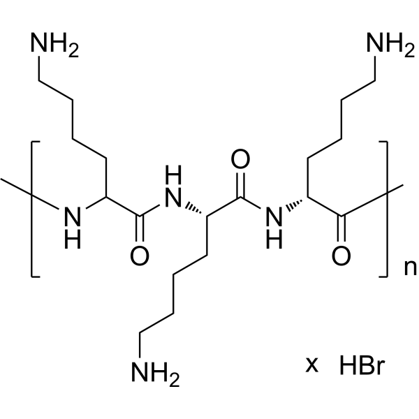
HY-139201
Poly-D-lysine hydrobromide (MW 30000-70000)CaSR Agonist -
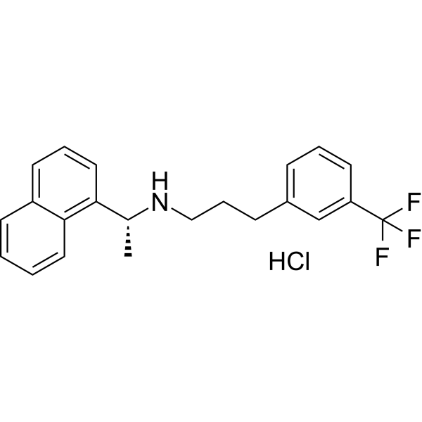
HY-70037A
Cinacalcet hydrochlorideCaSR Agonist -
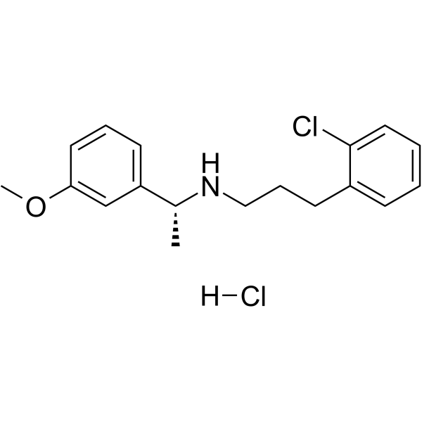
HY-10167A
Tecalcet HydrochlorideCaSR Positive Allosteric Modulator -

HY-22009A
Strontium chloride,anhydrous grade,99.99% metals basisStrontium Salt -
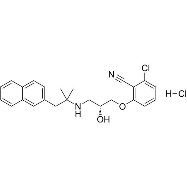
HY-10171
NPS-2143 hydrochlorideCaSR Antagonist -

HY-139201B
Poly-D-lysine hydrobromide (MW 84000)CaSR Agonist -
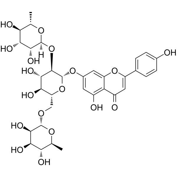
HY-N0546
LigustroflavoneFlavonoid Compound -
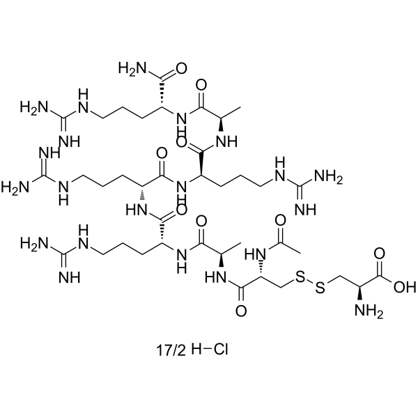
HY-P1955A
Etelcalcetide hydrochlorideCaSR Activator -

HY-139201E
Poly-D-lysine hydrobromide (MW 150000-300000)CaSR Agonist Peptide -

HY-139201D
Poly-D-lysine hydrobromide (MW 4000-15000)Polymer Matrix


Biological Activity
Description


Chemical Information
-
CAS No. 2649575-19-7
-
Appearance Solid
-
Molecular Weight 351.76
-
Formula C11H14ClN3O6S
-
Color White to off-white
-
SMILES
O=C(NCC(N)C(O)=O)NC1=CC(S(=O)(O)=O)=CC(Cl)=C1C
-
Synonyms
(Rac)-SK-1403 free acid; (Rac)-AJT240 free acid; (Rac)-PLS240 free acid
-
Shipping
Room temperature in continental US; may vary elsewhere.
-
Storage
Powder -20°C 3 years 4°C 2 years In solvent -80°C 6 months -20°C 1 month


Solvent & Solubility
In Vitro:
DMSO : 100 mg/mL (284.28 mM; Need ultrasonic; Hygroscopic DMSO has a significant impact on the solubility of product, please use newly opened DMSO)
Please refer to the solubility information to select the appropriate solvent. Once prepared, please aliquot and store the solution to prevent product inactivation from repeated freeze-thaw cycles.
Storage method and period of stock solution: -80°C, 6 months; -20°C, 1 month. When stored at -80°C, please use it within 6 months. When stored at -20°C, please use it within 1 month.
Please refer to the solubility information to select the appropriate solvent. Once prepared, please aliquot and store the solution to prevent product inactivation from repeated freeze-thaw cycles.
Storage method and period of stock solution: -80°C, 6 months; -20°C, 1 month. When stored at -80°C, please use it within 6 months. When stored at -20°C, please use it within 1 month.
Concentration (start) × Volume (start) = Concentration (final) × Volume (final)


Purity & Documentation
-
Data Sheet (271 KB)
-
SDS (252 KB)
- English - EN (252 KB)
- Français - FR (252 KB)
- Deutsch - DE (252 KB)
- Norwegian - NO (252 KB)
- Español - ES (252 KB)
- Swedish - SV (252 KB)
- Italian - IT (252 KB)
- Korean - KR (252 KB)
- Portuguese - PT (252 KB)
-
Handling Instructions (2659 KB)
References
[1]. Goto M, et al. Pharmacological profile of upacicalcet, a novel positive allosteric modulator of calcium-sensing receptor, in vitro and in vivo. Eur J Pharmacol. 2023 Oct 5;956:175936. [Content Brief]
[2]. Sato H, et al. Upacicalcet, a positive allosteric modulator of the calcium-sensing receptor, prevents vascular calcification and bone disorder in a rat adenine-induced secondary hyperparathyroidism model. Bone. 2023 Feb;167:116613. [Content Brief]


Complete Stock Solution Preparation Table
Please refer to the solubility information to select the appropriate solvent. Once prepared, please aliquot and store the solution to prevent product inactivation from repeated freeze-thaw cycles.
Storage method and period of stock solution: -80°C, 6 months; -20°C, 1 month. When stored at -80°C, please use it within 6 months. When stored at -20°C, please use it within 1 month.
| Optional Solvent | Concentration Solvent Mass | 1 mg | 5 mg | 10 mg | 25 mg |
|---|---|---|---|---|---|
| DMSO | 1 mM | 2.8428 mL | 14.2142 mL | 28.4285 mL | 71.0712 mL |
| 5 mM | 0.5686 mL | 2.8428 mL | 5.6857 mL | 14.2142 mL | |
| 10 mM | 0.2843 mL | 1.4214 mL | 2.8428 mL | 7.1071 mL | |
| 15 mM | 0.1895 mL | 0.9476 mL | 1.8952 mL | 4.7381 mL | |
| 20 mM | 0.1421 mL | 0.7107 mL | 1.4214 mL | 3.5536 mL | |
| 25 mM | 0.1137 mL | 0.5686 mL | 1.1371 mL | 2.8428 mL | |
| 30 mM | 0.0948 mL | 0.4738 mL | 0.9476 mL | 2.3690 mL | |
| 40 mM | 0.0711 mL | 0.3554 mL | 0.7107 mL | 1.7768 mL | |
| 50 mM | 0.0569 mL | 0.2843 mL | 0.5686 mL | 1.4214 mL | |
| 60 mM | 0.0474 mL | 0.2369 mL | 0.4738 mL | 1.1845 mL | |
| 80 mM | 0.0355 mL | 0.1777 mL | 0.3554 mL | 0.8884 mL | |
| 100 mM | 0.0284 mL | 0.1421 mL | 0.2843 mL | 0.7107 mL |


(Rac)-Upacicalcet Related Classifications
HY-109106B Related Classifications
 Powered by Bioz