



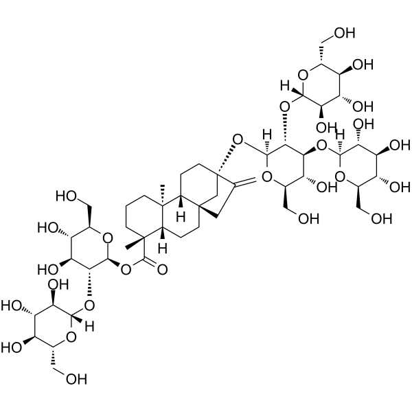
Rebaudioside D
Based on 1 publication(s) in Google Scholar
Rebaudioside D is an orally active sweetener that targets and activates FXR, modulates Acetyl-CoA Carboxylase, and inhibits 3-hydroxy-3-methylglutaryl-CoA reductase. Rebaudioside D regulates bile acid homeostasis and lipid metabolism, reduces the synthesis rates of fatty acids and cholesterol, and exerts multiple effects including anti-adipogenesis, hepatoprotection, anti-steatosis, gut microbiota modulation, enhancement of secondary bile acid metabolism, anti-endotoxin activity, regulation of bile acid transport, and inhibition of bile acid efflux. Rebaudioside D also reduces body weight gain, visceral fat accumulation, hepatic triglyceride and cholesterol accumulation, hepatic lipid peroxidation, and decreases the circulating level of lipopolysaccharide-binding protein. Rebaudioside D additionally enhances the secondary bile acid metabolic pathway of intestinal bacteria, upregulates the gene expression of ileal organic solute transporter α, and downregulates the gene expression of hepatic bile salt export pump. Rebaudioside D does not affect glucose homeostasis, alter total caloric intake or fecal energy excretion, induce weight gain, exacerbate obesity, promote hepatic steatosis, impair brown adipose tissue function, nor change skeletal muscle metabolism-related proteins. Rebaudioside D can be used in diet-induced obesity and obesity-related research.
For research use only. We do not sell to patients.
- Purity : 98.48%
- CAS No.: 63279-13-0
- Formula: C50H80O28
- Molecular Weight:1129.15
-
Storage:
4°C, protect from light
* In solvent : -80°C, 6 months; -20°C, 1 month (protect from light)

To place orders, for customer services and technical support, please contact: MedChemExpress USA
Tel: 609-228-6898 E-mail: [email protected] [email protected]


-
 Biological Activity
-
Chemical Information
-
Solvent & Solubility
- Protocols
- Purity & Documentation
- References
-
 Help & FAQs


-
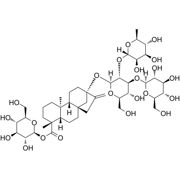
HY-N0467
Rebaudioside CTAS1R2/TAS1R3 Activator -
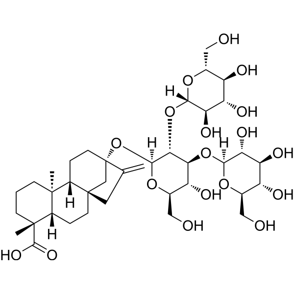
HY-N6808
Rebaudioside BSweetener -
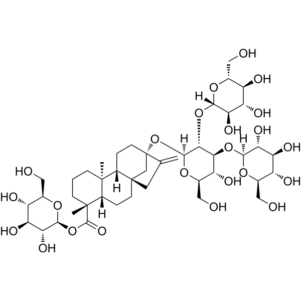
HY-N0466
Rebaudioside Aα-Glucosidase Inhibitor -
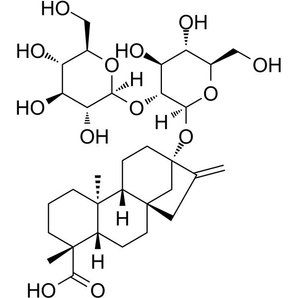
HY-N2547
SteviolbiosideSweetener -
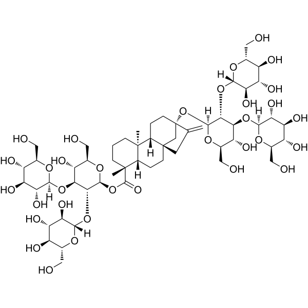
HY-N6833
Rebaudioside MSweetener -
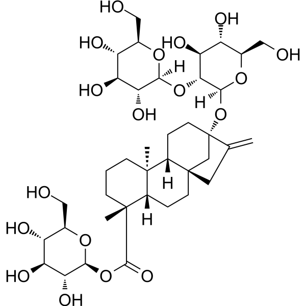
HY-N0669
Steviosidesweetener -
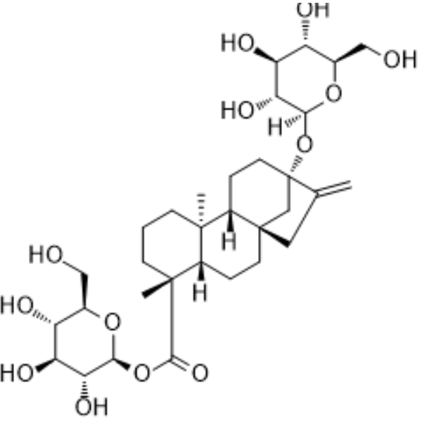
HY-N0668
RubusosideSolubilizing Agent -
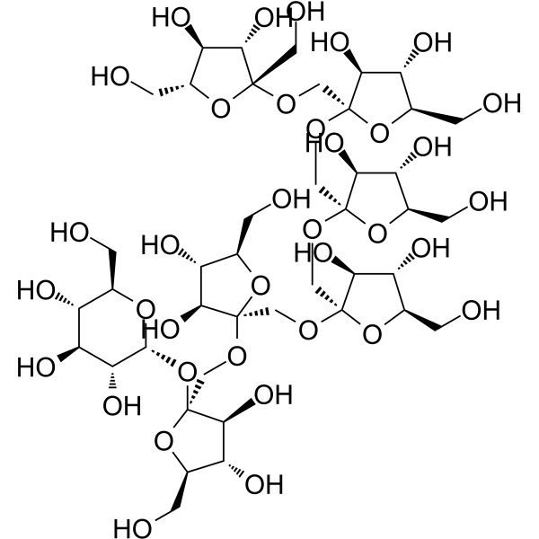
HY-N6837
Fructo-oligosaccharide DP7/GF6 -

HY-N0468R
Rebaudioside D (Standard)Sweetener -

HY-N6808R
Rebaudioside B (Standard)Sweetener -

HY-N0466R
Rebaudioside A (Standard)Standard of Rebaudioside A -

HY-N0467R
Rebaudioside C (Standard)Endogenous Metabolite
Publications Citing Use of MedChemExpress (MCE) Rebaudioside D
More

Biological Activity
Description
In Vivo
Rebaudioside D (0.2 g/L (0.02%); ad libitum access via drinking water; continuous administration for 16 weeks) does not induce obesity, hepatic steatosis or brown adipose tissue dysfunction, nor does it interfere with skeletal muscle metabolism in mice fed a regular diet[1].
Rebaudioside D (0.2 g/L (0.02%); free access via drinking water; ad libitum access; for 16 consecutive weeks) does not exacerbate obesity or hepatic steatosis in male mice fed a high-fat diet. It maintains the expression of thermogenic proteins in brown adipose tissue, reduces several serum lipid and blood glucose parameters, and shifts the gut microbiota toward a more beneficial profile[2].
Rebaudioside D (0.2 g/L (0.02%); ad libitum access via drinking water for 16 weeks) does not increase the expression of hepatic lipogenic proteins in female mice fed a high-fat diet[2].
MedChemExpress (MCE) has not independently confirmed the accuracy of these methods. They are for reference only.
-
Animal Model:C57BL/6 J (8-week-old male, high-fat/high-sucrose diet-induced obesity)[1]
-
Dosage:50 mg/kg
-
Administration:p.o.; daily; 12 weeks
-
Result:Reduced weight gain significantly at weeks 8 and 10 through 12 compared to HFHS-fed vehicle controls.
Decreased the sum of all white adipose tissues, with a specific reduction in epididymal adipose tissue weight.
Reduced liver triglyceride and cholesterol levels, and decreased hepatic lipid peroxidation (measured by TBARS).
-
Animal Model:C57BL/6J (male, 8-10 weeks old)[2]
-
Dosage:0.2 g/L (0.02%)
-
Administration:in drinking water; ad libitum; daily for 16 weeks
-
Result:Showed no significant difference in oral glucose tolerance test AUC, with a non-significant decreasing trend in fasting blood glucose levels compared to HFD controls.
Had a relative abundance of Bacillota phylum of 75.3%, relative abundance of Akkermansia of 8.07%, and relative abundance of Faecalibaculum of 13.57%.
Identified short-chain fatty acid-producing genera including Faecalibaculum, Acetatifactor, and Parabacteroides as biomarkers via LEfSe analysis.


Chemical Information
-
CAS No. 63279-13-0
-
Appearance Solid
-
Molecular Weight 1129.15
-
Formula C50H80O28
-
Color White to off-white
-
SMILES
O[C@H]1[C@H](O)[C@@H](CO)O[C@@H](OC([C@@]2(C)[C@](CC[C@]3(CC4=C)[C@@]5([H])CC[C@]4(O[C@]6([H])O[C@H](CO)[C@@H](O)[C@H](O[C@]7([H])O[C@H](CO)[C@@H](O)[C@H](O)[C@H]7O)[C@H]6O[C@@]8([H])[C@H](O)[C@@H](O)[C@H](O)[C@@H](CO)O8)C3)([H])[C@@]5(C)CCC2)=O)[C@@H]1O[C@@]9([H])[C@H](O)[C@@H](O)[C@H](O)[C@@H](CO)O9
-
Structure Classification
-
Initial Source
-
Shipping
Room temperature in continental US; may vary elsewhere.
-
Storage
4°C, protect from light
* In solvent : -80°C, 6 months; -20°C, 1 month (protect from light)


Publications (1)
-
Journal Impact Factor
-
Most Recent


Solvent & Solubility
In Vitro:
DMSO : 100 mg/mL (88.56 mM; Need ultrasonic; Hygroscopic DMSO has a significant impact on the solubility of product, please use newly opened DMSO)
H2O : < 0.1 mg/mL (insoluble)
Please refer to the solubility information to select the appropriate solvent. Once prepared, please aliquot and store the solution to prevent product inactivation from repeated freeze-thaw cycles.
Storage method and period of stock solution: -80°C, 6 months; -20°C, 1 month (protect from light). When stored at -80°C, please use it within 6 months. When stored at -20°C, please use it within 1 month.
Please refer to the solubility information to select the appropriate solvent. Once prepared, please aliquot and store the solution to prevent product inactivation from repeated freeze-thaw cycles.
Storage method and period of stock solution: -80°C, 6 months; -20°C, 1 month (protect from light). When stored at -80°C, please use it within 6 months. When stored at -20°C, please use it within 1 month.
Concentration (start) × Volume (start) = Concentration (final) × Volume (final)
In Vivo:
Select the appropriate dissolution method based on your experimental animal and administration route.
- For the following dissolution methods, please ensure to first prepare a clear stock solution using an In Vitro approach and then sequentially add co-solvents:
- To ensure reliable experimental results, the clarified stock solution can be appropriately stored based on storage conditions. As for the working solution for In Vivo experiments, it is recommended to prepare freshly and use it on the same day.
- The percentages shown for the solvents indicate their volumetric ratio in the final prepared solution. If precipitation or phase separation occurs during preparation, heat and/or sonication can be used to aid dissolution.
Add each solvent one by one: 10% DMSO 40% PEG300 5% Tween-80 45% Saline
Solubility: ≥ 2.5 mg/mL (2.21 mM); Clear solution
This protocol yields a clear solution of ≥ 2.5 mg/mL (saturation unknown).
Taking 1 mL working solution as an example, add 100 μL DMSO stock solution (25.0 mg/mL) to 400 μL PEG300, and mix evenly; then add 50 μL Tween-80 and mix evenly; then add 450 μL Saline to adjust the volume to 1 mL.
Preparation of Saline: Dissolve 0.9 g sodium chloride in ddH₂O and dilute to 100 mL to obtain a clear Saline solution.
Add each solvent one by one: 10% DMSO 90% (20% SBE-β-CD in Saline)
Solubility: ≥ 2.5 mg/mL (2.21 mM); Clear solution
This protocol yields a clear solution of ≥ 2.5 mg/mL (saturation unknown).
Taking 1 mL working solution as an example, add 100 μL DMSO stock solution (25.0 mg/mL) to 900 μL 20% SBE-β-CD in Saline, and mix evenly.
Preparation of 20% SBE-β-CD in Saline (4°C, storage for one week): 2 g SBE-β-CD powder is dissolved in 10 mL Saline, completely dissolve until clear.
In Vivo Dissolution Calculator
Please enter the basic information of animal experiments:
-
-
-
-
Recommended: Prepare an additional quantity of animals to account for potential losses during experiments.
Please enter your animal formula composition:
-
%DMSO +
Recommended: Keep the proportion of DMSO in working solution below 2% if your animal is weak.
-
%+
-
+%Tween-80 + +
-
%Saline +
The co-solvents required include: DMSO, . All of co-solvents are available by MedChemExpress (MCE). , Tween 80. All of co-solvents are available by MedChemExpress (MCE).
Working solution concentration: 0.22 mg/mL
Method for preparing stock solution: mg drug dissolved in μL DMSO. Stock solution concentration: mg/mL. * In solvent : -80°C, 6 months; -20°C, 1 month (protect from light)
1. Take μL DMSO stock solution;
2. Add μL .
μL , mix evenly;
3. Then add μL Tween 80, mix evenly;
4. Then add μL
Please ensure that the stock solution in the first step is dissolved to a clear state, and add co-solvents in sequence. You can use ultrasonic heating (ultrasonic cleaner, recommended frequency 20-40 kHz), vortexing, etc. to assist dissolution.

Protocols
-
RT-PCR
Reverse transcription technology uses RNA as a template to synthesize DNA. RT-PCR is simple, specific and sensitive, and can be used to detect gene expression levels and expression differences in cells; detect RNA virus content; clone cDNA sequences of specific genes.
-
3T3-L1 preadipocyte-to-adipocyte differentiation
3T3-L1 preadipocytes are induced to differentiate after growth arrest using adipogenic media containing insulin, dexamethasone, and IBMX; differentiation is assessed by lipid-droplet accumulation, triglyceride increase, Oil Red O staining, and adipocyte-marker induction such as PPARγ and C/EBPα.
-
RNA extraction experimental
By lysing cells, releasing RNA, and removing impurities such as proteins and DNA, high-purity RNA products are finally obtained. The commonly used traditional method is the guanidine isothiocyanate/phenol/chloroform method (Trizol), which is suitable for a variety of animal materials including animal tissues, microorganisms, cultured cells, etc., and most plant materials.
-
Large-size fat particle sorting
Large-size fat particle sorting is widely used to isolate cells up to 200 μm in diameter. Single-cell flow sorting will allow greater insight into adipocyte heterogeneity by identifying gene expression, protein composition, and metabolic signatures at the single-cell level.
-
Research Protocol for Microbiome Analysis
Microbiome analysis characterizes microbial communities in biological or environmental samples by measuring community composition, diversity, taxonomic structure, functional potential, and associations with host or environmental phenotypes. 16S rRNA gene amplicon sequencing is commonly used for bacterial and archaeal taxonomic profiling, while shotgun metagenomics provides higher taxonomic resolution and direct functional information, including microbial genes, pathways, viruses, fungi, and antimicrobial-resistance genes when sequencing depth and host-DNA contamination are adequately controlled. Microbiome results are strongly affected by sample collection, storage, DNA extraction, contamination, sequencing method, reference database, and bioinformatic pipeline; therefore, standardized protocols, negative controls, mock communities, and transparent analysis workflows are required. Unresolved issues include low-biomass contamination, compositional-data bias, inconsistent species-level c
-
Ferroptosis Solutions
Ferroptosis is an iron-dependent, non-apoptotic form of regulated cell death characterized by lethal lipid peroxidation and sensitivity to suppression by iron chelators or lipophilic radical-trapping antioxidants. The core pathway links cystine uptake through system Xc−, glutathione availability, GPX4-dependent detoxification of phospholipid hydroperoxides, iron-dependent oxidative reactions, and polyunsaturated-phospholipid metabolism into a cell-death program that is biochemically and morphologically distinct from apoptosis, necrosis, and autophagy. The ferroptosis pathway is experimentally linked to phenotype through chemical and genetic perturbation. Erastin induces ferroptosis by inhibiting cystine uptake through system Xc− and weakening antioxidant defenses, while GPX4 inhibition or depletion causes lipid peroxide accumulation and ferroptotic cancer-cell death. ACSL4 and oxidizable arachidonoyl- or adrenoyl-containing phosphatidylethanolamines shape ferroptosis sensitivity by con
-
Research Protocol for Metabolic Diseases
AMP-activated protein kinase, AMPK, is a conserved cellular energy sensor that responds to reduced cellular energy status and coordinates metabolism by increasing ATP-generating catabolic pathways while suppressing ATP-consuming anabolic processes. In metabolic disease research, the AMPK pathway is experimentally relevant because it regulates hepatic lipid synthesis, fatty acid oxidation, glucose production, skeletal-muscle glucose disposal, mTORC1-linked biosynthesis, autophagy, mitochondrial homeostasis, and whole-body energy balance. The central pathway logic is that energy stress, metformin, exercise-like stimulation, or direct AMPK activators increase AMPKα Thr172 phosphorylation and downstream substrate phosphorylation, including ACC and RAPTOR. Phosphorylation of ACC suppresses lipogenesis and supports fatty acid oxidation, whereas phosphorylation of RAPTOR suppresses mTORC1 signaling and links cellular energy status to growth and protein synthesis control. The pathway is linked
-
Real Time qPCR (Q-PCR)
Real-time quantitative PCR (qPCR) quantifies an amplifiable nucleic-acid target by monitoring fluorescence during PCR cycling rather than measuring product only after amplification. The increase in fluorescence tracks accumulation of PCR product, and the quantification cycle (Cq; historically also Ct/CP) is related to the initial amount of target: samples containing more starting target generally reach the defined fluorescence threshold in fewer cycles.
-
Lipid Droplets: Oil Red O/Sudan Dye Lipid Staining
Lipid droplets are intracellular organelles with a neutral-lipid core that stores triacylglycerols and sterol esters, and Oil Red O or Sudan dyes detect these hydrophobic lipid deposits by partitioning into retained lipids in fresh or frozen specimens. Oil Red O stains neutral triglycerides and lipids in frozen tissue sections or air-dried cytologic preparations, while Sudan Black B has also been used as a histochemical fat stain for lipid-rich tissue structures.
-
Liver Histomorphometry
Liver histomorphometry is a quantitative histological approach used to measure structural alterations in hepatic tissue, including parenchymal loss, steatosis, fibrosis, and vascular remodeling, by combining stained tissue section analysis with stereological or computerized image-based measurements. Classical morphometric frameworks quantify volume fractions of liver compartments and fibrotic regions using systematic sampling and image analysis, enabling objective comparison of pathological changes across experimental groups. These approaches are widely applied in liver cirrhosis and fibrosis studies to reduce subjectivity in histological scoring and improve reproducibility of tissue evaluation. Recent methodological advances integrate automated image analysis and radiomics-based extraction of histological features from standard liver stains (e. g. , H&E and fibrotic stains), enabling quantitative correlation between morphometric features and fibrosis stages in non-alcoholic fatty live


Purity & Documentation
-
Data Sheet (285 KB)
-
SDS (394 KB)
- English - EN (394 KB)
- Français - FR (394 KB)
- Deutsch - DE (394 KB)
- Norwegian - NO (394 KB)
- Español - ES (394 KB)
- Swedish - SV (394 KB)
- Italian - IT (394 KB)
- Korean - KR (394 KB)
- Portuguese - PT (394 KB)
-
Handling Instructions (2659 KB)
References
[1]. Morissette A, et al. Rebaudioside D decreases adiposity and hepatic lipid accumulation in a mouse model of obesity. Sci Rep. 2024;14(1):3077. Published 2024 Feb 6. [Content Brief]
[2]. Lim S, et al. Rebaudioside D and M, the next-generation sugar substitutes, do not exacerbate metabolic dysfunction in high-fat diet mice. Food Chem X. 2025;29:102679. Published 2025 Jun 19. [Content Brief]


Complete Stock Solution Preparation Table
Please refer to the solubility information to select the appropriate solvent. Once prepared, please aliquot and store the solution to prevent product inactivation from repeated freeze-thaw cycles.
Storage method and period of stock solution: -80°C, 6 months; -20°C, 1 month (protect from light). When stored at -80°C, please use it within 6 months. When stored at -20°C, please use it within 1 month.
| Optional Solvent | Concentration Solvent Mass | 1 mg | 5 mg | 10 mg | 25 mg |
|---|---|---|---|---|---|
| DMSO | 1 mM | 0.8856 mL | 4.4281 mL | 8.8562 mL | 22.1405 mL |
| 5 mM | 0.1771 mL | 0.8856 mL | 1.7712 mL | 4.4281 mL | |
| 10 mM | 0.0886 mL | 0.4428 mL | 0.8856 mL | 2.2141 mL | |
| 15 mM | 0.0590 mL | 0.2952 mL | 0.5904 mL | 1.4760 mL | |
| 20 mM | 0.0443 mL | 0.2214 mL | 0.4428 mL | 1.1070 mL | |
| 25 mM | 0.0354 mL | 0.1771 mL | 0.3542 mL | 0.8856 mL | |
| 30 mM | 0.0295 mL | 0.1476 mL | 0.2952 mL | 0.7380 mL | |
| 40 mM | 0.0221 mL | 0.1107 mL | 0.2214 mL | 0.5535 mL | |
| 50 mM | 0.0177 mL | 0.0886 mL | 0.1771 mL | 0.4428 mL | |
| 60 mM | 0.0148 mL | 0.0738 mL | 0.1476 mL | 0.3690 mL | |
| 80 mM | 0.0111 mL | 0.0554 mL | 0.1107 mL | 0.2768 mL |


Rebaudioside D Related Classifications
HY-N0468 Related Classifications
 Powered by Bioz
Keywords
- Rebaudioside D
- 63279-13-0
- FXR
- Acetyl-CoA Carboxylase
- acetyl-CoA carboxylase
- 3-hydroxy-3-methylglutaryl-CoA reductase
- lipid metabolism
- Faecalibaculum rodentium
- HFHS-fed obese mice
- diet-induced obesity
- high-fat diet-fed male mice
- chow diet-fed mice
- bile acid homeostasis
- farnesoid X receptor
- Inhibitor
- inhibitor
- inhibit